RB1 / Retinoblastoma-associated protein · IHC design guide

Design Immunohistochemistry for RB1

Plan chromogenic RB1 IHC in paraffin sections using a nuclear readout (HPA tissue IHC). Compare cell types with tissue controls, and start the catalog antibody at 2–5 μg/ml (HPA tissue IHC; datasheet A00039-3).

Evidence assembled Oct 2026 · For research use; verify linked source records and product datasheet before use
Immunohistochemistry protocol sheet for RB1 (IHC for RB1): expected localisation Predominantly nuclear in tissue (HPA tissue IHC), antibody A00039-3, validated IHC image, and IHC protocol steps
Printable RB1 IHC protocol sheet — expected localisation Predominantly nuclear in tissue (HPA tissue IHC), antibody A00039-3, controls and protocol steps. Open the full RB1 IHC guide →

RB1 Immunohistochemistry Experimental Design Guide

Expected localisation, validated protocols, controls and antibodies — the at-a-glance facts below, then the full design guide.

Must know before staining
Expected localisation Predominantly nuclear in tissue (HPA tissue IHC)
Staining pattern Nuclear staining in most tissues, including retina (HPA tissue IHC)
Antigen retrieval EDTA pH 8.0 HIER, heat-mediated (datasheet A00039-3)
Positive control ⓘ Adrenal gland+4 more · see all
Negative control ⓘ Adipose tissue+1 more · see all
Important caveats
Reasons your staining may differ from the expected pattern.
Fixation Keep fixation consistent across paraffin sections (standard IHC practice; not target-specific)
Caveat Some cell types have no detectable staining (HPA tissue IHC)
Regulation Phosphorylation may shift nuclear signal (UniProt)
Isoform / epitope 0 annotated isoforms; no extracellular segment (UniProt)
Section 1

Recommended RB1 IHC & IF Protocols

The catalog antibody’s IHC-P protocol (datasheet: A00039-3) is accompanied by four published RB1 IHC protocols (PMC10530403; PMC10253801; PMC3246124; PMC5992011).

Recommended immunohistochemistry (IHC-P) protocol parameters
SampleParaffin-embedded human colorectal adenocarcinoma tissue; fixative not specified (datasheet A00039-3)
FixationImage fixative and duration unreported (datasheet A00039-3); verify before use.
Sectioning4–5 µm sections on charged slides (standard)
DeparaffinisationXylene, graded ethanol series to water (standard)
Antigen retrievalHeat retrieval: EDTA pH 8.0 (datasheet A00039-3); 20 min, 95–100 °C (standard)
Peroxidase block3% H2O2, 10 min, room temperature (standard)
Blocking10% goat serum (datasheet A00039-3)
Primary antibodyRabbit anti-RB1, 2-5 μg/ml recommended; image 1:100 (datasheet A00039-3)
Primary incubationOvernight at 4 °C (datasheet A00039-3)
DetectionHRP-conjugated secondary, DAB chromogen (datasheet A00039-3)
CounterstainHematoxylin, blue, dehydrate and mount (standard)
Expected resultRB1-positive staining in glandular cells of adrenal gland (HPA tissue IHC: High). HPA tissue profile: Nuclear expression in most tissues, including retina. No signal in the no-primary control.
💡Decision noteStart with heat-mediated EDTA pH 8.0 retrieval for the catalog antibody (datasheet: A00039-3); the published protocols used their own retrieval conditions (PMC10530403; PMC10253801; PMC3246124).
Section 2

What Is the Expected RB1 Staining Pattern?

RB1 should show predominantly nuclear staining across many tissues, including retina (HPA tissue IHC: nuclear expression in most tissues). Strong examples include retinal ganglion cells, cerebellar Purkinje cells and breast glandular cells (HPA tissue IHC: High). HPA rates the tissue pattern Supported, with medium consistency between antibody staining and RNA data (HPA tissue IHC). RB1 has no transmembrane segment, consistent with an intracellular pattern (UniProt P06400 topology).

What am I looking at on my slide?
Distinct nuclear staining in retinal ganglion cells, with little surrounding background.This matches a high expressing cell population and the predominant tissue compartment (HPA tissue IHC: High in retinal ganglion cells; nuclear expression in most tissues). Judge the stained cells within their tissue context: HPA also reports high staining in several other cell populations, while expression is lower or undetected in some populations (HPA tissue IHC).
Predominantly cytoplasmic staining with weak or absent nuclei.Recheck specificity and detection because the expected tissue pattern is nuclear (HPA tissue IHC). Cytoplasmic RB1 remains biologically possible: UniProt lists cytoplasm and reports cytoplasmic localization when hyperphosphorylated, by similarity (UniProt P06400 subcellular location). A routine RB1 stain alone cannot establish phosphorylation state (general IHC interpretation practice).
Strong staining in adipocytes or hippocampal glial cells.These populations are reported as Not detected (HPA tissue IHC). Unexpected signal warrants checks for antibody cross-reactivity, endogenous detection activity or pigment before assigning it to RB1 (general IHC interpretation practice). HPA's Supported rating and medium RNA–staining consistency make a single discordant cell population worth investigating, not proof that every stained cell is false positive (HPA tissue IHC).
Broad, diffuse color across cells and extracellular areas, without clear nuclear contrast.This is less convincing than the predominantly nuclear pattern (HPA tissue IHC). Inspect a no-primary control and review blocking, washing and detection conditions for nonspecific or endogenous signal (general chromogenic IHC practice). Interpret the result against cell-specific morphology and a known positive region rather than scoring diffuse color as RB1 expression (general IHC interpretation practice).
No nuclear signal in retinal ganglion cells or another HPA high population.An absent signal in a documented high population raises concern about assay performance (HPA tissue IHC: High in retinal ganglion cells, Purkinje cells and several other listed populations). Check a validated positive control, antibody dilution, retrieval, detection reagents and counterstain visibility (general IHC practice). An individual negative field does not, by itself, establish RB1 loss throughout a specimen (general IHC interpretation practice).
💡Expected RB1 appearanceCall a result positive when identifiable cells show clear, predominantly nuclear chromogenic staining, especially in an HPA high population such as retinal ganglion cells (HPA tissue IHC); diffuse background or dominant staining in HPA Not detected populations warrants investigation before scoring (HPA tissue IHC; general IHC interpretation practice).
How each factor affects the staining
Cell population selected for the positive controlHPA reports High staining in retinal ganglion cells, Purkinje cells, melanocytes, decidual cells and several glandular populations; adipocytes and hippocampal glial cells are Not detected (HPA tissue IHC). Select and score the documented cell population, since a whole tissue label can obscure differences among its cells (general IHC interpretation practice).
Expected compartment and RB1 stateNuclear expression predominates in tissue IHC (HPA tissue IHC). UniProt also lists cytoplasm and links hyperphosphorylation to cytoplasmic localization by similarity; acetylation by KAT2B/PCAF is required for nuclear localization during keratinocyte differentiation (UniProt P06400 subcellular location). These annotations support cautious review of cytoplasmic signal, without identifying its cause in a stained section.
Antibody validationHPA lists IHC Supported for HPA050082 and CAB016687, while CAB000095 has no IHC status listed (HPA antibody validation). ICC Supported status for an antibody does not supply an IHC validation result (HPA antibody validation). Compare a new assay's distribution with the documented tissue pattern and its own controls (HPA tissue IHC; general IHC practice).
IF/ICC Q: Where should RB1 appear in cells?A: Mainly in the nucleoplasm (HPA subcellular ICC-IF: supported). An additional primary cilium transition-zone location is marked uncertain (HPA subcellular ICC-IF). Treat that additional location as tentative; the tissue IHC call remains predominantly nuclear (HPA tissue IHC). IF/ICC images describe subcellular localization and do not define a paraffin-section staining condition (HPA subcellular ICC-IF; general IHC practice).
Target-specific preparation evidenceTarget-specific fixation effects are not established by the supplied assay evidence. Verify with a matched IHC source before attributing a result to fixation.
Why is my staining missing, weak or wrong?
SituationLikely causeNext action
Nuclei are blank in a known positive population.The observed absence conflicts with a documented High cell population, but does not identify which assay step failed (HPA tissue IHC).Run a documented positive population alongside the specimen; verify primary-antibody preparation, retrieval, chromogenic detection and counterstain readability (HPA tissue IHC; general IHC practice). Interpret a failed positive control as an unresolved assay result (general IHC practice).
All compartments stain, including areas without identifiable cells.A diffuse pattern does not match predominantly nuclear tissue expression and may reflect nonspecific or endogenous signal (HPA tissue IHC; general IHC practice).Examine a no-primary control, reduce background through appropriate blocking and washing, and review detection conditions (general chromogenic IHC practice). Reassess nuclear contrast only after the control distinguishes tissue signal from background.
Adipocytes or hippocampal glial cells appear strongly positive.Those cell populations are Not detected in HPA tissue IHC; cross-reactivity or endogenous chromogenic activity is possible, but the image alone cannot identify the cause (HPA tissue IHC; general IHC practice).Confirm cell identity, compare a no-primary control and check a documented high population in the same run (HPA tissue IHC; general IHC practice). If discordance persists, compare staining with an independently IHC-supported antibody (HPA antibody validation; general IHC practice).
Cytoplasmic color overwhelms the nuclei.This departs from the usual nuclear tissue profile (HPA tissue IHC). UniProt's cytoplasmic assignment and hyperphosphorylation note, marked by similarity, leave a biological explanation possible (UniProt P06400 subcellular location).Check nuclear counterstain, morphology and no-primary background; compare the distribution with a documented high population (HPA tissue IHC; general IHC practice). Do not label the cytoplasmic signal as hyperphosphorylated RB1 from this stain alone (general IHC interpretation practice).
Signal varies sharply between neighboring cell types.Cell-specific variation may be expected: HPA reports High, Low and Not detected populations, with medium consistency between antibody staining and RNA data (HPA tissue IHC).Score identifiable cell types separately and record compartment and intensity for each (general IHC interpretation practice). Use HPA's cell-level observations as reference points, then check controls before calling an unexpected population positive or negative (HPA tissue IHC; general IHC practice).
A proposed ciliary focus appears in an IF/ICC image.HPA marks the primary cilium transition-zone location uncertain, while its supported main IF/ICC location is nucleoplasm (HPA subcellular ICC-IF).Treat the focus as tentative and assess whether the nucleoplasmic pattern is present (HPA subcellular ICC-IF). Keep its interpretation separate from paraffin-section chromogenic scoring, whose expected tissue pattern is predominantly nuclear (HPA tissue IHC; general IHC interpretation practice).

Sample controls for RB1 IHC & IF

🧪Run breast first and expect staining in its glandular cells (High; HPA: breast glandular cells). Use adipose tissue as the negative and assess adipocytes on the breast slide as internal negatives if present (HPA: adipose adipocytes Not detected).
Positive control tissue: Adrenal gland (Glandular cells, HPA High)
Negative control tissue: Adipose tissue (HPA Not detected)
ICC-IF cell lines (HPA subcellular resource): HPA ICC-IF images show RB1 in A-431, U-251MG, U2OS, ASC52telo, RPTEC/TERT1, hTERT-RPE1 (serum starved), with annotated localisation: Nucleoplasm (supported) (HPA subcellular).
Technical controls: Include a no-primary, secondary-only control, a matched rabbit IgG isotype control, and an RB1-knockout sample as a biological negative (hero caption: rabbit primary; standard IHC practice). Quench endogenous peroxidase and check background in the breast section before interpreting DAB staining (hero caption: HRP/DAB detection; standard IHC practice).
⚠️Feasibility: A target-specific fixation window or fixation effect is unreported, and the selected A00039-3 paraffin-section caption does not state the fixative (hero caption: fixative not stated). That caption uses heat-mediated EDTA retrieval at pH 8.0, but does not establish that retrieval is required for all RB1 antibodies (hero caption: EDTA retrieval). Frozen sections are not established as easier; ICC-IF images support predominantly nucleoplasmic localization, while peroxidase background remains a practical consideration for the breast IHC slide (HPA: nucleoplasm supported; hero caption: HRP/DAB detection; standard IHC practice).

HPA tissue IHC evidence for RB1

Comprehensive Human Protein Atlas IHC scoring per tissue (reliability: Supported — Medium consistency between antibody staining and RNA expression data.). Rows are taken directly from the HPA tissue chart — click any row's HPA link to view the source.

Positive expression · recommended positive controls

TissueCell typeLevelEvidenceSource
Adrenal gland Glandular cells High Protein (IHC) HPA →
Breast Glandular cells High Protein (IHC) HPA →
Cerebellum Purkinje cells High Protein (IHC) HPA →
Cervix Glandular cells High Protein (IHC) HPA →
Lymph node Non-germinal center cells High Protein (IHC) HPA →

Undetected expression · recommended negative controls

TissueCell typeLevelEvidenceSource
Adipose tissue Adipocytes Not detected Protein (IHC) HPA →
Hippocampus Glial cells Not detected Protein (IHC) HPA →
Section 3

Advanced RB1 IHC Tips

Troubleshoot RB1 staining in paraffin sections by checking retrieval, nuclear localisation, controls, and scoring before interpreting loss or redistribution.

Which retrieval conditions should I use if RB1 nuclear staining is weak?
Use heat-mediated retrieval in EDTA at pH 8.0 for this paraffin-section assay (datasheet A00039-3). The selected image used that retrieval before an overnight primary incubation at 4°C, but its caption does not report heating time or temperature (datasheet A00039-3). If nuclear staining remains weak, vary retrieval duration in small steps while keeping buffer and detection conditions fixed, and compare a known-positive section alongside each run (standard IHC practice; HPA: nuclear expression in most tissues). Check tissue integrity and background after each adjustment, because excessive heating can make apparent signal harder to interpret (standard IHC practice).
How should I investigate variable RB1 staining across differently fixed sections?
Target-specific RB1 sensitivity to fixation is unknown from the supplied evidence; the selected paraffin-section caption does not state a fixative (datasheet A00039-3). Record each specimen’s fixative, fixation duration, processing history, and section age before comparing nuclear staining across batches (standard IHC practice). Keep section thickness, EDTA retrieval at pH 8.0, primary incubation, and chromogenic development consistent while assessing a known-positive control with each batch (datasheet A00039-3; standard IHC practice). If results still differ, treat fixation as a possible technical variable requiring a controlled comparison, rather than assigning the difference to RB1 biology (standard IHC practice).
What should I check when RB1 staining appears cytoplasmic instead of nuclear?
First check whether nuclei in an expected-positive control stain, since tissue IHC shows predominantly nuclear RB1 expression across most tissues (HPA: tissue IHC profile). RB1 is annotated in both nucleus and cytoplasm, and cytoplasmic localisation when hyperphosphorylated is reported by similarity, so a cytoplasmic pattern alone cannot establish phosphorylation state (UniProt P06400). Compare the suspect cells with adjacent morphology, a nuclear counterstain, and a section lacking primary antibody to assess compartment boundaries and diffuse chromogen (standard IHC practice). Document nuclear and cytoplasmic staining separately, and investigate a reproducible shift with an independent phospho-specific assay if that distinction matters (standard IHC practice; UniProt P06400).
Could phosphorylation or an unknown epitope explain inconsistent RB1 staining?
The supplied record lists 0 annotated isoforms and multiple modified residues, including phosphorylation sites, but does not identify this antibody’s epitope (UniProt P06400; datasheet A00039-3). Therefore, a change in chromogenic staining cannot by itself distinguish protein loss from altered epitope accessibility or modification (standard IHC interpretation; UniProt P06400). Keep EDTA retrieval at pH 8.0 and the same antibody conditions when comparing specimens, then assess whether nuclear counterstaining and positive controls remain intact (datasheet A00039-3; standard IHC practice). If the biological question concerns phosphorylation, use a separately validated modification-specific reagent and controls rather than inferring it from total-RB1 staining (standard IHC practice).
How can I corroborate an RB1 IHC pattern with multiplex immunofluorescence?
For a separate IF/ICC assay, pair RB1 with a marker validated for the cell population being examined and inspect nuclear overlap with a DNA counterstain (standard IF practice; HPA: mainly nucleoplasmic RB1). Choose spectrally separated fluorophores after checking unstained tissue autofluorescence, and reserve a brighter, cleaner channel for a weak RB1 signal (standard IF practice). Because RB1 has no transmembrane segment and is mainly nucleoplasmic, test permeabilisation sufficient to expose an intracellular nuclear epitope while preserving morphology (UniProt P06400 topology; HPA: subcellular localisation; standard IF practice). Validate the IF antibody and conditions independently; the selected antibody evidence describes paraffin-section chromogenic IHC (datasheet A00039-3).
How do I separate weak nuclear RB1 staining from chromogenic background?
Compare the sample with a section lacking primary antibody and with a consistently stained positive control, then judge signal within intact nuclei (standard IHC practice; HPA: predominantly nuclear tissue profile). The selected paraffin-section image used 10% goat serum blocking, primary antibody at 1:100 overnight at 4°C, and peroxidase-based DAB detection (datasheet A00039-3). Apply an endogenous peroxidase block and check DAB development time as general controls for diffuse brown staining, especially when background spans nuclei and cytoplasm (standard IHC practice). Titrate antibody and detection conditions against nuclear contrast without treating the image’s settings as universal optima (standard IHC practice; datasheet A00039-3).
How should I score heterogeneous RB1 staining in paraffin sections? ⚠ ANSWER MARKED FOR VERIFICATION
Score the percentage of evaluable cells with nuclear RB1 staining and record nuclear intensity on a consistent scale; an H-score can combine percentage and intensity (standard IHC practice; HPA: predominantly nuclear tissue profile). Define the cell population and exclude folds, necrosis, and poorly preserved areas before counting (standard IHC practice). Normalise positive nuclei or H-score to the number of evaluable nuclei within the same annotated compartment, and use area-normalised positive-cell density only when reporting cells per mm² (standard IHC practice). Hold retrieval, DAB development, imaging, and scoring thresholds constant across samples, with a control section in each staining run (datasheet A00039-3; standard IHC practice).
When does absent or misplaced RB1 staining indicate a real biological difference?
Interpret absent nuclear staining only after an on-run positive control retains nuclear signal and the specimen has preserved, countable nuclei (standard IHC practice; HPA: nuclear expression in most tissues). Evaluate the relevant cell type rather than assuming uniform staining: HPA reports high staining in retinal ganglion cells but no detected staining in adipocytes (HPA: tissue IHC). Discount staining confined to section edges, necrotic regions, or sites of endogenous peroxidase activity when the pattern lacks convincing nuclear localisation (standard IHC practice). Cytoplasmic RB1 is annotated, but total-protein IHC alone cannot establish hyperphosphorylation or explain loss of nuclear signal (UniProt P06400; standard IHC interpretation).
Boster reagents

Best RB1 / Retinoblastoma-associated protein IHC Antibodies

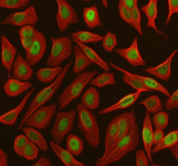
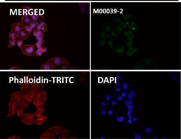
Catalog anti-RB1 antibodies have IHC images from human and rat paraffin sections and IF images from SIHA and SH-SY5Y cells (catalog IHC and IF image captions).

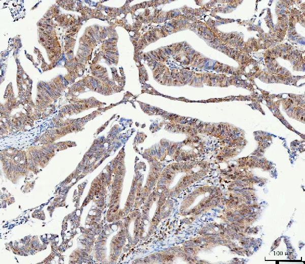
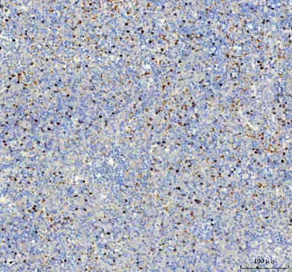
Real IHC data IHC analysis of RB1 using anti-RB1 antibody (A00039-3). RB1 was detected in a paraffin-embedded section of human colorectal adenocarcinoma tissue. Heat mediated antigen retrieval was performed in EDTA buffer (pH 8.0, epitope retrieval solution). The tissue section was blocked with 10% goat serum. The tissue section was then incubated with 1:100 rabbit anti-RB1 Antibody (A00039-3) overnight at 4°C. Peroxidase Conjugated Goat Anti-rabbit IgG was used as secondary antibody and incubated for 30 minutes at 37°C. The tissue section was developed using HRP Conjugated Rabbit IgG Super Vision Assay Kit (Catalog # SV0002) with DAB as the chromogen.
Anti-RB1 Antibody ®
Cat # A00039-3
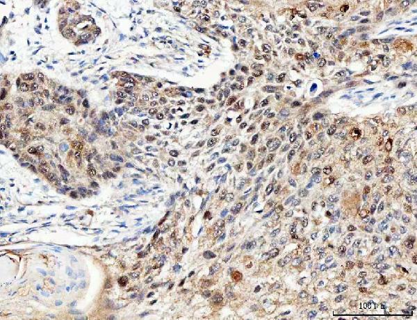
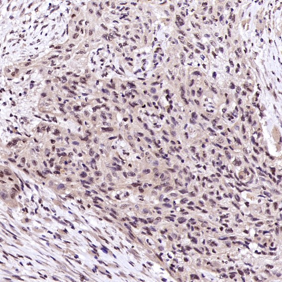
Real IHC data IHC analysis of Retinoblastoma using anti-Retinoblastoma antibody (P00039-1). Retinoblastoma was detected in a paraffin-embedded section of human colorectal adenocarcinoma tissue. Heat mediated antigen retrieval was performed in EDTA buffer (pH 8.0, epitope retrieval solution). The tissue section was blocked with 10% goat serum. The tissue section was then incubated with 1:50 rabbit anti-Retinoblastoma Antibody (P00039-1) overnight at 4°C. Peroxidase Conjugated Goat Anti-rabbit IgG was used as secondary antibody and incubated for 30 minutes at 37°C. The tissue section was developed using HRP Conjugated Rabbit IgG Super Vision Assay Kit (Catalog # SV0002) with DAB as the chromogen.
Anti-Phospho-Rb (S807) RB1 Rabbit Monoclonal Antibody
Cat # P00039-1
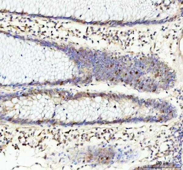
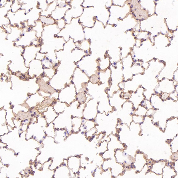
Real IHC data Immunohistochemical analysis of paraffin-embedded Rat lung, using the Antibody at 1:250 dilution.
Anti-Rb RB1 Rabbit Monoclonal Antibody
Cat # M00039-2

A00039-3 shows IHC in human colorectal adenocarcinoma paraffin sections and IF in SIHA cells; P00039-1 shows IHC in human colorectal adenocarcinoma paraffin sections (respective image captions). M00039-2 shows IHC in rat lung paraffin sections and IF in SH-SY5Y cells (M00039-2 image captions).

Which to pick: For tissue IHC, start with A00039-3, which has a human paraffin-section image and a listed dilution of 2–5 μg/ml (A00039-3 IHC image caption; catalog dilution); P00039-1 also has a human paraffin-section image, but its caption does not establish S807 specificity (P00039-1 title and IHC image caption). For IF/ICC, A00039-3 has a SIHA-cell IF image, while monoclonal M00039-2 has a SH-SY5Y-cell IF image (respective IF image captions; M00039-2 catalog clone). For cross-species selection, A00039-3 lists human, mouse, and rat reactivity; M00039-2 lists human and mouse despite its rat-lung IHC caption, and the paraffin-section captions do not report a fixative (catalog reactivity; respective IHC image captions).

Each figure is that product's own IHC / IF validation image from its datasheet.

References

  1. UniProt Consortium. UniProt entry P06400 (RB_HUMAN, Retinoblastoma-associated protein).
  2. Human Protein Atlas. RB1 tissue IHC expression (reliability: Supported).
  3. Human Protein Atlas. RB1 subcellular location (ICC-IF): Mainly localized to the nucleoplasm. In addition localized to the primary cilium transition zone..
  4. Human Protein Atlas. RB1 antibody validation summary (3 antibodies).
  5. Lipoblastoma-Like Tumor and Fibrosarcoma-Like Lipomatous Neoplasm Represent the Same Entity: A Clinicopathologic and Molecular Genetic Study of 23 Cases Occurring in Both Men and Women at Diverse Locations. Modern pathology : an official journal of the United States and Canadian Academy of Pathology, Inc 2023 — PMC10530403.
  6. Diagnostic Utility of TSSC3 and RB1 Immunohistochemistry in Hydatidiform Mole. International journal of molecular sciences 2023 — PMC10253801.
  7. Clinical stratification of glioblastoma based on alterations in retinoblastoma tumor suppressor protein (RB1) and association with the proneural subtype. Journal of neuropathology and experimental neurology 2012 — PMC3246124.
  8. Well differentiated grade 3 pancreatic neuroendocrine tumors compared with related neoplasms: A morphologic study. Cancer cytopathology 2018 — PMC5992011.
  9. PubMed PMID:3657987 — UniProt-cited evidence.
  10. PubMed PMID:3823889 — UniProt-cited evidence.
  11. PubMed PMID:3480530 — UniProt-cited evidence.