This website uses cookies to ensure you get the best experience on our website.


- Table of Contents
Plan RB1CC1 chromogenic IHC around cytoplasmic staining in most tissues, including epithelial cells (HPA tissue IHC). Start with the catalog antibody's 2–5 μg/ml IHC range (datasheet A02414-1) and assess staining with appropriate controls (standard IHC practice).

Expected localisation, validated protocols, controls and antibodies — the at-a-glance facts below, then the full design guide.
| Expected localisation | Cytoplasmic in most tissues (HPA tissue IHC) | |
| Staining pattern | Cytoplasmic staining in most tissues, including epithelial cells (HPA tissue IHC) | |
| Antigen retrieval | EDTA pH 8.0 HIER, heat-mediated (datasheet A02414-1) | |
| Positive control | Bronchus+4 more · see all | |
| Negative control | Bone marrow+4 more · see all |
| Fixation | Keep fixation consistent across sections (standard IHC practice; not target-specific) | |
| Caveat | Staining has medium consistency with RNA expression (HPA tissue IHC) | |
| Regulation | Expression correlates with RB1 levels (UniProt) | |
| Isoform / epitope | Two isoforms; confirm epitope coverage for both (UniProt) |
The catalog antibody’s IHC-P protocol is accompanied by three published RB1CC1 protocols using human breast cancer sections (PMC2894861; PMC3008740; PMC3291565).
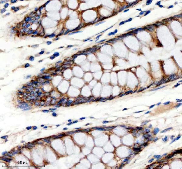
| Sample | Paraffin-embedded human colon tissue; fixative not specified (datasheet A02414-1) |
| Fixation | Image fixative and duration unreported (datasheet A02414-1); verify before use. |
| Sectioning | 4–5 µm sections on charged slides (standard) |
| Deparaffinisation | Xylene, graded ethanol series to water (standard) |
| Antigen retrieval | Heat retrieval: EDTA pH 8.0 (datasheet A02414-1); 20 min, 95–100 °C (standard) |
| Peroxidase block | 3% H2O2, 10 min, room temperature (standard) |
| Blocking | 10% goat serum (datasheet A02414-1) |
| Primary antibody | Rabbit anti-RB1CC1, 2-5 μg/ml (datasheet A02414-1) |
| Primary incubation | Overnight at 4 °C (datasheet A02414-1) |
| Detection | HRP-conjugated secondary, DAB chromogen (datasheet A02414-1) |
| Counterstain | Hematoxylin, blue, dehydrate and mount (standard) |
| Expected result | RB1CC1-positive staining in ciliated cells (cilia axoneme) of bronchus (HPA tissue IHC: High). HPA tissue profile: Cytoplasmic expression in most tissues, including epithelial cells. No signal in the no-primary control. |
Expect predominantly cytoplasmic RB1CC1 staining across many tissues, including epithelial cells (HPA tissue IHC). Ciliated cells in bronchus, fallopian tube and nasopharynx show High axonemal staining, while several glandular cell populations show Medium staining (HPA tissue IHC). RB1CC1 has no transmembrane segment (UniProt Q8TDY2 topology). Interpret tissue patterns with caution: HPA rates its IHC evidence Approved, with medium consistency between staining and RNA expression (HPA tissue IHC).
| Cytoplasmic staining in epithelial cells, with strong ciliary axoneme staining in bronchus or fallopian tube (HPA tissue IHC). | This fits the reported tissue pattern: cytoplasmic expression in most tissues and High signal in those ciliated cells (HPA tissue IHC). Score the cell compartment and ciliary signal separately so a strong axoneme does not imply uniformly strong cytoplasm. |
| Predominantly nuclear-only staining, without the expected cytoplasmic pattern (HPA tissue IHC). | Treat this as a discordant result and examine controls and detection conditions. UniProt also lists the nucleus, so nuclear signal alone cannot prove an artifact (UniProt Q8TDY2 subcellular location); its dominance still differs from HPA's usual tissue pattern (HPA tissue IHC). |
| Strong signal in a cell population HPA reports as Not detected, such as bone-marrow hematopoietic cells (HPA tissue IHC). | Investigate cross-reactivity or endogenous detection activity before scoring it as RB1CC1. An HPA Not detected result is a comparator for that cell population, not proof that every specimen must be negative (HPA tissue IHC). |
| Diffuse chromogen covers tissue, extracellular space and regions lacking recognizable cell boundaries. | This prevents reliable cytoplasmic or ciliary scoring. Check the negative detection control and review blocking, washes and chromogen development as general IHC practice; the supplied HPA and UniProt records do not identify a target-specific cause. |
| No signal in bronchial or fallopian-tube ciliated cells, where HPA reports High staining (HPA tissue IHC). | A negative run there calls for a technical review before interpreting other tissues as negative. Check section integrity, primary antibody, detection and counterstain using routine IHC controls; HPA's Approved rating still allows specimen-to-specimen variation (HPA tissue IHC). |
| Cell and compartment choice | HPA reports cytoplasmic expression in most tissues, including epithelial cells, but High signal specifically at ciliary axonemes in bronchus, fallopian tube and nasopharynx (HPA tissue IHC). Document which structure was scored rather than pooling all visible signal. |
| Strength of tissue evidence | The tissue profile has Approved reliability and medium consistency with RNA expression (HPA tissue IHC). Both listed antibodies have IHC Approved status (HPA antibodies). Use the profile as a pattern reference, not an absolute pass/fail threshold for an individual section. |
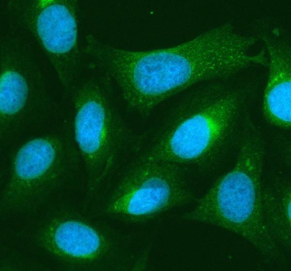
| What should IF/ICC show? | Mainly cytosolic signal is supported; nuclear-membrane localisation is uncertain (HPA subcellular ICC-IF). Only HPA053049 has ICC Supported status among the listed antibodies (HPA antibodies). IF/ICC localisation is context for interpretation, not an IHC-P protocol option. |
| Isoforms and antibody epitope | UniProt lists 2 isoforms (UniProt Q8TDY2 isoforms). The supplied records do not locate the catalog antibody's epitope or establish isoform-specific staining. Avoid assigning different cell patterns to isoforms from these data alone. |
| Situation | Likely cause | Next action |
|---|---|---|
| Expected positive ciliated epithelium is blank (HPA tissue IHC). | The run may have failed at primary incubation, detection or chromogen development; the records do not identify which step. | Review the IHC-validated antibody's IHC-P instructions and run a known-positive section alongside a negative detection control. Confirm cilia are present on the section before treating absent axonemal color as biological. |
| Only nuclei stain intensely while epithelial cytoplasm is blank (HPA tissue IHC). | The pattern conflicts with HPA's predominant cytoplasmic profile, although UniProt also lists nuclear localisation (HPA tissue IHC; UniProt Q8TDY2 subcellular location). | Compare with a known-positive tissue and a negative detection control. Record nuclear and cytoplasmic scores separately; do not call all nuclear color false solely from the HPA pattern. |
| Bone-marrow hematopoietic cells or heart cardiomyocytes stain strongly despite HPA Not detected entries (HPA tissue IHC). | Cross-reactivity or endogenous detection activity is possible; a discordant specimen alone cannot distinguish them. | Inspect the negative detection control and cellular morphology. Recheck the pattern with an independently validated antibody when available, then report the discrepancy against the specific HPA cell population. |
| Brown deposit appears throughout the section, including spaces between cells. | Nonspecific background or endogenous detection activity can obscure cell-associated staining under standard chromogenic IHC practice. | Compare a negative detection control; review blocking, wash steps and chromogen development. Score RB1CC1 only where the signal follows identifiable cellular structures. |
| A weak glandular section is called negative because it lacks the ciliary intensity. | The comparator is mismatched: HPA reports Medium glandular staining in several tissues and High axonemal staining in specified ciliated cells (HPA tissue IHC). | Score each reported cell type and structure against its own HPA level. Include a positive reference section in the run and document intensity and localisation separately. |
| Puncta are interpreted as a required RB1CC1 pattern in routine tissue IHC. | UniProt describes punctate preautophagosomal structures under starvation, while HPA's general tissue IHC profile emphasizes cytoplasmic expression (UniProt Q8TDY2 subcellular location; HPA tissue IHC). | Record puncta if present, but do not require them for a positive tissue result. Confirm the experimental context before linking puncta to starvation-associated localisation. |


Comprehensive Human Protein Atlas IHC scoring per tissue (reliability: Approved — Medium consistency between antibody staining and RNA expression data.). Rows are taken directly from the HPA tissue chart — click any row's HPA link to view the source.
| Tissue | Cell type | Level | Evidence | Source |
|---|---|---|---|---|
| Bronchus | Ciliated cells (cilia axoneme) | High | Protein (IHC) | HPA → |
| Fallopian tube | Ciliated cells (cilia axoneme) | High | Protein (IHC) | HPA → |
| Nasopharynx | Ciliated cells (cilia axoneme) | High | Protein (IHC) | HPA → |
| Adipose tissue | Adipocytes | Medium | Protein (IHC) | HPA → |
| Adrenal gland | Glandular cells | Medium | Protein (IHC) | HPA → |
| Tissue | Cell type | Level | Evidence | Source |
|---|---|---|---|---|
| Bone marrow | Hematopoietic cells | Not detected | Protein (IHC) | HPA → |
| Caudate | Glial cells | Not detected | Protein (IHC) | HPA → |
| Cervix | Glandular cells | Not detected | Protein (IHC) | HPA → |
| Heart muscle | Cardiomyocytes | Not detected | Protein (IHC) | HPA → |
| Hippocampus | Glial cells | Not detected | Protein (IHC) | HPA → |
Troubleshoot RB1CC1 staining by comparing matched paraffin sections and judging signal against documented cell types and compartments (datasheet A02414-1; HPA tissue IHC; HPA subcellular).
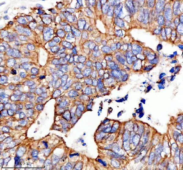
A02414-1 has real IHC data from paraffin-embedded human colon, colon cancer, breast cancer, and mouse heart sections, plus IF data from U2OS cells and human breast cancer sections (catalog image captions).
A02414-1 has IHC images from paraffin-embedded human colon, colon cancer, and breast cancer sections and mouse heart sections (A02414-1 IHC image captions). Its IF images show U2OS cells and paraffin-embedded human breast cancer sections; the catalog lists human, mouse, and rat reactivity (A02414-1 IF image captions; catalog reactivity).
Which to pick: Choose A02414-1 for chromogenic tissue IHC: its paraffin-section captions specify EDTA retrieval at pH 8.0, 2 μg/ml primary antibody, and peroxidase/DAB detection; the fixative is unreported (A02414-1 IHC image captions). For IF/ICC, A02414-1 is listed for both applications and has IF images from U2OS cells and human breast cancer sections at 5 μg/ml (catalog applications; A02414-1 IF image captions). For cross-species IHC, A02414-1 lists human, mouse, and rat reactivity, with IHC images from human and mouse sections; its host is rabbit and clonality is unreported (catalog reactivity and host; A02414-1 IHC image captions; catalog clone field).