This website uses cookies to ensure you get the best experience on our website.


- Table of Contents
Plan RBBP6 staining in paraffin sections around the low nuclear tissue pattern (HPA tissue IHC). Use the catalog antibody’s 2–5 μg/ml IHC range (datasheet A04382-2), and interpret intensity with the reported staining–RNA inconsistency in mind (HPA tissue IHC).

Expected localisation, validated protocols, controls and antibodies — the at-a-glance facts below, then the full design guide.
| Expected localisation | Low nuclear staining in several tissues (HPA tissue IHC) | |
| Staining pattern | Low nuclear staining across several tissue cell types (HPA tissue IHC) | |
| Antigen retrieval | EDTA pH 8.0 HIER, heat-mediated (datasheet A04382-2) | |
| Positive control | Appendix+4 more · see all | |
| Negative control | Adipose tissue+4 more · see all |
| Fixation | Matched tissue-IHC evidence does not establish the fixation claim. Validate the specimen-specific method before use. (selected-SKU IHC image A04382-2) | |
| Caveat | Antibody staining has low consistency with RNA expression (HPA tissue IHC) | |
| Regulation | Overexpressed in esophageal cancer (UniProt) | |
| Isoform / epitope | 4 isoforms; epitope coverage is unspecified (UniProt) |
The catalog antibody protocol is accompanied by four published RBBP6 IHC protocols covering tumor specimens and tissue sections (PMC6828677; PMC3684577; PMC7068609; PMC6138973).
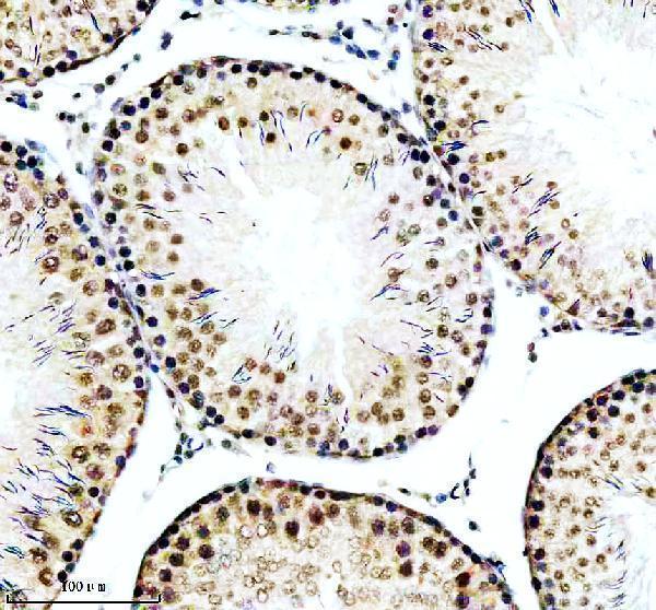
| Sample | Paraffin-embedded rat testis tissue; fixative not specified (datasheet A04382-2) |
| Fixation | Image fixative and duration unreported (datasheet A04382-2); verify before use. |
| Sectioning | 4–5 µm sections on charged slides (standard) |
| Deparaffinisation | Xylene, graded ethanol series to water (standard) |
| Antigen retrieval | Heat retrieval: EDTA pH 8.0 (datasheet A04382-2); 20 min, 95–100 °C (standard) |
| Peroxidase block | 3% H2O2, 10 min, room temperature (standard) |
| Blocking | 10% goat serum (datasheet A04382-2) |
| Primary antibody | Rabbit anti-RBBP6, 2-5 μg/ml (datasheet A04382-2) |
| Primary incubation | Overnight at 4 °C (datasheet A04382-2) |
| Detection | HRP-conjugated secondary, DAB chromogen (datasheet A04382-2) |
| Counterstain | Hematoxylin, blue, dehydrate and mount (standard) |
| Expected result | RBBP6-positive staining in glandular cells of appendix (HPA tissue IHC: Medium). HPA tissue profile: Low nuclear expression in several tissues. No signal in the no-primary control. |
RBBP6 is primarily nuclear, with nucleolar localisation also reported; UniProt additionally lists mitotic chromosomes and the centrosome (UniProt Q7Z6E9 subcellular location). In paraffin-section IHC, expect nuclear staining in selected cell populations, including appendix and duodenal glandular cells and bronchial respiratory epithelial cells (HPA tissue IHC). HPA rates tissue IHC “Approved” but reports low consistency between antibody staining and RNA expression (HPA tissue IHC). RBBP6 has no transmembrane segment (UniProt Q7Z6E9 topology).
| Nuclear staining in appendix or duodenal glandular cells, or bronchial respiratory epithelial cells (HPA tissue IHC). | This fits the reported medium staining in those cell types and RBBP6’s nuclear localisation (HPA tissue IHC; UniProt Q7Z6E9 subcellular location). Judge intensity against cells on the same section and the controls; “medium” is an HPA observation, not a required score for every specimen. |
| Predominantly cytoplasmic staining with little nuclear signal. | Treat this as a localisation mismatch requiring validation: HPA describes low nuclear tissue expression (HPA tissue IHC). UniProt also lists the centrosome and mitotic chromosomes, so an occasional nonnuclear structure alone does not establish an artefact (UniProt Q7Z6E9 subcellular location). |
| Strong staining in adipocytes, bone-marrow hematopoietic cells or pancreatic exocrine cells. | HPA reports these populations as not detected in its tissue IHC survey (HPA tissue IHC). Check for cross-reactivity or endogenous chromogenic activity before calling them RBBP6-positive (general IHC practice). A different result should be verified rather than dismissed solely from the HPA survey. |
| Diffuse colour across cells, stroma or empty areas without a readable nuclear pattern. | Background limits localisation and scoring (general IHC practice). Review the no-primary control, blocking and detection conditions; diffuse colour cannot be interpreted as the low nuclear expression described by HPA (HPA tissue IHC; general IHC practice). |
| No signal in an appendix or bronchus section expected to provide a positive population. | HPA reports medium staining in appendix glandular cells and bronchial respiratory epithelial cells (HPA tissue IHC). First confirm those cells are present and that controls worked; a blank section may reflect assay failure, while HPA’s low RNA–staining consistency limits certainty for any individual specimen (HPA tissue IHC; general IHC practice). |
| Tissue and cell selection | HPA reports medium staining in several specified populations, low staining in others, and no detection in some (HPA tissue IHC). Select and score by cell type, not by whole-section colour; bone marrow RNA enrichment does not establish hematopoietic-cell IHC positivity (HPA tissue IHC). |
| Subcellular interpretation | Nucleus and nucleolus are listed by UniProt; HPA tissue IHC summarises low nuclear expression (UniProt Q7Z6E9 subcellular location; HPA tissue IHC). The reported chromosome and centrosome associations call for care when interpreting unusual puncta (UniProt Q7Z6E9 subcellular location). |
| Antibody evidence | Two listed antibodies have “Approved” IHC status, while HPA still reports low consistency with RNA expression (HPA antibodies; HPA tissue IHC). Approval supports use of the observed pattern as a reference, not automatic confirmation of every stained cell. |
| IF/ICC Q&A: what pattern should an IF image show? | HPA reports enhanced nuclear-speckle localisation in ICC-IF (HPA subcellular; HPA antibodies). This is a separate imaging observation; do not require individually resolved speckles in chromogenic paraffin IHC (HPA subcellular; general IHC practice). |
| Isoforms and processing | UniProt lists four RBBP6 isoforms, no signal peptide or propeptide, and no transmembrane segment (UniProt Q7Z6E9 isoforms, processing and topology). The payload gives no antibody epitope, so it cannot establish which isoforms a given stain detects. |
| Situation | Likely cause | Next action |
|---|---|---|
| Expected positive cells are blank. | The reference population may be absent from the section, or an IHC step may have failed (HPA tissue IHC; general IHC practice). | Confirm the relevant cells by morphology, inspect a known-positive section, then check retrieval and detection using the antibody’s IHC instructions (general IHC practice). No RBBP6-specific retrieval requirement is supplied. |
| All tissue compartments are uniformly brown. | Nonspecific reagent binding or endogenous detection activity can obscure localisation (general IHC practice). | Compare with a no-primary control; review blocking, washes and endogenous-activity quenching appropriate to the chromogen (general IHC practice). Interpret only a resolved cell-associated pattern. |
| Colour is mainly cytoplasmic. | The result differs from HPA’s nuclear tissue profile, although UniProt lists additional locations (HPA tissue IHC; UniProt Q7Z6E9 subcellular location). | Check counterstained nuclei and controls, then confirm with an independently validated antibody if the cytoplasmic result persists (general IHC practice). |
| A population listed as undetected stains strongly. | Cross-reactivity or endogenous activity is possible; HPA’s undetected call is specific to its survey (HPA tissue IHC; general IHC practice). | Identify the stained cell type, compare no-primary and known-positive controls, and verify the result independently before assigning RBBP6 expression (general IHC practice). |
| Signal varies across replicate sections. | Section composition or inconsistent processing may change the apparent score (general IHC practice); HPA also reports low RNA–staining consistency (HPA tissue IHC). | Compare the same cell population across sections and review section handling, retrieval and detection records (general IHC practice). Do not infer a RBBP6-specific fixation effect from this variation. |
| IF shows speckles, but IHC looks broadly nuclear. | HPA’s speckle call comes from ICC-IF, while its tissue IHC summary is nuclear (HPA subcellular; HPA tissue IHC). | Assess each image against its own assay reference and controls; score paraffin IHC by interpretable nuclear staining in the specified cells (HPA tissue IHC; general IHC practice). |


Comprehensive Human Protein Atlas IHC scoring per tissue (reliability: Approved — Low consistency between antibody staining and RNA expression data.). Rows are taken directly from the HPA tissue chart — click any row's HPA link to view the source.
| Tissue | Cell type | Level | Evidence | Source |
|---|---|---|---|---|
| Appendix | Glandular cells | Medium | Protein (IHC) | HPA → |
| Bronchus | Respiratory epithelial cells | Medium | Protein (IHC) | HPA → |
| Cerebellum | Cells in granular layer | Medium | Protein (IHC) | HPA → |
| Cerebral cortex | Neuronal cells | Medium | Protein (IHC) | HPA → |
| Cervix | Squamous epithelial cells | Medium | Protein (IHC) | HPA → |
| Tissue | Cell type | Level | Evidence | Source |
|---|---|---|---|---|
| Adipose tissue | Adipocytes | Not detected | Protein (IHC) | HPA → |
| Adrenal gland | Glandular cells | Not detected | Protein (IHC) | HPA → |
| Bone marrow | Hematopoietic cells | Not detected | Protein (IHC) | HPA → |
| Hippocampus | Glial cells | Not detected | Protein (IHC) | HPA → |
| Lymph node | Germinal center cells | Not detected | Protein (IHC) | HPA → |
Troubleshoot RBBP6 staining in paraffin sections by checking retrieval, cellular localisation and controls before comparing chromogenic scores (datasheet A04382-2; UniProt Q7Z6E9).
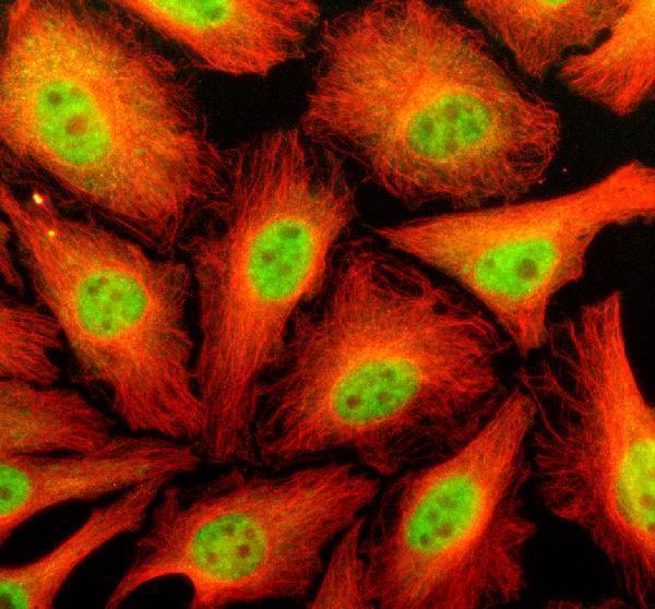
A04382-2 has IHC data from rat testis paraffin sections and IF/ICC data from HeLa cells; the catalog lists Human, Mouse and Rat reactivity (A04382-2 image captions; catalog reactivity).
A04382-2 was demonstrated by chromogenic IHC on a rat testis paraffin section (A04382-2 IHC image caption). The same SKU was demonstrated by IF/ICC in HeLa cells and is listed for IHC, ICC and IF with Human, Mouse and Rat reactivity (A04382-2 IF image caption; catalog applications and reactivity).
Which to pick: For tissue IHC, choose A04382-2: its rat testis paraffin-section image used EDTA retrieval at pH 8.0 and 2 μg/ml primary antibody; the fixative is unreported (A04382-2 IHC image caption). For IF/ICC, A04382-2 has a HeLa-cell image using 5 μg/ml primary antibody (A04382-2 IF image caption). For cross-species work, A04382-2 lists Human, Mouse and Rat reactivity and a rabbit host; clonality is unspecified, and the supplied tissue IHC image demonstrates Rat only (A04382-2 catalog reactivity, host and clone fields; IHC image caption).