This website uses cookies to ensure you get the best experience on our website.


- Table of Contents
Plan RBFOX2 IHC in paraffin sections using the catalog antibody’s validated staining conditions. Compare nuclear and cytoplasmic staining with the tissue profile, while accounting for the antibody specificity caution (HPA tissue IHC).

Expected localisation, validated protocols, controls and antibodies — the at-a-glance facts below, then the full design guide.
| Expected localisation | Nuclear and cytoplasmic staining (HPA tissue IHC) | |
| Staining pattern | Nuclear and cytoplasmic signal in diverse cell types (HPA tissue IHC) | |
| Antigen retrieval | EDTA pH 8.0 HIER, heat-mediated (datasheet A05389-2) | |
| Positive control | Caudate+4 more · see all | |
| Negative control | Heart muscle+1 more · see all |
| Fixation | Keep fixation consistent across paraffin sections. (standard IHC practice; not target-specific) | |
| Caveat | Antibody may detect proteins from more than one gene (HPA tissue IHC) | |
| Regulation | Low tissue specificity reported for RNA expression (HPA tissue RNA) | |
| Isoform / epitope | 10 isoforms; check epitope coverage across variants (UniProt) |
The catalog antibody protocol (datasheet: A05389-2) is followed by published chromogenic IHC protocols for tissue sections (PMC13576891 methods; PMC10730836 methods).
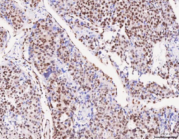
| Sample | Paraffin-embedded human liver cancer tissue; fixative not specified (datasheet A05389-2) |
| Fixation | Image fixative and duration unreported (datasheet A05389-2); verify before use. |
| Sectioning | 4–5 µm sections on charged slides (standard) |
| Deparaffinisation | Xylene, graded ethanol series to water (standard) |
| Antigen retrieval | Heat retrieval: EDTA pH 8.0 (datasheet A05389-2); 20 min, 95–100 °C (standard) |
| Peroxidase block | 3% H2O2, 10 min, room temperature (standard) |
| Blocking | 10% goat serum (datasheet A05389-2) |
| Primary antibody | Rabbit anti-RBFOX2, 2-5 μg/ml (datasheet A05389-2) |
| Primary incubation | Overnight at 4 °C (datasheet A05389-2) |
| Detection | HRP-conjugated secondary, DAB chromogen (datasheet A05389-2) |
| Counterstain | Hematoxylin, blue, dehydrate and mount (standard) |
| Expected result | RBFOX2-positive staining in neuronal cells of caudate (HPA tissue IHC: High). HPA tissue profile: General nuclear and cytoplasmic expression. No signal in the no-primary control. |
RBFOX2 should stain nuclei and may stain cytoplasm (UniProt O43251: nucleus and cytoplasm; HPA tissue IHC: general nuclear and cytoplasmic expression). Expect stronger staining in HPA High cell populations, including neuronal cells in caudate and glomerular cells in kidney (HPA tissue IHC). HPA rates the tissue IHC profile Approved, with medium consistency against RNA data and a warning that the antibodies may recognize proteins from more than one gene (HPA tissue IHC). RBFOX2 has no transmembrane segment (UniProt O43251 topology).
| Nuclear staining, with or without cytoplasmic staining, in the expected cell population. | This fits the reported compartments (UniProt O43251: nucleus and cytoplasm; HPA tissue IHC: general nuclear and cytoplasmic expression). For a tissue check, compare the named cell population rather than judging every cell in the section as equally positive (HPA tissue IHC: cell-specific levels). |
| Staining appears chiefly at the cell membrane or extracellularly. | That is outside the reported nuclear and cytoplasmic pattern (UniProt O43251; HPA tissue IHC). RBFOX2 has no transmembrane segment (UniProt O43251 topology). Review morphology and detection background before treating this distribution as RBFOX2. |
| A cell population reported as Not detected stains strongly. | Cardiomyocytes in heart muscle and glandular cells in parathyroid are reported as Not detected (HPA tissue IHC). Strong staining there warrants checking cell identity, antibody specificity and endogenous detection activity; the HPA profile cautions that antibodies may recognize proteins from more than one gene (HPA tissue IHC). |
| Chromogen covers cells broadly without a clear nuclear or cytoplasmic pattern. | Diffuse staining cannot establish the reported RBFOX2 distribution (UniProt O43251; HPA tissue IHC). Compare a no-primary control and inspect tissue edges and counterstain; nonspecific binding or detection background can obscure compartments (standard IHC practice). |
| No staining appears in an expected positive cell population. | Check that the scored cells match the reference: HPA reports High staining in caudate neuronal cells, kidney glomerular cells and lung alveolar cells (HPA tissue IHC). A blank result in such cells prompts assay troubleshooting; it alone does not establish absent RBFOX2. |
| Cell population and tissue | HPA reports High staining in caudate neurons, endometrial stromal cells, kidney glomerular cells and ovarian stromal cells, but Not detected in cardiomyocytes and parathyroid glandular cells (HPA tissue IHC). Score the named cells, since whole-section intensity can conceal that distinction. |
| Antibody validation | The tissue IHC profile is Approved, with medium staining–RNA consistency and a warning that antibodies may recognize proteins from more than one gene (HPA tissue IHC). HPA006240 and HPA075862 have IHC Approved status; this does not establish specificity for the catalog antibody (HPA antibodies). |
| Compartment | UniProt reports nuclear and cytoplasmic localization, while HPA tissue IHC describes general nuclear and cytoplasmic expression (UniProt O43251; HPA tissue IHC). HPA ICC-IF supports nucleoplasm as the main location (HPA subcellular); use that observation as an IF comparison, not an IHC intensity rule. |
| Isoforms and antibody coverage | UniProt lists 10 RBFOX2 isoforms (UniProt O43251). The payload gives no epitope map or isoform coverage for the catalog antibody, so a staining difference cannot be assigned to a particular isoform from these records. |
| Target-specific fixation sensitivity | Neither supplied source reports how fixation changes RBFOX2 staining (UniProt O43251; HPA tissue IHC). Record fixation and retrieval conditions during optimization, but do not infer an RBFOX2-specific fixation effect from tissue intensity or topology. |
| Situation | Likely cause | Next action |
|---|---|---|
| Expected positive cells are blank. | Cell identity, antibody performance or detection may be at issue; HPA reports High staining in caudate neuronal cells and kidney glomerular cells (HPA tissue IHC). | Verify the scored cell population and run an appropriate positive section with the same IHC conditions. Check primary incubation, retrieval and detection steps systematically (standard IHC practice); the payload supplies no RBFOX2-specific retrieval condition. |
| Signal is diffuse across the section. | Nonspecific binding or chromogenic detection background can obscure cell boundaries (standard IHC practice). The expected distribution is nuclear and cytoplasmic (UniProt O43251; HPA tissue IHC). | Compare a no-primary control, inspect morphology, and optimize blocking, washing and primary-antibody concentration one variable at a time (standard IHC practice). |
| Membrane or extracellular staining dominates. | This conflicts with the reported nuclear and cytoplasmic pattern and lack of a transmembrane segment (UniProt O43251; HPA tissue IHC). The image alone does not identify the source of staining. | Check a no-primary control and review whether staining follows cells or section artefacts. Reassess antibody specificity before scoring that signal as RBFOX2 (standard IHC practice; HPA tissue IHC: antibody caveat). |
| Cardiomyocytes stain strongly. | HPA reports RBFOX2 as Not detected in cardiomyocytes and warns that tissue antibodies may recognize proteins from more than one gene (HPA tissue IHC). Endogenous detection activity is another assay possibility (standard IHC practice). | Confirm the cells are cardiomyocytes; compare no-primary and positive-section controls. Investigate background and specificity before reporting a new tissue pattern (standard IHC practice). |
| High and low regions look similar. | The named populations differ: lung alveolar cells are High, while bronchus respiratory epithelial cells are Low (HPA tissue IHC). Broad chromogen deposits can make intensity comparisons unreliable (standard IHC practice). | Score the specified cells separately using consistent exposure to chromogen and counterstain. Review controls and reduce excessive background before interpreting intensity (standard IHC practice). |
| Does IF/ICC need the same protocol or readout? | HPA supports nucleoplasmic localization in ICC-IF, whereas its tissue IHC profile reports general nuclear and cytoplasmic expression (HPA subcellular; HPA tissue IHC). | Use nucleoplasmic signal as the IF/ICC localization comparison (HPA subcellular). Consult the separate IF/ICC guide for its workflow; this IHC section supplies no IF/ICC protocol. |
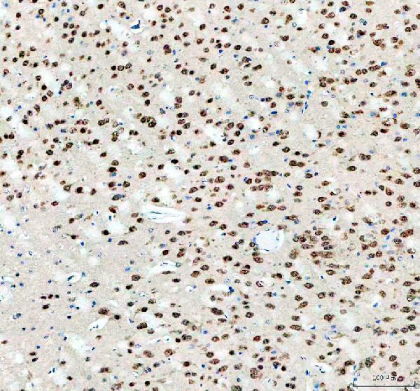
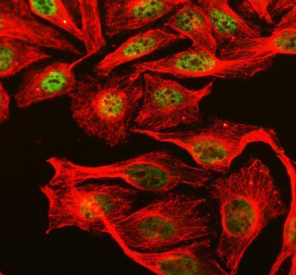
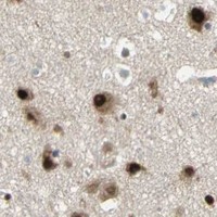


Comprehensive Human Protein Atlas IHC scoring per tissue (reliability: Approved — Medium consistency between antibody staining and RNA expression data. Caution, targets protein from more than one gene.). Rows are taken directly from the HPA tissue chart — click any row's HPA link to view the source.
| Tissue | Cell type | Level | Evidence | Source |
|---|---|---|---|---|
| Caudate | Neuronal cells | High | Protein (IHC) | HPA → |
| Cerebellum | Cells in molecular layer | High | Protein (IHC) | HPA → |
| Cerebral cortex | Neuronal cells | High | Protein (IHC) | HPA → |
| Endometrium | Cells in endometrial stroma | High | Protein (IHC) | HPA → |
| Hippocampus | Neuronal cells | High | Protein (IHC) | HPA → |
Use the IHC-validated antibody's paraffin-section result as a starting point, then check compartment, cell type and controls when interpreting RBFOX2 staining.
Rabbit anti-RBFOX2 antibodies have IHC images from human paraffin sections and mouse and rat brain, plus IF images from human tissue and cells and mouse and rat brain (catalog image captions).
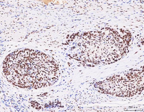
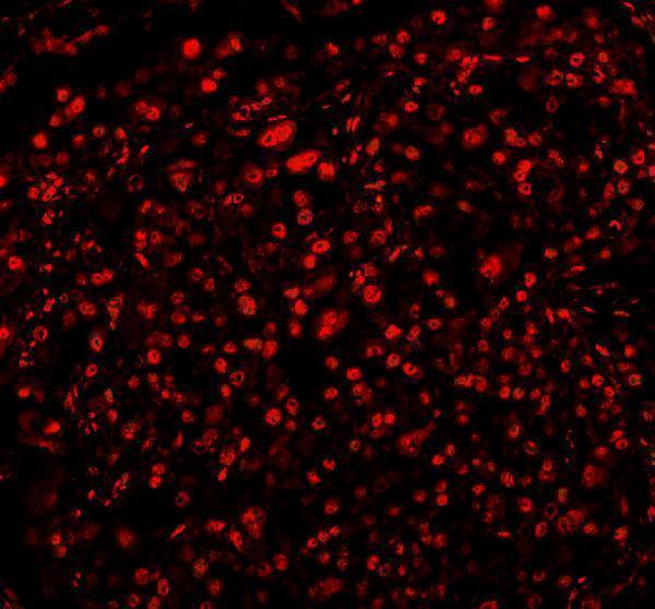
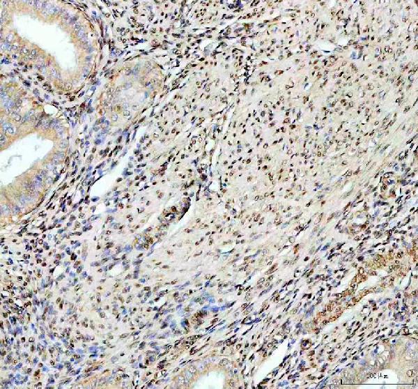
A05389-1 has IHC images from human endometrial cancer and mouse and rat brain, plus IF images from U2OS cells and mouse and rat brain (A05389-1 image captions). A05389-2 has IHC images from human liver cancer, esophageal squamous carcinoma, lung cancer and cortical adenoma, plus an IF image from human lung cancer (A05389-2 image captions).
Which to pick: For human tissue IHC, choose A05389-2 when its paraffin-section examples fit your study; its IHC caption reports EDTA retrieval at pH 8.0 and 2 μg/ml primary antibody overnight at 4°C, while the fixative is unreported (A05389-2 IHC captions). Choose A05389-1 for IF/ICC or cross-species tissue IHC: ICC is listed, and its images show human cell IF and mouse and rat brain IHC; its own IHC captions report EDTA retrieval at pH 8.0 and 2 μg/ml primary antibody overnight at 4°C, with fixative unreported (A05389-1 applications; A05389-1 IF and IHC captions). Both have rabbit hosts, and clonality is unreported (catalog host and clone fields).