RBM25 / RNA-binding protein 25 · IHC design guide

Design Immunohistochemistry for RBM25

Plan RBM25 IHC in paraffin sections around general nuclear staining, with cytoplasmic signal in several tissues (HPA tissue IHC). The catalog antibody has an IHC concentration range of 2–5 μg/ml (datasheet A07494-1); assess staining by cell type.

Evidence assembled Oct 2026 · For research use; verify linked source records and product datasheet before use
Immunohistochemistry protocol sheet for RBM25 (IHC for RBM25): expected localisation General nuclear staining; cytoplasmic signal in several tissues (HPA tissue IHC), antibody A07494-1, validated IHC image, and IHC protocol steps
Printable RBM25 IHC protocol sheet — expected localisation General nuclear staining; cytoplasmic signal in several tissues (HPA tissue IHC), antibody A07494-1, controls and protocol steps. Open the full RBM25 IHC guide →

RBM25 Immunohistochemistry Experimental Design Guide

Expected localisation, validated protocols, controls and antibodies — the at-a-glance facts below, then the full design guide.

Must know before staining
Expected localisation General nuclear staining; cytoplasmic signal in several tissues (HPA tissue IHC)
Staining pattern Nuclear staining across tissues; cytoplasmic staining in several (HPA tissue IHC)
Antigen retrieval EDTA pH 8.0 HIER, heat-mediated (datasheet A07494-1)
Positive control ⓘ Bone marrow+4 more · see all
Negative control ⓘ None in HPA (detected in all 44 tissues); use no-primary + isotype controls
Important caveats
Reasons your staining may differ from the expected pattern.
Fixation Keep fixation consistent across sections. (standard IHC practice; not target-specific)
Caveat Cytoplasmic signal can complicate nuclear scoring (HPA tissue IHC)
Regulation No abundance regulator annotated (UniProt)
Isoform / epitope 4 isoforms; antibody epitope coverage is unknown (UniProt; datasheet A07494-1)
Section 1

Recommended RBM25 IHC & IF Protocols

The catalog antibody protocol uses EDTA retrieval (datasheet: A07494-1). Published RBM25 IHC methods provide additional conditions for liver cancer tissue (PMC13583231) and myocardial sections (PMC12707302).

Recommended immunohistochemistry (IHC-P) protocol parameters
SampleParaffin-embedded mouse brain tissue; fixative not specified (datasheet A07494-1)
FixationImage fixative and duration unreported (datasheet A07494-1); verify before use.
Sectioning4–5 µm sections on charged slides (standard)
DeparaffinisationXylene, graded ethanol series to water (standard)
Antigen retrievalHeat retrieval: EDTA pH 8.0 (datasheet A07494-1); 20 min, 95–100 °C (standard)
Peroxidase block3% H2O2, 10 min, room temperature (standard)
Blocking10% goat serum (datasheet A07494-1)
Primary antibodyRabbit anti-RBM25, 2-5 μg/ml (datasheet A07494-1)
Primary incubationOvernight at 4 °C (datasheet A07494-1)
DetectionHRP-conjugated secondary, DAB chromogen (datasheet A07494-1)
CounterstainHematoxylin, blue, dehydrate and mount (standard)
Expected resultRBM25-positive staining in hematopoietic cells of bone marrow (HPA tissue IHC: High). HPA tissue profile: General nuclear expression, additional cytoplasmic expression in several tissues. No signal in the no-primary control.
💡Decision noteStart with heat-mediated EDTA retrieval at pH 8.0 for the catalog antibody (datasheet: A07494-1); the published protocols used citrate retrieval with different antibodies (PMC13583231; PMC12707302).
Section 2

What Is the Expected RBM25 Staining Pattern?

RBM25 should show predominantly nuclear staining, consistent with its enrichment in nuclear speckles; faint cytoplasmic signal is also possible (UniProt P49756: subcellular location). HPA reports general nuclear IHC staining, with additional cytoplasmic staining in several tissues, and rates the tissue pattern Supported, with medium consistency between staining and RNA data (HPA: tissue IHC). RBM25 has no transmembrane segment (UniProt P49756: topology), so a membrane-only pattern is unexpected.

What am I looking at on my slide?
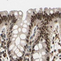
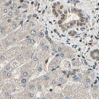
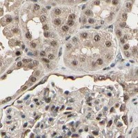
Predominantly nuclear staining in glandular or hematopoietic cells, with some cytoplasmic signal.This fits the reported IHC pattern. HPA scores glandular cells in cervix, duodenum, endometrium and parathyroid gland, and bone marrow hematopoietic cells, High (HPA: tissue IHC). Assess nuclei against the counterstain; chromogenic IHC may not resolve individual speckles.
Strong membrane-only staining, or intense cytoplasmic staining with consistently unstained nuclei.Treat this as discordant with the expected predominant nuclear distribution (UniProt P49756: subcellular location; HPA: tissue IHC). Check background and detection controls before calling it RBM25. Some cytoplasmic staining alone does not invalidate a slide (HPA: tissue IHC).
A cell population outside the expected distribution stains more strongly than adjacent known-positive cells.The pattern warrants a cross-reactivity or endogenous detection activity check; cell identity alone cannot establish specificity. HPA reports broad expression and low tissue specificity, so an unlisted cell type is not automatically negative (HPA: tissue IHC).
Diffuse color across nuclei, cytoplasm and surrounding tissue obscures cellular boundaries.Interpretation is unreliable when the background masks the nuclear pattern. Compare a no-primary control and inspect blocking, washes and detection chemistry as general IHC checks; HPA’s tissue profile does not identify the cause of background.
No nuclear signal in an HPA High tissue, despite intact tissue and a visible counterstain.First treat the run as potentially failed, then assess the antibody and detection controls. HPA’s High calls identify useful comparison cells, but its Supported rating and medium RNA–staining consistency do not make every section a guaranteed positive (HPA: tissue IHC).
💡Expected RBM25 appearanceCall a result consistent with RBM25 when staining is predominantly nuclear in HPA High cells, such as duodenal glandular cells or bone marrow hematopoietic cells; cytoplasmic signal may accompany it, while strong membrane-only color or widespread cell-free color should prompt a false-positive check (HPA: tissue IHC; UniProt P49756: topology).
How each factor affects the staining
Compartment and optical resolutionUniProt places RBM25 predominantly with splicing factors in nuclear speckles and describes faint cytoplasmic localization (UniProt P49756: subcellular location); HPA reports a broader nuclear IHC pattern with some cytoplasmic expression (HPA: tissue IHC). Use nuclear predominance for chromogenic scoring; do not require visible speckles.
Tissue and cell selectionHPA scores exocrine glandular cells in pancreas and respiratory epithelial cells in nasopharynx High, alongside several other listed populations; smooth muscle cells and skeletal myocytes are Low (HPA: tissue IHC). Low is a weaker-signal comparison, not a confirmed negative control.
Antibody evidenceHPA003025 is IHC Supported, while HPA070713 has no IHC status in the supplied record; both are ICC Enhanced (HPA: antibody validation). The tissue profile itself is Supported, with medium consistency against RNA data (HPA: tissue IHC). Keep each validation label tied to its assay.
Isoforms and epitope coverageUniProt lists 4 RBM25 isoforms and RRM and PWI domains (UniProt P49756: isoforms; domains). The supplied records give no antibody epitope, so isoform-specific staining or differential retrieval cannot be predicted from them.
IF/ICC: what pattern should I expect?Expect enhanced nuclear speckle localization in ICC-IF images (HPA: subcellular ICC-IF). UniProt also notes faint cytoplasmic localization (UniProt P49756: subcellular location). This is an IF interpretation cue, not an IHC protocol or evidence that speckles must resolve in a chromogenic section.
Why is my staining missing, weak or wrong?
SituationLikely causeNext action
Nuclei appear negative in a selected HPA High population.A failed staining or detection run is possible; the HPA High designation is observational, not a guarantee for every specimen (HPA: tissue IHC).Confirm the expected cells are present, then review the IHC-validated antibody’s instructions, antigen retrieval conditions, detection reagents and positive run control as general IHC checks.
Signal is mainly cytoplasmic with little nuclear color.This conflicts with nuclear predominance, although additional cytoplasmic IHC staining is reported in several tissues (HPA: tissue IHC).Compare matched positive tissue and no-primary controls; score the nuclear component separately before attributing cytoplasmic color to RBM25.
Sharp membrane staining dominates the field.A membrane-only pattern conflicts with the absence of a transmembrane segment and the reported nuclear localization (UniProt P49756: topology; HPA: tissue IHC).Check no-primary and detection controls, then reassess whether the signal follows cell boundaries rather than counterstained nuclei.
Smooth muscle or skeletal myocytes stain weakly.HPA lists these cells as Low, not negative (HPA: tissue IHC).Compare them with HPA High cells on the same run; avoid using weak muscle staining alone to label the assay nonspecific.
Broad haze hides nuclei and tissue structure.Background from blocking, washing or detection may obscure interpretation; the supplied RBM25 sources do not assign a target-specific cause.Inspect a no-primary control and repeat with the detection system’s standard blocking and wash checks; judge specificity only after nuclear boundaries are readable.
IF shows speckles, but IHC shows a smoother nuclear stain.ICC-IF resolves nuclear speckles (HPA: subcellular ICC-IF), whereas HPA describes general nuclear tissue IHC staining (HPA: tissue IHC).Use the assay-specific expected pattern: assess nuclear predominance in IHC and speckle localization in IF. Do not reject chromogenic staining solely because distinct speckles are absent.

Sample controls for RBM25 IHC & IF

🧪Run duodenum first: glandular cells should show RBM25 staining (HPA: High in duodenal glandular cells). HPA detects RBM25 in all 44 scored tissues, so there is no supported negative tissue; no cell population on the positive slide is validated as negative, and cells lacking nuclear signal should be treated only as within-slide background references (HPA: no negative tissue rows; HPA: detected in all 44 scored tissues).
Positive control tissue: Bone marrow (Hematopoietic cells, HPA High)
Negative control tissue: None in HPA: RBM25 is detected in all 44 scored tissues. Use a no-primary (secondary-only) and an isotype control instead.
ICC-IF cell lines (HPA subcellular resource): HPA ICC-IF images show RBM25 in A-431, U-251MG, U2OS, HaCaT, with annotated localisation: Nuclear speckles (enhanced) (HPA subcellular).
Technical controls: Include a no-primary, secondary-only section, a matched rabbit IgG isotype control, and RBM25-knockout material as a biological negative if available (caption: rabbit anti-RBM25 antibody; standard IHC controls). Block endogenous peroxidase and inspect any residual DAB staining in the duodenal section (standard chromogenic IHC practice).
⚠️Feasibility: A target-specific fixation window or fixation effect is unreported, and the selected paraffin-section caption does not state its fixative (caption: fixative not stated). The caption reports heat retrieval in EDTA at pH 8.0 for mouse brain, but it does not establish that retrieval is required for duodenum (caption: EDTA retrieval, pH 8.0; HPA: High in duodenal glandular cells). ICC-IF images show nuclear speckles, which can guide localization checks; the supplied evidence does not establish that IF or frozen sections are easier than paraffin IHC, and duodenal endogenous peroxidase can complicate chromogenic interpretation (HPA: nuclear speckles; standard IHC practice).

HPA tissue IHC evidence for RBM25

Comprehensive Human Protein Atlas IHC scoring per tissue (reliability: Supported — Medium consistency between antibody staining and RNA expression data.). Rows are taken directly from the HPA tissue chart — click any row's HPA link to view the source.

Positive expression · recommended positive controls

TissueCell typeLevelEvidenceSource
Bone marrow Hematopoietic cells High Protein (IHC) HPA →
Cerebral cortex Endothelial cells High Protein (IHC) HPA →
Cervix Glandular cells High Protein (IHC) HPA →
Duodenum Glandular cells High Protein (IHC) HPA →
Endometrium Glandular cells High Protein (IHC) HPA →

Undetected expression · recommended negative controls

TissueCell typeLevelEvidenceSource
None in HPA: RBM25 is detected in all 44 scored tissues. Use a no-primary (secondary-only) and an isotype control instead.
Section 3

Advanced RBM25 IHC Tips

Troubleshoot RBM25 staining in paraffin sections by checking nuclear localisation, retrieval, controls and cell-level scoring before interpreting chromogenic signal.

What retrieval conditions should I start with for RBM25 in paraffin sections?
Start with heat-mediated antigen retrieval in EDTA buffer at pH 8.0 for paraffin sections (datasheet A07494-1). The selected mouse brain IHC image used this retrieval before overnight incubation at 4°C with 2 μg/ml catalog antibody, followed by peroxidase detection with DAB (caption A07494-1). Keep retrieval temperature and heating time consistent across the comparison set, then inspect whether nuclear detail remains intact (standard IHC practice). If staining is weak, optimise heating duration on adjacent sections before considering a different retrieval buffer as a fallback (standard IHC practice). Compare those sections with a matched no-primary control so stronger DAB deposition is not mistaken for recovered antigen (standard IHC practice).
How should I troubleshoot fixation-dependent changes in RBM25 staining?
Target-specific fixation sensitivity for RBM25 is unknown from the supplied evidence; the selected tissue caption describes a paraffin section but does not state its fixative (caption A07494-1). Record fixative, fixation duration and processing history for each specimen before comparing staining intensity (standard IHC practice). If a batch loses nuclear signal, compare sections with matched processing and the same EDTA pH 8.0 retrieval before changing antibody concentration (datasheet A07494-1; standard IHC practice). Examine nuclear morphology and no-primary controls to distinguish damaged sections or nonspecific deposition from apparent loss of RBM25 (standard IHC practice). Do not attribute a fixation effect to RBM25 without a controlled fixation comparison.
Should RBM25 staining appear in nuclei, cytoplasm, or both?
Prioritise nuclear staining when assessing RBM25 IHC: it is enriched in nuclear speckles, with faint cytoplasmic localisation reported separately (UniProt P49756 localisation; HPA subcellular). HPA tissue IHC describes general nuclear expression and additional cytoplasmic expression in several tissues, with Supported reliability and medium agreement with RNA data (HPA tissue IHC). At the microscope, score nuclear and cytoplasmic DAB separately rather than treating total brown area as equivalent target signal (standard IHC practice). Check a haematoxylin counterstain to confirm that apparent nuclear puncta sit within intact nuclei (standard IHC practice). Diffuse cytoplasmic staining without corresponding nuclear signal merits review of retrieval, controls and section quality before biological interpretation (UniProt P49756 localisation; standard IHC practice).
Could RBM25 isoforms or modifications change my IHC result?
RBM25 has 4 annotated isoforms, an RRM at residues 87–164 and a PWI domain at 750–843 (UniProt P49756). Annotated modifications include acetylation at residue 135 and phosphorylation at 226, 229, 583 and 677 (UniProt P49756). The supplied caption does not identify the catalog antibody's epitope, so these annotations alone cannot establish which isoforms or modified forms it detects (caption A07494-1; UniProt P49756). When patterns differ across specimens, check the antibody's documented immunogen and isoform coverage before assigning the difference to splicing or modification (standard IHC practice). Compare adjacent sections under identical retrieval and detection conditions to test whether the staining difference is reproducible (standard IHC practice).
How can I assess RBM25 by IF alongside my IHC observations?
Treat IF/ICC as a separate application when comparing its pattern with chromogenic RBM25 IHC in paraffin sections (standard IHC/IF practice). For multiplexing, pair RBM25 with a marker for the expected cell type and a nuclear counterstain, then ask whether RBM25 signal falls within those cells' nuclei (UniProt P49756 localisation; standard IF practice). Select a fluorophore channel with low tissue autofluorescence and check single-stain controls for spectral bleed-through (standard IF practice). RBM25 has no transmembrane segment and is enriched in nuclear speckles, so permeabilisation must permit antibody access to intracellular nuclear antigen; optimise it for the IF specimen and antibody (UniProt P49756 topology and localisation; standard IF practice). HPA reports nuclear-speckle IF localisation, which can guide pattern review without establishing conditions for this catalog antibody (HPA subcellular).
What causes diffuse or patchy DAB background in RBM25 sections?
First compare a no-primary section with the RBM25-stained section; DAB that persists without primary antibody points to detection or tissue background (standard IHC practice). The selected caption used 10% goat serum blocking, 2 μg/ml primary antibody overnight at 4°C, and a peroxidase-conjugated secondary for 30 minutes at 37°C (caption A07494-1). Check peroxidase blocking and wash consistency, and shorten chromogen development if both control and test sections darken broadly (standard chromogenic IHC practice). Inspect folds, edges and damaged regions separately because concentrated reagent or trapped chromogen can mimic cellular staining there (standard IHC practice). Confirm that an improved background still leaves interpretable nuclear staining, the principal expected RBM25 pattern (UniProt P49756 localisation).
How should I quantify RBM25 staining across paraffin sections? ⚠ ANSWER MARKED FOR VERIFICATION
Define the scored cell population and compartment before measuring: RBM25 is mainly nuclear, while some tissue IHC shows cytoplasmic staining (UniProt P49756 localisation; HPA tissue IHC). For nuclear DAB, report the percentage of positive nuclei and an H-score using intensity categories 0–3, with a prespecified positivity threshold (standard IHC scoring practice). If spatial distribution matters, report positive-cell density per mm² of viable analysed tissue as a separate measure (standard IHC quantification practice). Normalise counts to all evaluable nuclei in the same defined cell population, or density to measured tissue area, rather than to whole-slide brown area (standard IHC quantification practice). Keep staining batches, counterstaining and image-analysis thresholds consistent across groups (standard IHC practice).
How can I distinguish genuine RBM25 signal from staining artefacts?
A credible RBM25 result has interpretable nuclear staining in intact cells, consistent with its nuclear-speckle enrichment; faint cytoplasmic signal can also occur (UniProt P49756 localisation). HPA reports high staining in bone-marrow hematopoietic cells and cerebral-cortex endothelial cells, and low staining in smooth-muscle cells and skeletal-muscle myocytes; use these observations as context when those cell types are present (HPA tissue IHC). Review cell identity on the counterstain before calling an unexpected population positive (standard IHC practice). Exclude section edges and necrotic or disrupted areas from interpretation, and check a no-primary control for endogenous peroxidase-related DAB or other detection background (standard IHC practice). A cytoplasmic-only or edge-restricted pattern needs controlled replication before it supports a localisation claim (UniProt P49756 localisation; standard IHC practice).
Boster reagents

Best RBM25 / RNA-binding protein 25 IHC Antibodies

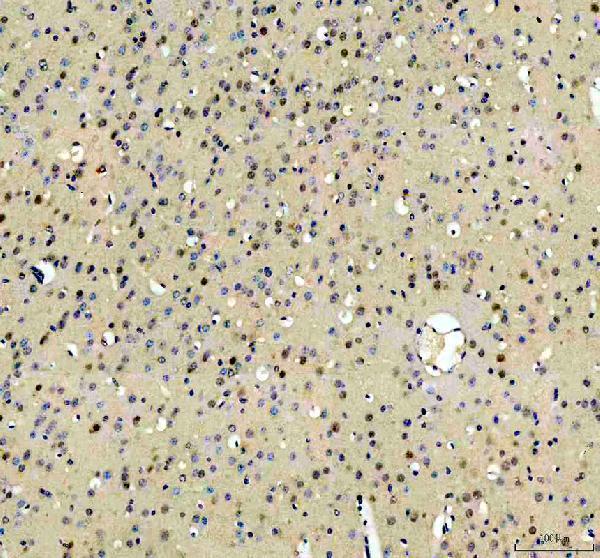
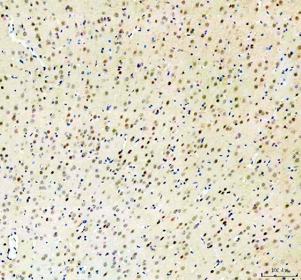
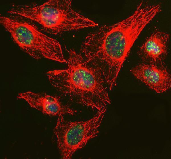
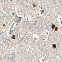
The IHC-validated anti-RBM25 antibody has images from paraffin brain sections of human, mouse, and rat, plus IF data from U2OS cells (catalog IHC and IF captions).

Real IHC data IHC analysis of RBM25 using anti-RBM25 antibody (A07494-1). RBM25 was detected in a paraffin-embedded section of mouse brain tissue. Heat mediated antigen retrieval was performed in EDTA buffer (pH 8.0, epitope retrieval solution). The tissue section was blocked with 10% goat serum. The tissue section was then incubated with 2 μg/ml rabbit anti-RBM25 Antibody (A07494-1) overnight at 4°C. Peroxidase Conjugated Goat Anti-rabbit IgG was used as secondary antibody and incubated for 30 minutes at 37°C. The tissue section was developed using HRP Conjugated Rabbit IgG Super Vision Assay Kit (Catalog # SV0002) with DAB as the chromogen.
Anti-RBM25 Antibody ®
Cat # A07494-1

A07494-1 has IHC images from paraffin brain sections of human, mouse, and rat (catalog IHC captions). A07494-1 lists IF and ICC applications and has an IF image from U2OS cells (catalog applications; catalog IF caption).

Which to pick: Choose A07494-1 for chromogenic tissue IHC: its paraffin brain captions document EDTA retrieval, 2 μg/ml primary antibody, and HRP/DAB detection (catalog IHC captions). For IF/ICC, A07494-1 has a U2OS IF image using 5 μg/ml primary antibody (catalog IF caption). The same SKU has human, mouse, and rat paraffin IHC images; its clone type and the sections’ fixative are unreported (catalog record; catalog IHC captions).

Each figure is that product's own IHC / IF validation image from its datasheet.

References

  1. UniProt Consortium. UniProt entry P49756 (RBM25_HUMAN, RNA-binding protein 25).
  2. Human Protein Atlas. RBM25 tissue IHC expression (reliability: Supported).
  3. Human Protein Atlas. RBM25 subcellular location (ICC-IF): Localized to the nuclear speckles..
  4. Human Protein Atlas. RBM25 antibody validation summary (2 antibodies).
  5. RBM25 Drives Hepatocellular Carcinoma Progression by Stabilizing YAP Through Regulating Oncogenic Splicing-switch of MYPT1. Advanced science (Weinheim, Baden-Wurttemberg, Germany) 2026 — PMC13583231.
  6. RBM25 Regulates p38 MAPK Pathway Activation via Exon 16 Skipping of MAP4K4 in a Rat Model of Post-Infarction Heart Failure. FASEB bioAdvances 2025 — PMC12707302.
  7. Prognostic alternative splicing regulatory network of RBM25 in hepatocellular carcinoma. Bioengineered 2021 — PMC8806338.
  8. PubMed PMID:14702039 — UniProt-cited evidence.
  9. PubMed PMID:12508121 — UniProt-cited evidence.
  10. PubMed PMID:15489334 — UniProt-cited evidence.