This website uses cookies to ensure you get the best experience on our website.


- Table of Contents
Plan RBM3 paraffin section IHC around the nuclear staining seen in most tissues (HPA tissue IHC). Use nasopharyngeal basal cells as a positive reference (HPA tissue IHC) and titrate the catalog antibody within 1:50–1:200 (datasheet M03104).

Expected localisation, validated protocols, controls and antibodies — the at-a-glance facts below, then the full design guide.
| Expected localisation | Nuclear staining in most tissues (HPA tissue IHC) | |
| Staining pattern | Nuclear staining in most tissue cell types (HPA tissue IHC) | |
| Antigen retrieval | EDTA pH 8.0 HIER, heat-mediated (datasheet M03104) | |
| Positive control | Nasopharynx+4 more · see all | |
| Negative control | Adipose tissue+4 more · see all |
| Fixation | Keep fixation consistent across sections. (standard IHC practice; not target-specific) | |
| Caveat | Staining and RNA expression show medium consistency (HPA tissue IHC) | |
| Regulation | Cold inducible (UniProt) | |
| Isoform / epitope | No annotated isoforms; chain spans residues 1–157 (UniProt) |
Compare the catalog antibody’s IHC-P protocol (datasheet M03104) with published RBM3 protocols for prostate, breast, melanoma, and esophageal tissue (PMC3195697; PMC6158158; PMC3156749; PMC6233549).
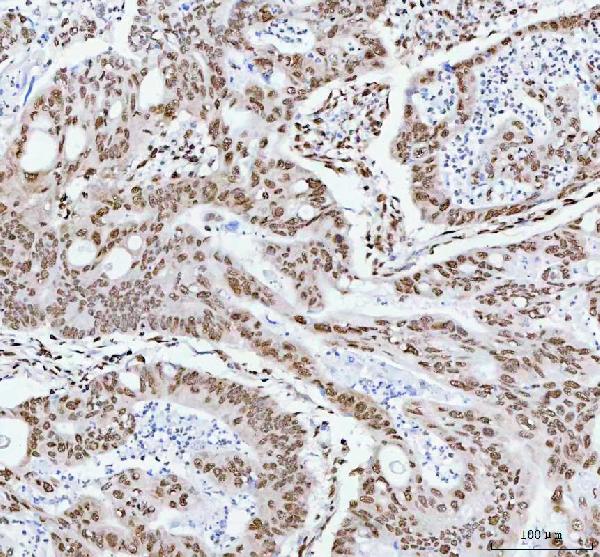
| Sample | Paraffin-embedded human colorectal adenocarcinoma tissue; fixative not specified (datasheet M03104) |
| Fixation | Image fixative and duration unreported (datasheet M03104); verify before use. |
| Sectioning | 4–5 µm sections on charged slides (standard) |
| Deparaffinisation | Xylene, graded ethanol series to water (standard) |
| Antigen retrieval | Heat retrieval: EDTA pH 8.0 (datasheet M03104); 20 min, 95–100 °C (standard) |
| Peroxidase block | 3% H2O2, 10 min, room temperature (standard) |
| Blocking | 10% goat serum (datasheet M03104) |
| Primary antibody | Rabbit monoclonal (clone 29R08) anti-RBM3, 1:50-1:200 (datasheet M03104) |
| Primary incubation | Overnight at 4 °C (datasheet M03104) |
| Detection | HRP-conjugated secondary, DAB chromogen (datasheet M03104) |
| Counterstain | Hematoxylin, blue, dehydrate and mount (standard) |
| Expected result | RBM3-positive staining in basal cells of nasopharynx (HPA tissue IHC: High). HPA tissue profile: Nuclear expression in most tissues. No signal in the no-primary control. |
RBM3 is predominantly nuclear in tissue IHC (HPA: nuclear expression in most tissues), with nucleoplasmic localisation in ICC-IF (HPA: enhanced). Expect strong staining in nasopharyngeal basal cells and moderate staining in several epithelial and lymphoid cell types (HPA: High; Medium). UniProt also records cytoplasm and dendritic mRNA granules, so localisation can vary by context (UniProt P98179). RBM3 has no transmembrane segment (UniProt P98179 topology).
| Nuclear signal is strongest in nasopharyngeal basal cells, with weaker or patchier staining in other cells. | This fits the tissue reference: basal cells are High, while appendix glandular cells, cervical squamous cells and lymph-node germinal-center cells are Medium (HPA: tissue IHC). Judge intensity within identified cell types; HPA reports low tissue RNA specificity and only medium consistency between antibody staining and RNA expression (HPA: reliability Enhanced). |
| Staining is predominantly cytoplasmic, membranous or extracellular, with little nuclear signal. | This departs from the expected tissue pattern (HPA: nuclear expression in most tissues). Check morphology, counterstain, detection controls and whether the signal follows tissue edges or deposits (general IHC practice). Cytoplasmic RBM3 is biologically possible, so cytoplasmic signal alone is not proof of artefact (UniProt P98179: cytoplasm). Membrane localisation is unsupported by the supplied record (UniProt P98179 topology). |
| Strong staining appears in an HPA-listed undetected cell type, such as adipocytes or cardiomyocytes. | Treat this as a discordance to investigate, not an automatic biological negative: those specific cell types were Not detected in the cited tissue samples (HPA: adipose tissue; heart muscle). Review cell identification, antibody-specific staining and endogenous detection activity with appropriate controls (general IHC practice). HPA's Enhanced tissue reliability still allows sample-level variation (HPA: medium staining–RNA consistency). |
| A broad haze covers nuclei, cytoplasm and blank areas, obscuring cell boundaries. | Diffuse signal without a cell-type or nuclear pattern is difficult to score against HPA's nuclear reference (HPA: tissue IHC). Uneven reagent coverage, insufficient blocking or washing, excess detection reagent, or chromogen precipitate are possible workflow causes (general IHC practice). Compare a no-primary control and inspect unstained spaces before calling RBM3 positive (general IHC practice). |
| Nasopharyngeal basal-cell nuclei show no signal in a section expected to be positive. | The result conflicts with the High reference staining for that cell type (HPA: nasopharynx basal cells), but one absent result cannot identify the failed step. Confirm that basal cells are present, then check the antibody's IHC-P instructions, retrieval and detection controls (general IHC practice). The supplied sources do not establish RBM3-specific fixation sensitivity. |
| Tissue and cell-type reference | Use the named cell type, not the organ alone, for comparison: nasopharyngeal basal cells are High; lung type-I alveolar cells and lymph-node germinal-center cells are Medium; adipocytes are Not detected (HPA: tissue IHC). These are observed examples, not universal thresholds; HPA reports low tissue RNA specificity and medium staining–RNA consistency (HPA: reliability Enhanced). |
| Compartment and molecular topology | The primary tissue expectation is nuclear (HPA: tissue IHC), and ICC-IF places RBM3 in the nucleoplasm (HPA: enhanced subcellular location). UniProt also lists cytoplasm and dendritic mRNA granules, so assess unusual cytoplasmic staining with controls rather than rejecting it outright (UniProt P98179). No transmembrane segment or signal peptide supports a routine membrane or secreted pattern (UniProt P98179 topology; processing). |
| Antibody validation | HPA lists IHC as Enhanced for CAB062559 and Supported for HPA003624; ICC is Supported for both (HPA: antibody validation). These grades are antibody-specific and do not validate every staining condition or unexpected compartment. Interpret a discordant result alongside the chosen antibody's validation status and control sections (HPA: antibody validation; general IHC practice). |
| IF/ICC Q: What pattern is expected? | A: Nucleoplasmic staining is the reported main location; HPA has ICC-IF images in A-431, U2OS and BJ cells (HPA: subcellular). That observation helps interpret localisation but does not supply an IHC-P retrieval, dilution or detection protocol (HPA: subcellular). |
| Processing and modifications | UniProt lists one 1–157 chain, no signal peptide, no propeptide, no annotated glycosylation sites and no isoforms (UniProt P98179). It also records arginine methylation, but the supplied sources do not connect those modifications to a particular antibody epitope or staining intensity; avoid using them to explain a weak section (UniProt P98179). |
| Situation | Likely cause | Next action |
|---|---|---|
| No nuclear signal in nasopharyngeal basal cells (HPA: High). | The expected cells may be absent from the field, or an IHC step may have failed; the source data do not identify a specific failure (HPA: nasopharynx; general IHC practice). | Verify basal-cell morphology and section integrity, then review the catalog antibody's IHC-P instructions and run an appropriate positive control through the same retrieval and detection workflow (general IHC practice). |
| Signal is present but too weak to score in an expected-positive cell type (HPA: tissue IHC). | Weak detection may reflect section or workflow performance; HPA's Medium categories are observed levels, not guaranteed intensity in every specimen (HPA: tissue IHC; general IHC practice). | Compare a validated positive section in the same run and check antibody preparation, retrieval execution, incubation and chromogen development against the chosen IHC-P instructions (general IHC practice). Avoid assigning an RBM3-specific fixation effect from these sources. |
| Nuclei stain uniformly across cells expected to differ (HPA: High, Medium and Not detected examples). | Overdeveloped chromogen, excessive detection signal or background may obscure cell-type differences (general IHC practice); HPA's categories do not define a universal quantitative cutoff (HPA: tissue IHC). | Review development and wash steps, examine a no-primary control, and score only clearly identified cells with interpretable nuclear morphology (general IHC practice). Compare the same cell type across control and test sections. |
| Staining is concentrated at tissue edges or in empty spaces (general IHC practice). | Edge effects, drying or precipitate can produce spatial patterns unrelated to the nuclear tissue reference (general IHC practice; HPA: tissue IHC). | Inspect reagent coverage and chromogen preparation, repeat with even section wetting, and use a no-primary control to identify detection background (general IHC practice). Do not count acellular deposits as RBM3-positive cells. |
| Only cytoplasmic staining appears, without convincing nuclear staining (HPA: tissue IHC). | The result is discordant with HPA's predominant nuclear pattern, although UniProt permits cytoplasmic localisation (HPA: tissue IHC; UniProt P98179). Compartment misidentification or background is also possible (general IHC practice). | Check the nuclear counterstain and cell boundaries, compare a known-positive section, and assess no-primary staining (general IHC practice). Report a reproducible cytoplasmic pattern separately from the expected nuclear score. |
| A HPA-listed undetected cell type stains strongly (HPA: adipocytes, cardiomyocytes or bone-marrow hematopoietic cells). | The finding may reflect cell-identification error, antibody cross-reactivity, endogenous detection activity or sample variation; HPA provides an observed reference, not proof of absence in every specimen (HPA: tissue IHC; general IHC practice). | Recheck morphology, examine a no-primary control and compare the positive control in the same run (general IHC practice). If the signal persists, describe the cell type and compartment rather than calling the HPA reference disproven. |


Comprehensive Human Protein Atlas IHC scoring per tissue (reliability: Enhanced — Medium consistency between antibody staining and RNA expression data.). Rows are taken directly from the HPA tissue chart — click any row's HPA link to view the source.
| Tissue | Cell type | Level | Evidence | Source |
|---|---|---|---|---|
| Nasopharynx | Basal cells | High | Protein (IHC) | HPA → |
| Appendix | Glandular cells | Medium | Protein (IHC) | HPA → |
| Cervix | Squamous epithelial cells | Medium | Protein (IHC) | HPA → |
| Epididymis | Glandular cells | Medium | Protein (IHC) | HPA → |
| Esophagus | Squamous epithelial cells | Medium | Protein (IHC) | HPA → |
| Tissue | Cell type | Level | Evidence | Source |
|---|---|---|---|---|
| Adipose tissue | Adipocytes | Not detected | Protein (IHC) | HPA → |
| Adrenal gland | Glandular cells | Not detected | Protein (IHC) | HPA → |
| Bone marrow | Hematopoietic cells | Not detected | Protein (IHC) | HPA → |
| Caudate | Glial cells | Not detected | Protein (IHC) | HPA → |
| Cerebellum | Cells in granular layer | Not detected | Protein (IHC) | HPA → |
Troubleshoot RBM3 staining in paraffin sections by checking retrieval, nuclear localisation, controls and scoring before interpreting chromogenic signal.
Anti-RBM3 M03104 has IHC images from paraffin-embedded human tumor sections (M03104 IHC captions) and IF data from A549 cells (M03104 IF caption); human and mouse reactivity is listed (catalog: M03104).
M03104 is listed for IHC and IF/ICC (catalog: M03104 applications), with IHC images from paraffin-embedded human colorectal adenocarcinoma, testicular germ cell tumor, liver cancer, and lung adenocarcinoma sections (M03104 IHC captions). Its IF image shows A549 cells (M03104 IF caption), and the catalog lists human and mouse reactivity (catalog: M03104 reactivity).
Which to pick: Choose M03104 for paraffin-section IHC: it is a rabbit monoclonal listed for IHC (catalog: M03104), and its own captions document EDTA retrieval at pH 8.0 and 1:50 primary dilution (M03104 IHC captions). The IHC captions do not report the fixative (M03104 IHC captions). Choose M03104 for IF/ICC based on its application listing and A549 IF image (catalog: M03104 applications; M03104 IF caption); for cross-species work, it lists human and mouse reactivity, while the supplied IHC images show human tissue (catalog: M03104 reactivity; M03104 IHC captions).