This website uses cookies to ensure you get the best experience on our website.


- Table of Contents
Plan RBMX paraffin-section IHC around its nuclear staining across most tissues (HPA tissue IHC). Use high-staining adrenal glandular cells and low-staining adipocytes as reference patterns, while accounting for the atlas warning that staining may detect products from more than one gene (HPA tissue IHC).

Expected localisation, validated protocols, controls and antibodies — the at-a-glance facts below, then the full design guide.
| Expected localisation | Nuclear staining in most tissues (HPA tissue IHC) | |
| Staining pattern | Nuclear staining across most tissue cell types (HPA tissue IHC) | |
| Antigen retrieval | Tris-EDTA pH 9.0 HIER, heat-mediated (datasheet A04738) | |
| Positive control | Adrenal gland+4 more · see all | |
| Negative control | None in HPA (detected in all 45 tissues); use no-primary + isotype controls |
| Fixation | Keep fixation consistent across paraffin sections. (standard IHC practice; not target-specific) | |
| Caveat | Staining may detect products from more than one gene (HPA tissue IHC) | |
| Regulation | Weaker in oral squamous carcinomas (UniProt) | |
| Isoform / epitope | 3 isoforms; N-terminal processing may affect epitopes near residue 1 (UniProt) |
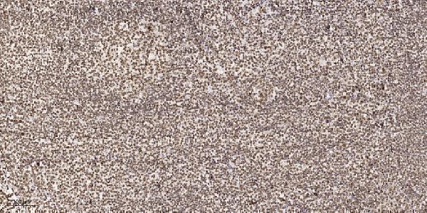
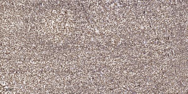
The catalog antibody’s IHC-P protocol is paired with published chromogenic RBMX IHC methods for lymphoma, esophageal carcinoma, and paraffin-embedded tissue (PMC8507920; PMC10583014; PMC10442647).
| Sample | Paraffin-embedded human tonsil tissue; fixative not specified (datasheet A04738) |
| Fixation | Image fixative and duration unreported (datasheet A04738); verify before use. |
| Sectioning | 4–5 µm sections on charged slides (standard) |
| Deparaffinisation | Xylene, graded ethanol series to water (standard) |
| Antigen retrieval | Heat retrieval: Tris-EDTA pH 9.0 (datasheet A04738); 20 min, 95–100 °C (standard) |
| Peroxidase block | 3% H2O2, 10 min, room temperature (standard) |
| Blocking | 10% normal serum of the secondary host, 30 min, room temperature (standard) |
| Primary antibody | Rabbit anti-RBMX, 1:100-1:300 (datasheet A04738) |
| Primary incubation | Overnight at 4 °C (standard) |
| Detection | HRP-polymer secondary, DAB chromogen 5–10 min (standard) |
| Counterstain | Hematoxylin, blue, dehydrate and mount (standard) |
| Expected result | RBMX-positive staining in glandular cells of adrenal gland (HPA tissue IHC: High). HPA tissue profile: Ubiquitous nuclear expression in most tissues. No signal in the no-primary control. |
RBMX is nuclear, with numerous small nuclear granules, and has no transmembrane segment (UniProt P38159). In paraffin-section IHC, expect nuclear staining across many tissues, including respiratory epithelial, glandular and neuronal cells (HPA: ubiquitous nuclear profile; High in listed cells). HPA rates tissue IHC Supported, with medium agreement with RNA data, pending external verification, and cautions that staining may include proteins from more than one gene (HPA: reliability description).
| Clear nuclear staining in bronchial respiratory epithelial cells, with nuclei identifiable by counterstain. | This fits the expected compartment and a documented High-staining cell type (UniProt P38159: nucleus; HPA: High in bronchial respiratory epithelial cells). Record the proportion and intensity of stained nuclei while keeping tissue structure visible. This supports an interpretable staining pattern, although HPA cautions that the antibody evidence may include proteins from more than one gene (HPA: tissue IHC reliability description). |
| Predominantly cytoplasmic staining, with little or no nuclear signal in cells expected to stain. | Treat this as a compartment mismatch and investigate an artefact before assigning it to RBMX (UniProt P38159: nuclear localization; HPA: ubiquitous nuclear IHC profile). UniProt also describes a role in cytoplasmic TNFR1 trafficking, but that functional annotation does not establish predominantly cytoplasmic IHC staining (UniProt P38159: function). Check the counterstain and detection controls, then compare nuclear and cytoplasmic signal in the same cell type (general IHC practice). |
| Strong signal is confined to an unexpected cell population while documented High-staining cells lack nuclear signal. | Compare the actual cell identities with the tissue-specific HPA entry before calling the pattern unexpected: HPA reports High staining in colon endothelial cells, for example, and describes expression as broadly nuclear (HPA: colon entry; tissue profile). A persistent mismatch can indicate cross-reactivity or endogenous detection activity; morphology alone cannot distinguish those causes (general IHC practice). HPA's multiple-gene caution makes target attribution especially qualified (HPA: reliability description). |
| Diffuse colour across nuclei, cytoplasm and tissue spaces obscures cell boundaries. | This is difficult to score as RBMX because the supported pattern is nuclear (HPA: tissue IHC profile; UniProt P38159: localization). Assess whether background also appears in a no-primary control, and review blocking, washing and chromogen development as general IHC checks (general IHC practice). Diffuse background does not establish that RBMX is absent from the section. |
| No discernible nuclear signal in bronchial respiratory epithelium or another documented High-staining population. | Do not immediately interpret this as biological absence: these populations are reference points for an expected positive pattern (HPA: High in bronchial respiratory epithelial cells and other listed cells). Confirm that the expected cells are present, examine a concurrently stained positive section, and review the IHC-validated antibody and detection workflow (general IHC practice). HPA's Supported rating describes agreement, not a guarantee that every specimen will stain (HPA: reliability description). |
| Tissue and cell selection | Bronchial respiratory epithelial, cerebral cortical neuronal and adrenal glandular cells are documented High-staining examples; adipocytes are listed Low (HPA: tissue IHC entries). Use cell identity when comparing sections: a weak adipocyte signal is less informative than absent nuclear signal in a documented High-staining population (HPA: tissue IHC entries). |
| Antibody interpretation | The listed antibody HPA057707 has Supported IHC status, while HPA reports medium staining-to-RNA consistency, pending external verification, and possible detection of proteins from multiple genes (HPA: antibody status; reliability description). A plausible nuclear pattern alone therefore cannot prove RBMX-specific recognition. |
| Isoforms and processing | UniProt lists 3 isoforms and chains beginning at residue 1 or 2 (UniProt P38159: isoforms; processing). No antibody epitope is supplied, so the record cannot establish which isoforms or processed forms this IHC stain detects; avoid assigning a difference in intensity to one form. |
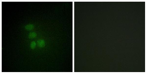
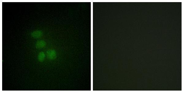
| IF/ICC Q&A: where should signal appear? | In IF/ICC, expect nucleoplasmic signal; UniProt also describes numerous small nuclear granules (HPA: supported nucleoplasm; UniProt P38159: localization). HPA lists HEK293, MCF-7 and U2OS image sets but cautions that its subcellular assignment uses antibodies targeting proteins from multiple genes (HPA: subcellular record). |
| Situation | Likely cause | Next action |
|---|---|---|
| A documented High-staining cell type has no nuclear signal. | The expected cells may be absent from the section, or the staining run may have failed; this observation alone cannot distinguish either possibility from biological variation (HPA: High tissue entries; general IHC practice). | Locate the expected cells by morphology and counterstain, then compare a concurrently processed positive section. Review antibody use and detection against the available IHC protocol before interpreting the result (general IHC practice). |
| Signal appears mainly outside nuclei. | The compartment conflicts with the supported nuclear pattern; non-specific staining or a detection artefact is possible (HPA: ubiquitous nuclear IHC profile; UniProt P38159: nucleus; general IHC practice). | Compare signal with the counterstained nuclei and check a no-primary control. If extranuclear colour persists, report the compartment mismatch rather than scoring it as the expected RBMX pattern (general IHC practice). |
| Broad tissue colour makes positive nuclei hard to identify. | Background from the staining or detection workflow may obscure localized signal; the record provides no RBMX-specific cause for diffuse background (general IHC practice). | Check a no-primary control and review blocking, washing and development time. Score nuclear staining only where nuclei and cell boundaries remain interpretable (general IHC practice). |
| Unexpected cells stain more strongly than the documented reference population. | Cell identification, cross-reactivity or endogenous detection activity could contribute; HPA also cautions about proteins from multiple genes (general IHC practice; HPA: reliability description). | Verify cell identity on the section, consult the matching HPA tissue entry, and inspect a detection control. Describe the observed cells and compartment without assigning target specificity from intensity alone (HPA: tissue IHC entries; general IHC practice). |
| Adipocytes stain weakly while nearby cells show nuclear signal. | Low adipocyte staining is documented and need not indicate a failed run (HPA: Low in adipocytes; ubiquitous nuclear tissue profile). | Judge run performance against a documented High-staining cell population on a suitable section. Record adipocyte staining separately by cell type and intensity (HPA: tissue IHC entries; general IHC practice). |
| A nuclear pattern looks convincing, but RBMX specificity is the deciding question. | Correct localization supports pattern agreement but does not resolve HPA's warning about detection of proteins from more than one gene (UniProt P38159: nucleus; HPA: reliability description). | Report the result as pattern-consistent and retain the specificity caveat. If a target-specific conclusion is required, seek an independent validation approach appropriate to the experiment (general IHC practice; HPA: reliability description). |

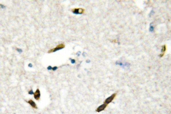


Comprehensive Human Protein Atlas IHC scoring per tissue (reliability: Supported — Medium consistency between antibody staining and RNA expression data. Pending external verification. Caution, targets protein from more than one gene.). Rows are taken directly from the HPA tissue chart — click any row's HPA link to view the source.
| Tissue | Cell type | Level | Evidence | Source |
|---|---|---|---|---|
| Adrenal gland | Glandular cells | High | Protein (IHC) | HPA → |
| Appendix | Glandular cells | High | Protein (IHC) | HPA → |
| Bronchus | Respiratory epithelial cells | High | Protein (IHC) | HPA → |
| Caudate | Neuronal cells | High | Protein (IHC) | HPA → |
| Cerebellum | Cells in granular layer | High | Protein (IHC) | HPA → |
| Tissue | Cell type | Level | Evidence | Source |
|---|---|---|---|---|
| None in HPA: RBMX is detected in all 45 scored tissues. Use a no-primary (secondary-only) and an isotype control instead. | ||||
Use nuclear staining as the main readout for RBMX in paraffin section IHC, while checking controls and tissue context before interpreting intensity (UniProt P38159; HPA tissue IHC).
The catalog includes RBMX antibodies with human tissue IHC images for tonsil, brain and testis, plus IF images in HepG2 cells (catalog image captions). Human, mouse and rat reactivity is listed for selected SKUs (catalog reactivity).
A04738 and A04738-1 each show IHC in paraffin-embedded human tonsil and IF in HepG2 cells (catalog image captions); A04738-2 shows IHC in paraffin-embedded human brain (catalog IHC image caption). M04738 shows IHC in formaldehyde-fixed, paraffin-embedded human testis (catalog IHC image caption), and its application list includes IHC-P but excludes IF/ICC (catalog applications).
Which to pick: For tissue IHC, choose A04738 or A04738-1 for paraffin-embedded tonsil examples, A04738-2 for a paraffin-embedded brain example, or M04738 for a paraffin-section, paraffin-embedded testis example (each SKU’s IHC image caption); the fixative is unreported for A04738, A04738-1 and A04738-2 (their IHC image captions). For IF/ICC, A04738-1 lists both applications, while A04738 lists IF; both have HepG2 IF images and are listed as polyclonal (catalog applications, IF image captions and dilution data). For work across human, mouse and rat samples, consider A04738, A04738-1 or A04738-2 based on listed reactivity, while treating their tissue IHC images as human examples (catalog reactivity and IHC image captions). The selected A04738 tissue-IHC caption documents paraffin sections, but does not specify the fixative (selected-SKU IHC image A04738).