This website uses cookies to ensure you get the best experience on our website.


- Table of Contents
Plan chromogenic IHC on paraffin sections with the catalog antibody at a 1:50–1:200 starting dilution (datasheet). Compare cell type and staining intensity with the cytoplasmic tissue profile (HPA tissue IHC).

Expected localisation, validated protocols, controls and antibodies — the at-a-glance facts below, then the full design guide.
| Expected localisation | Cytoplasmic tissue staining (HPA tissue IHC); ER lumen (UniProt) | |
| Staining pattern | Cytoplasmic signal in neurons, placenta and epididymis (HPA tissue IHC) | |
| Antigen retrieval | Citrate pH 6.0 HIER, 95–98 °C, 20 min (rule: cytoplasmic / membrane antigen) | |
| Positive control | Caudate+4 more · see all | |
| Negative control | Adipose tissue+4 more · see all |
| Fixation | Keep fixation consistent across sections (standard IHC practice; not target-specific); Matched tissue-IHC evidence does not establish the fixation claim. Validate the specimen-specific method before use. (selected-SKU IHC image A10286) | |
| Caveat | Some cell types show little or no detectable staining (HPA tissue IHC) | |
| Regulation | Tissue enhanced RNA in epididymis (HPA tissue RNA) | |
| Isoform / epitope | 2 isoforms; signal peptide cleaved, no TM split; epitope unknown (UniProt) |
The catalog antibody protocol is paired with published RCN2 IHC methods for arterial tissue (PMC8997427) and mouse xenograft tumors (PMC10356900).
| Sample | Paraffin-embedded human gastric tissue; fixative not specified (datasheet A10286) |
| Fixation | Image fixative and duration unreported (datasheet A10286); verify before use. |
| Sectioning | 4–5 µm sections on charged slides (standard) |
| Deparaffinisation | Xylene, graded ethanol series to water (standard) |
| Antigen retrieval | Heat-induced epitope retrieval in citrate buffer, pH 6.0, 20 min at 95–98 °C (standard rule: cytoplasmic / membrane antigen) |
| Peroxidase block | 3% H2O2, 10 min, room temperature (standard) |
| Blocking | 10% normal serum of the secondary host, 30 min, room temperature (standard) |
| Primary antibody | Rabbit anti-RCN2, 1:50-1:200 (datasheet A10286) |
| Primary incubation | Overnight at 4 °C (standard) |
| Detection | HRP-polymer secondary, DAB chromogen 5–10 min (standard) |
| Counterstain | Hematoxylin, blue, dehydrate and mount (standard) |
| Expected result | RCN2-positive staining in neuronal cells of caudate (HPA tissue IHC: High). HPA tissue profile: Cytoplasmic expression in selected tissues including neurons, seminiferous duct cells, placenta and epididymis. No signal in the no-primary control. |
RCN2 is a soluble endoplasmic reticulum lumen protein with no transmembrane segment (UniProt Q14257 topology). In paraffin-section IHC, expect predominantly cytoplasmic staining in selected cells, especially epididymal glandular cells, neurons and pachytene spermatocytes (HPA tissue IHC: High). HPA rates the tissue pattern Enhanced because staining agrees with RNA expression, while noting that external verification is pending (HPA tissue IHC: reliability).
| Clear cytoplasmic staining in epididymal glandular cells, with little staining in adjacent negative cells. | This fits a high-expression tissue and cell population (HPA tissue IHC: Epididymis, glandular cells, High). Chromogenic IHC cannot by itself resolve the endoplasmic reticulum, so score the cell and broad compartment rather than requiring a visible reticular network (UniProt Q14257: ER lumen; general IHC practice). |
| Cytoplasmic staining in neuronal cells or pachytene spermatocytes. | This agrees with reported High staining in caudate, cerebral cortex and hippocampal neurons, and in testicular pachytene spermatocytes (HPA tissue IHC). Compare the named cell population, not overall tissue darkness; surrounding cells can have different reported levels (HPA tissue IHC: cell-specific annotations). |
| Predominantly nuclear staining or a crisp cell-surface rim without convincing cytoplasmic signal. | Treat an isolated nuclear or surface pattern as suspect: the annotated location is the ER lumen, and RCN2 has no transmembrane segment (UniProt Q14257 topology; HPA subcellular: ER). Review morphology and controls before calling it positive. A cytoplasm/membrane annotation for cerebellar granular cells does not establish exclusive cell-surface localization (HPA tissue IHC). |
| Strong stain in a cell population reported as Not detected, especially with staining in blank areas. | Adipocytes in adipose tissue and bronchial basal cells are reported Not detected (HPA tissue IHC). Such a result calls for checking nonspecific binding and, with chromogenic detection, endogenous detection activity using appropriate controls (general IHC practice). A negative HPA annotation is a comparison point, not proof that every specimen must be unstained. |
| Diffuse staining across many structures, or no stain in a known-positive control section. | Diffuse signal without cell boundaries is hard to assign to the HPA cell-specific pattern (HPA tissue IHC). Absent signal in epididymal glandular cells or a listed high-staining neuronal population fails the expected positive-control check (HPA tissue IHC: High). Investigate the assay and section before interpreting the experimental sample as RCN2 negative (general IHC practice). |
| Cell population and tissue selection | RCN2 is described as ubiquitous at the tissue level (UniProt Q14257), while HPA reports High staining in selected cell populations and Not detected staining in others (HPA tissue IHC). Select and score a named cell population; the word “ubiquitous” does not predict uniform chromogenic intensity across a section. |
| Compartment and optical resolution | The mature protein is annotated in the ER lumen, with no transmembrane segment (UniProt Q14257 topology). HPA reports mainly ER localization by ICC-IF (HPA subcellular). In paraffin-section IHC, a cytoplasmic pattern can be consistent with that location, but chromogenic staining alone does not prove ER colocalization (general IHC practice). |
| Antibody evidence | HPA030694 and HPA030695 each have Enhanced IHC validation (HPA antibodies). The tissue profile also has Enhanced reliability because staining is consistent with RNA data, with external verification pending (HPA tissue IHC: reliability). These ratings support comparison with the reported pattern; they do not establish that every specimen or assay condition will reproduce it. |
| Protein processing and isoforms | RCN2 has a signal peptide at residues 1–22, a mature chain at 23–317 and two listed isoforms (UniProt Q14257). The supplied sources give no antibody epitope or isoform-specific staining result, so they cannot justify assigning a changed IHC pattern to signal-peptide processing or one isoform. |
| Fixation and antigen retrieval | Target-specific fixation sensitivity and retrieval conditions are unreported in the supplied UniProt and HPA records. If staining fails, use the IHC-validated antibody’s documented paraffin-section procedure and its controls when available (general IHC practice); do not infer an RCN2-specific retrieval requirement from the ER annotation or tissue staining levels. |
| Situation | Likely cause | Next action |
|---|---|---|
| No signal in epididymal glandular cells on the positive-control section. | The section, detection sequence or antibody conditions may have failed; these cells are reported High (HPA tissue IHC). The supplied sources do not identify an RCN2-specific fixation or retrieval failure. | Verify the control section and IHC workflow, including retrieval, antibody incubation and chromogenic detection, against the IHC-validated antibody’s documented procedure (general IHC practice). |
| Staining appears mainly nuclear or confined to the outer cell border. | That distribution conflicts with the ER-lumen annotation and absence of a transmembrane segment (UniProt Q14257 topology; HPA subcellular). Morphology or nonspecific signal could mislead compartment assignment (general IHC practice). | Recheck focus and cellular boundaries, compare a known-positive population, and examine a no-primary control before scoring RCN2 (general IHC practice). |
| Adipocytes or bronchial basal cells stain as strongly as the intended positive cells. | Both populations are reported Not detected (HPA tissue IHC). Nonspecific antibody binding or endogenous chromogenic activity is possible (general IHC practice); the HPA comparison alone cannot identify which mechanism applies. | Check the no-primary and detection controls, then assess blocking and wash conditions using standard IHC practice. Score the intended positive and negative cell populations separately (general IHC practice). |
| Brown precipitate or diffuse color obscures cell boundaries. | Excess background or endogenous detection activity may make a cell-specific pattern unreadable (general IHC practice). HPA describes cytoplasmic expression in selected cells, not a uniform tissue-wide signal (HPA tissue IHC). | Inspect a no-primary control, review chromogen development and washes, and repeat under the documented IHC procedure if needed (general IHC practice). |
| Neuronal or testicular signal is present, but neighboring cells vary in intensity. | HPA assigns High staining to specified neuronal populations and pachytene spermatocytes, not every cell in those tissues (HPA tissue IHC). Heterogeneity alone is therefore insufficient to call assay failure. | Identify the annotated cells by morphology and score intensity within those cells; compare the same cell type across sections and include a suitable control (general IHC practice). |
| IF/ICC question: should a paraffin-section IHC pattern look reticular? | ICC-IF reports mainly ER localization and Enhanced ER assignment (HPA subcellular), whereas paraffin-section chromogenic IHC has different spatial resolution (general IHC practice). | For this IHC guide, assess cytoplasmic staining in the HPA-reported cell populations (HPA tissue IHC). Use the separate IF/ICC guide for fluorescence localization; do not treat a missing visible ER network in chromogenic IHC as failure (general IHC practice). |
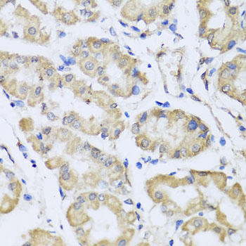
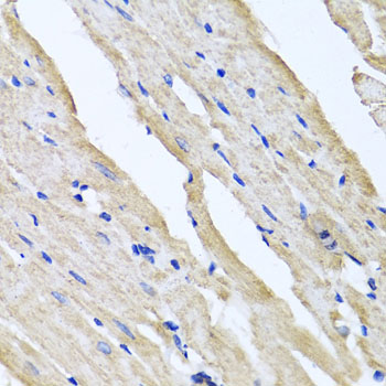
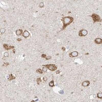


Comprehensive Human Protein Atlas IHC scoring per tissue (reliability: Enhanced — High consistency between antibody staining and RNA expression data. Pending external verification.). Rows are taken directly from the HPA tissue chart — click any row's HPA link to view the source.
| Tissue | Cell type | Level | Evidence | Source |
|---|---|---|---|---|
| Caudate | Neuronal cells | High | Protein (IHC) | HPA → |
| Cerebellum | Granular cells - cytoplasm/membrane | High | Protein (IHC) | HPA → |
| Cerebral cortex | Neuronal cells | High | Protein (IHC) | HPA → |
| Epididymis | Glandular cells | High | Protein (IHC) | HPA → |
| Hippocampus | Neuronal cells | High | Protein (IHC) | HPA → |
| Tissue | Cell type | Level | Evidence | Source |
|---|---|---|---|---|
| Adipose tissue | Adipocytes | Not detected | Protein (IHC) | HPA → |
| Adrenal gland | Glandular cells | Not detected | Protein (IHC) | HPA → |
| Bone marrow | Hematopoietic cells | Not detected | Protein (IHC) | HPA → |
| Breast | Adipocytes | Not detected | Protein (IHC) | HPA → |
| Bronchus | Basal cells | Not detected | Protein (IHC) | HPA → |
Use RCN2’s expected endoplasmic reticulum pattern and cell type controls to troubleshoot chromogenic staining in paraffin sections (UniProt Q14257; HPA tissue IHC).
A10286 has IHC images of paraffin-embedded human gastric tissue and rat heart (A10286 image captions). Its listed reactivity covers human, mouse, and rat (A10286 catalog).
A10286 lists IHC among its applications and human, mouse, and rat reactivity (A10286 catalog). Its IHC images show paraffin-embedded human gastric tissue and rat heart at 1:100 (A10286 image captions).
Which to pick: For tissue IHC, choose A10286 for paraffin sections; the image captions do not report a fixative (A10286 image captions). No listed SKU has IF/ICC application or image evidence, so there is no supported IF/ICC pick here (A10286 catalog). For cross-species IHC, A10286 lists human, mouse, and rat reactivity, while its IHC images cover human and rat; clonality is unreported (A10286 catalog; A10286 image captions).