This website uses cookies to ensure you get the best experience on our website.


- Table of Contents
Plan RCOR1 paraffin IHC around its general nuclear staining pattern (HPA tissue IHC). Compare high staining in bone marrow hematopoietic cells with low staining in parathyroid glandular cells (HPA tissue IHC), using 2–5 μg/ml as the catalog antibody's starting range (datasheet A02799-3).

Expected localisation, validated protocols, controls and antibodies — the at-a-glance facts below, then the full design guide.
| Expected localisation | Predominantly nuclear in tissue sections (HPA tissue IHC) | |
| Staining pattern | General nuclear staining across tissue cell types (HPA tissue IHC) | |
| Antigen retrieval | EDTA pH 8.0 HIER, heat-mediated (datasheet A02799-3) | |
| Positive control | Adipose tissue+4 more · see all | |
| Negative control | None in HPA (detected in all 44 tissues); use no-primary + isotype controls |
| Fixation | Keep fixation conditions consistent across sections. (standard IHC practice; not target-specific) | |
| Caveat | Parathyroid glandular cells show low staining (HPA tissue IHC) | |
| Regulation | No induction specified in the record (UniProt) | |
| Isoform / epitope | No isoforms annotated; one 1–485 chain (UniProt) |
The catalog antibody IHC-P protocol (datasheet A02799-3) is paired with a published RCOR1 protocol for HCC tissue microarrays (PMC10277624).
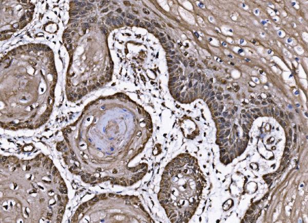
| Sample | Paraffin-embedded human esophageal squamous carcinoma tissue; fixative not specified (datasheet A02799-3) |
| Fixation | Image fixative and duration unreported (datasheet A02799-3); verify before use. |
| Sectioning | 4–5 µm sections on charged slides (standard) |
| Deparaffinisation | Xylene, graded ethanol series to water (standard) |
| Antigen retrieval | Heat retrieval: EDTA pH 8.0 (datasheet A02799-3); 20 min, 95–100 °C (standard) |
| Peroxidase block | 3% H2O2, 10 min, room temperature (standard) |
| Blocking | 10% goat serum (datasheet A02799-3) |
| Primary antibody | Rabbit anti-RCOR1, 2-5 μg/ml (datasheet A02799-3) |
| Primary incubation | Overnight at 4 °C (datasheet A02799-3) |
| Detection | Streptavidin-biotin complex (SABC), DAB chromogen (datasheet A02799-3) |
| Counterstain | Hematoxylin, blue, dehydrate and mount (standard) |
| Expected result | RCOR1-positive staining in adipocytes of adipose tissue (HPA tissue IHC: High). HPA tissue profile: General nuclear expression. No signal in the no-primary control. |
RCOR1 should stain nuclei across many cell types: HPA reports general nuclear expression, high staining in several listed tissues, and low tissue specificity (HPA tissue IHC: Supported). Its nuclear localisation and lack of a transmembrane segment support a nuclear reading of the stain (UniProt Q9UKL0: subcellular location; topology). Compare compartments and cell populations within each section, since broadly distributed expression does not imply equal intensity in every cell (HPA tissue IHC).
| Nuclear chromogen appears in hematopoietic cells of bone marrow and glandular cells of adrenal gland or appendix (HPA tissue IHC: High). | This fits the reported general nuclear pattern; judge signal in identifiable cells against the counterstain and background, allowing intensity to vary between cells (HPA tissue IHC: Supported; General IHC practice). |
| Cytoplasmic or membranous staining dominates while nuclei remain pale. | Treat this as discordant with the expected nuclear pattern and review staining specificity and detection controls (UniProt Q9UKL0: nucleus; HPA tissue IHC: general nuclear expression). UniProt reports partial cytoplasmic translocation during HSV-1 infection, so infection context matters before assigning artefact (UniProt Q9UKL0: subcellular location). |
| Strong chromogen marks an unexpected cell population or acellular material, without matching nuclei. | Check for cross-reactivity or endogenous detection activity before calling it RCOR1 (General IHC practice). HPA reports broad expression, so cell identity alone cannot establish a negative population; compare its compartment and an appropriate control (HPA tissue IHC: general nuclear expression; General IHC practice). |
| Diffuse colour fills stroma, lumina, or most of the section and obscures nuclear boundaries. | This is background rather than interpretable nuclear localisation (General IHC practice). Review blocking, reagent concentration, detection chemistry, and washes using a control section; HPA's tissue profile does not identify the procedural cause (HPA tissue IHC; General IHC practice). |
| No nuclear signal is visible in hematopoietic cells of bone marrow or adipocytes of adipose tissue (HPA tissue IHC: High). | A missing signal in these reported high-staining populations calls for a run check before interpreting other tissue as negative (HPA tissue IHC; General IHC practice). Inspect control performance, antibody use, retrieval, and detection; the supplied sources do not establish RCOR1-specific fixation sensitivity. |
| Reference tissue and cell population | HPA reports high staining in adipocytes of adipose tissue and breast; glandular cells of adrenal gland and appendix; marrow hematopoietic cells; bronchial respiratory epithelial cells; caudate glial cells; and cortical endothelial cells (HPA tissue IHC: High). Parathyroid glandular cells are reported low, so use a listed high-staining population for a positive run check (HPA tissue IHC: Low; General IHC practice). |
| Antibody evidence | HPA lists IHC support for HPA054241 and CAB001956; HPA049327 has no listed IHC status (HPA antibodies). The tissue profile is Supported because antibody staining is consistent with RNA expression; this does not validate every antibody or every protocol (HPA tissue IHC: reliability description; HPA antibodies). |
| Compartment and protein context | RCOR1 is nuclear, has no transmembrane segment or signal peptide, and is recorded as one chain of residues 1–485 (UniProt Q9UKL0: subcellular location; topology; processing). These facts support evaluating nuclear staining; they do not predict retrieval conditions or a fixation response. |
| Modified residues and epitope limits | UniProt records phosphoserines at positions 127, 260, and 460 and no glycosylation sites (UniProt Q9UKL0: modified residues; glycosylation). Their presence alone does not show whether a particular antibody's epitope or IHC signal changes; no epitope location is supplied. |
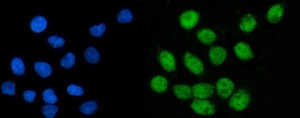
| IF/ICC Q&A: Where should RCOR1 appear? | In the nucleoplasm: HPA calls this its enhanced main location and lists ICC-IF images in A-431, MCF-7, U2OS, and NIH 3T3 (HPA subcellular). That imaging evidence supports a localisation comparison, while the primary interpretation here concerns paraffin-section IHC. |
| Situation | Likely cause | Next action |
|---|---|---|
| A listed high-staining control tissue has no nuclear chromogen (HPA tissue IHC: High). | An unsuccessful staining run or weak detection is possible; the tissue result alone does not identify the failed step (General IHC practice). | Review the control section, catalog antibody instructions, retrieval run, reagent activity, and chromogenic detection before scoring samples (General IHC practice). Do not infer RCOR1-specific fixation sensitivity from this result. |
| Nuclei are pale, but cytoplasm is strongly stained. | The compartment conflicts with the usual pattern (HPA tissue IHC: general nuclear expression; UniProt Q9UKL0: nucleus). HSV-1 infection is a documented exception (UniProt Q9UKL0: subcellular location). | Check infection context, antibody specificity evidence, and control sections; call the cytoplasmic signal unexplained if the exception is inapplicable (UniProt Q9UKL0: subcellular location; General IHC practice). |
| Colour appears in an unexpected cell population or on acellular material. | Cross-reactivity or endogenous detection activity may contribute (General IHC practice). Broad RCOR1 expression means an unexpected cell identity alone is inconclusive (HPA tissue IHC: low tissue specificity). | Compare nuclear localisation with a listed high-staining control; use appropriate reagent and detection controls to test background before assigning cellular positivity (HPA tissue IHC: general nuclear expression; General IHC practice). |
| The section has diffuse colour that hides nuclear detail. | Nonspecific reagent binding or excessive chromogenic background may obscure the expected pattern (General IHC practice; HPA tissue IHC: general nuclear expression). | Review blocking, antibody concentration against its supplied instructions, washes, and detection timing with a control section (General IHC practice). Avoid assigning a target-specific cause without supporting evidence. |
| Parathyroid glandular cells stain less than a positive control. | Low staining in that population is reported by HPA; lower intensity there need not indicate run failure (HPA tissue IHC: Low). | Judge run performance in a listed high-staining population, then assess parathyroid nuclei against local background and counterstain (HPA tissue IHC: High; General IHC practice). |
| Different antibody preparations give conflicting nuclear patterns. | Their IHC evidence differs: HPA054241 and CAB001956 are IHC Supported, while HPA049327 has no listed IHC status (HPA antibodies). | Review each antibody's stated IHC validation and instructions, then compare staining in the same listed high-staining cell population and with the same scoring criteria (HPA antibodies; HPA tissue IHC: High; General IHC practice). |
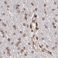


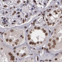


Comprehensive Human Protein Atlas IHC scoring per tissue (reliability: Supported — High consistency between antibody staining and RNA expression data.). Rows are taken directly from the HPA tissue chart — click any row's HPA link to view the source.
| Tissue | Cell type | Level | Evidence | Source |
|---|---|---|---|---|
| Adipose tissue | Adipocytes | High | Protein (IHC) | HPA → |
| Adrenal gland | Glandular cells | High | Protein (IHC) | HPA → |
| Appendix | Glandular cells | High | Protein (IHC) | HPA → |
| Bone marrow | Hematopoietic cells | High | Protein (IHC) | HPA → |
| Breast | Adipocytes | High | Protein (IHC) | HPA → |
| Tissue | Cell type | Level | Evidence | Source |
|---|---|---|---|---|
| None in HPA: RCOR1 is detected in all 44 scored tissues. Use a no-primary (secondary-only) and an isotype control instead. | ||||
Troubleshoot RCOR1 staining in paraffin sections by checking retrieval, nuclear localisation, tissue controls and scoring consistency before interpreting chromogenic signal.
A02799-3 has IHC data from a human paraffin-embedded tissue section and IF/ICC data from SiHa cells (catalog image captions); its listed reactivity is human (catalog applications/reactivity).
A02799-3 is the SKU that will render with an IHC figure from a human esophageal squamous carcinoma paraffin section (catalog IHC image caption). Its IF/ICC figure shows SiHa cells (catalog IF image caption), and its listed reactivity is human (catalog reactivity).
Which to pick: Choose A02799-3 for human tissue IHC: its own image caption documents staining in a paraffin section, with the fixative unreported (catalog IHC image caption). Choose A02799-3 for IF/ICC when image-backed evidence matters: its own caption documents SiHa cells (catalog IF image caption). For mouse or rat work, M02799-1 is the listed rabbit monoclonal option with IHC and IF/ICC applications, but the payload supplies no IHC or IF image for it (catalog clone, reactivity, applications and image captions).