This website uses cookies to ensure you get the best experience on our website.


- Table of Contents
Plan RDX IHC in paraffin sections using adrenal glandular cells and kidney proximal tubules as strong-staining references (HPA tissue IHC). Start with the catalog antibody at 1:50 (datasheet M01926), then score cytoplasmic and membranous staining (HPA tissue IHC).

Expected localisation, validated protocols, controls and antibodies — the at-a-glance facts below, then the full design guide.
| Expected localisation | Cytoplasmic and membranous staining (HPA tissue IHC) | |
| Staining pattern | Variable cytoplasmic/membranous staining; highest in adrenal gland and renal tubules (HPA tissue IHC) | |
| Antigen retrieval | EDTA pH 8.0 HIER, heat-mediated (datasheet M01926) | |
| Positive control | Adrenal gland+4 more · see all | |
| Negative control | Adipose tissue+4 more · see all |
| Fixation | Keep fixation consistent across sections (standard IHC practice; not target-specific) | |
| Caveat | Antibody may detect proteins from more than one gene (HPA tissue IHC) | |
| Regulation | Stimulus-dependent regulation is unreported (UniProt) | |
| Isoform / epitope | 5 isoforms; epitope coverage is unspecified (UniProt; datasheet M01926) |
Compare the catalog antibody’s IHC-P protocol (datasheet M01926) with one published paraffin-section, chromogenic RDX protocol (PMC2766609).
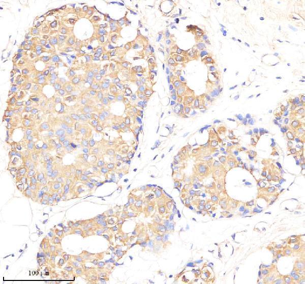
| Sample | Paraffin-embedded human breast cancer tissue; fixative not specified (datasheet M01926) |
| Fixation | Image fixative and duration unreported (datasheet M01926); verify before use. |
| Sectioning | 4–5 µm sections on charged slides (standard) |
| Deparaffinisation | Xylene, graded ethanol series to water (standard) |
| Antigen retrieval | Heat retrieval: EDTA pH 8.0 (datasheet M01926); 20 min, 95–100 °C (standard) |
| Peroxidase block | 3% H2O2, 10 min, room temperature (standard) |
| Blocking | 10% goat serum (datasheet M01926) |
| Primary antibody | Rabbit monoclonal (clone FBC-18) anti-RDX, 1:50 (datasheet M01926) |
| Primary incubation | Overnight at 4 °C (datasheet M01926) |
| Detection | HRP-conjugated secondary, DAB chromogen (datasheet M01926) |
| Counterstain | Hematoxylin, blue, dehydrate and mount (standard) |
| Expected result | RDX-positive staining in glandular cells of adrenal gland (HPA tissue IHC: High). HPA tissue profile: Cytoplasmic and membranous expression at variable levels in several different cell types. Most abundant in adrenal gland and renal tubules. No signal in the no-primary control. |
RDX should show cytoplasmic and membranous staining, with prominent signal in adrenal glandular cells and renal proximal tubule microvilli (HPA tissue IHC). Its localisation at the cell membrane, cytoskeleton and cell projections supports a peripheral or apical pattern (UniProt P35241 localisation). RDX has no transmembrane segment (UniProt P35241 topology). HPA rates tissue IHC reliability as Enhanced but cautions that the staining may include protein from more than one gene (HPA tissue IHC).
| Strong apical staining outlines renal proximal tubule microvilli, with relatively little signal away from the brush border. | This matches the reported High proximal tubule microvillus signal (HPA tissue IHC) and RDX localisation to microvilli and the membrane-associated cytoskeleton (UniProt P35241 localisation). Evaluate the apical edge separately from general cytoplasmic colour; the two patterns carry different localisation information (general IHC practice). |
| Adrenal glandular cells stain strongly, while endocrine cells in colon or duodenum also show clear signal. | These are reported High cell-specific observations (HPA tissue IHC). Variable cytoplasmic and membranous staining elsewhere is compatible with the broader tissue profile (HPA tissue IHC). Judge intensity within the stated cell population rather than treating every cell in a positive organ as an expected positive (HPA tissue IHC; general IHC practice). |
| Predominantly nuclear staining replaces the expected peripheral, apical or cytoplasmic pattern. | Review this as a possible artefact or nonspecific result against the membrane and cytoskeletal localisation (UniProt P35241 localisation). HPA ICC-IF reports additional nucleoplasmic localisation with uncertain support, so nuclear signal alone cannot establish a specific RDX IHC pattern (HPA subcellular ICC-IF). |
| Strong colour appears in a cell type listed as not detected, or colour covers the section without cell boundaries. | Compare the named cell type with HPA's negative observations before calling a mismatch (HPA tissue IHC). Cross-reactivity or endogenous detection activity can contribute to unexpected colour (general IHC practice). HPA also cautions that its tissue staining may detect protein from more than one gene, so staining alone does not prove RDX identity (HPA tissue IHC). |
| A known-positive adrenal or renal proximal tubule section has little or no staining. | The missing expected High signal makes the run or reagent performance worth checking before interpreting test sections as negative (HPA tissue IHC; general IHC practice). Confirm that the relevant glandular cells or proximal tubule microvilli are present and assess the control alongside the test slide (HPA tissue IHC; general IHC practice). |
| Subcellular distribution | RDX is associated with the cell membrane, cytoskeleton, microvilli and adherens junction undercoat (UniProt P35241 localisation). Its lack of a transmembrane segment means membrane-associated staining need not form a uniform outline of every cell (UniProt P35241 topology; general IHC interpretation). |
| Cell-specific tissue pattern | HPA reports High staining at the tips of fallopian tube cilia and in renal proximal tubule microvilli, alongside high adrenal glandular staining (HPA tissue IHC). These are compartment and cell-specific reference patterns; organ-wide intensity alone is a less precise comparison (HPA tissue IHC; general IHC practice). |
| Isoforms and epitope | UniProt lists 5 RDX isoforms and a FERM domain at residues 5–295 (UniProt P35241). The supplied record gives no epitope for the IHC-validated antibody, so isoform coverage and domain-specific staining cannot be inferred from the tissue pattern (UniProt P35241; HPA antibody records). |
| Evidence strength | HPA marks the tissue profile Enhanced while describing medium agreement between staining and RNA expression and cautioning that the signal may include protein from more than one gene (HPA tissue IHC). Antibody-level IHC ratings differ: HPA000263 is Supported; HPA000763 and CAB037312 are Enhanced (HPA antibody records). |
| Retrieval and detection | Target-specific fixation effects are not established by the supplied assay evidence. Verify with a matched IHC source before attributing a result to fixation. |
| Situation | Likely cause | Next action |
|---|---|---|
| Renal proximal tubules stain, but the apical microvillus edge is indistinct. | Diffuse colour or limited structural resolution can obscure the cell-specific High microvillus pattern (HPA tissue IHC; general IHC practice). | Inspect a well-preserved proximal tubule field and compare the apical edge with adjacent cytoplasm; check background and detection conditions before scoring localisation (HPA tissue IHC; general IHC practice). |
| A listed negative cell population stains strongly. | The result conflicts with that population's HPA observation; cross-reactivity or endogenous detection activity is possible (HPA tissue IHC; general IHC practice). | Verify cell identity, compare the positive control and run an appropriate detection control. Treat unexpected colour cautiously because HPA warns that staining may include protein from more than one gene (HPA tissue IHC; general IHC practice). |
| Colour is diffuse across tissue, including areas without a recognisable cell-associated pattern. | Background from blocking, antibody concentration or chromogenic detection can obscure interpretable staining (general IHC practice). | Review negative detection controls and optimise blocking, antibody concentration and wash steps using the antibody's IHC instructions; score only localised cellular signal (general IHC practice). |
| The known-positive adrenal or renal control is negative. | The control does not reproduce the reported High glandular or proximal tubule microvillus pattern, so a run-level problem is possible (HPA tissue IHC; general IHC practice). | Confirm the expected cells are present, then review retrieval, antibody application and detection against the antibody's IHC instructions before interpreting test-slide negatives (HPA tissue IHC; general IHC practice). |
| The only convincing signal is nuclear. | Nucleoplasmic localisation has uncertain support in HPA ICC-IF, while membrane localisation is supported there and peripheral sites are listed by UniProt (HPA subcellular ICC-IF; UniProt P35241 localisation). | Check the positive tissue and detection controls and report the compartment explicitly; avoid calling nuclear-only chromogenic staining a confirmed RDX pattern (HPA tissue IHC; HPA subcellular ICC-IF; general IHC practice). |
| Can IF/ICC settle an ambiguous IHC localisation? | HPA ICC-IF supports plasma membrane localisation and lists nucleoplasmic localisation as uncertain; it is a separate application from tissue IHC (HPA subcellular ICC-IF; HPA tissue IHC). | Use the separate IF/ICC guide for that experiment. Compare any IF/ICC result with its own controls and report it separately from the chromogenic tissue result (HPA subcellular ICC-IF; general IHC/IF practice). |
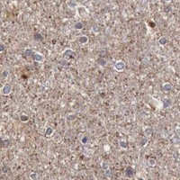
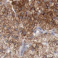
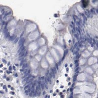
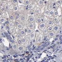

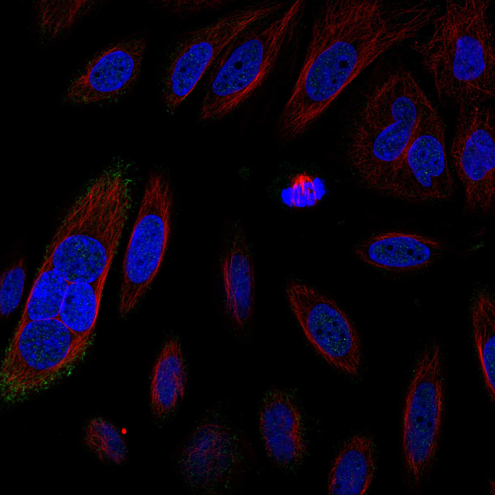
Comprehensive Human Protein Atlas IHC scoring per tissue (reliability: Enhanced — Medium consistency between antibody staining and RNA expression data. Caution, targets protein from more than one gene.). Rows are taken directly from the HPA tissue chart — click any row's HPA link to view the source.
| Tissue | Cell type | Level | Evidence | Source |
|---|---|---|---|---|
| Adrenal gland | Glandular cells | High | Protein (IHC) | HPA → |
| Colon | Endocrine cells | High | Protein (IHC) | HPA → |
| Duodenum | Endocrine cells | High | Protein (IHC) | HPA → |
| Fallopian tube | Ciliated cells (tip of cilia) | High | Protein (IHC) | HPA → |
| Kidney | Proximal tubules (microvilli) | High | Protein (IHC) | HPA → |
| Tissue | Cell type | Level | Evidence | Source |
|---|---|---|---|---|
| Adipose tissue | Adipocytes | Not detected | Protein (IHC) | HPA → |
| Bone marrow | Hematopoietic cells | Not detected | Protein (IHC) | HPA → |
| Bronchus | Basal cells | Not detected | Protein (IHC) | HPA → |
| Cerebellum | Cells in granular layer | Not detected | Protein (IHC) | HPA → |
| Cervix | Glandular cells | Not detected | Protein (IHC) | HPA → |
Troubleshoot chromogenic RDX staining in paraffin sections by checking retrieval, compartment, cell identity and controls before comparing staining intensity.
Catalog anti-RDX antibodies list human, mouse, and rat reactivity; the supplied figures cover a paraffin-embedded human tissue section and HeLa-cell IF (catalog: reactivity; image captions).
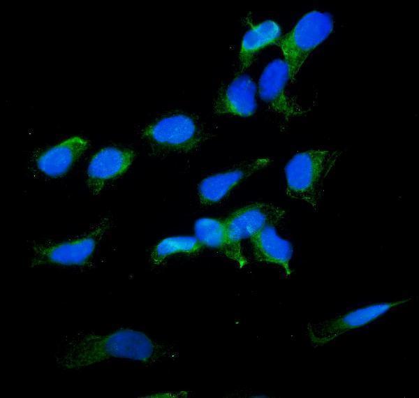
M01926 lists IHC and IF, but its paraffin-embedded human breast cancer IHC caption identifies the stained target as smc4, so the figure does not establish RDX staining (catalog: applications; M01926 image caption). A01926-1 lists IF/ICC and shows RDX staining in HeLa cells at 2 μg/mL (catalog: applications; A01926-1 image caption).
Which to pick: For paraffin-section IHC, M01926 is the listed rabbit monoclonal option (catalog: applications/clone); its caption describes EDTA retrieval at pH 8.0 and primary antibody at 1:50, but names smc4 and does not report the fixative (M01926 image caption). For IF/ICC, A01926-1 is the listed rabbit polyclonal option with an RDX HeLa-cell IF figure (catalog: applications/dilution_raw; A01926-1 image caption). Both list human, mouse, and rat reactivity, while the supplied figures cover human samples only (catalog: reactivity; image captions).