REC8 / Meiotic recombination protein REC8 homolog · IHC design guide

Design Immunohistochemistry for REC8

Plan REC8 paraffin IHC around nuclear staining in testis and ciliary-rootlet staining in ciliated cells (HPA tissue IHC). The guide covers fixation, chromogenic detection and interpretation of a tissue staining profile with low concordance with RNA expression (HPA tissue IHC).

Evidence assembled Oct 2026 · For research use; verify linked source records and product datasheet before use
Immunohistochemistry protocol sheet for REC8 (IHC for REC8): expected localisation Nuclear in testis; cytoplasmic in several tissues (HPA tissue IHC), antibody A04915-2, validated IHC image, and IHC protocol steps
Printable REC8 IHC protocol sheet — expected localisation Nuclear in testis; cytoplasmic in several tissues (HPA tissue IHC), antibody A04915-2, controls and protocol steps. Open the full REC8 IHC guide →

REC8 Immunohistochemistry Experimental Design Guide

Expected localisation, validated protocols, controls and antibodies — the at-a-glance facts below, then the full design guide.

Must know before staining
Expected localisation Nuclear in testis; cytoplasmic in several tissues (HPA tissue IHC)
Staining pattern Testis nuclei; ciliary rootlets in ciliated cells (HPA tissue IHC)
Antigen retrieval EDTA pH 8.0 HIER, heat-mediated (datasheet A04915-2)
Positive control ⓘ Bronchus+4 more · see all
Negative control ⓘ Adipose tissue+4 more · see all
Important caveats
Reasons your staining may differ from the expected pattern.
Fixation Keep fixation consistent across sections. (standard IHC practice; not target-specific)
Caveat Staining has low concordance with RNA expression (HPA tissue IHC)
Regulation Chromosome signal shifts with meiotic stage (UniProt)
Isoform / epitope 2 isoforms; epitope differences are unreported (UniProt)
Section 1

Recommended REC8 IHC & IF Protocols

The catalog antibody’s IHC-P protocol uses EDTA pH 8.0 retrieval (datasheet A04915-2). Published REC8 IHC methods cover gastric cancer tissue and human testis (PMC7507279; PMC5241426; PMC9673495).

Recommended immunohistochemistry (IHC-P) protocol parameters
SampleParaffin-embedded human placenta tissue; fixative not specified (datasheet A04915-2)
FixationImage fixative and duration unreported (datasheet A04915-2); verify before use.
Sectioning4–5 µm sections on charged slides (standard)
DeparaffinisationXylene, graded ethanol series to water (standard)
Antigen retrievalHeat retrieval: EDTA pH 8.0 (datasheet A04915-2); 20 min, 95–100 °C (standard)
Peroxidase block3% H2O2, 10 min, room temperature (standard)
Blocking10% goat serum (datasheet A04915-2)
Primary antibodyRabbit anti-REC8, 0.5-1μg/ml (datasheet A04915-2)
Primary incubationOvernight at 4 °C (datasheet A04915-2)
DetectionStreptavidin-biotin complex (SABC), DAB chromogen (datasheet A04915-2)
CounterstainHematoxylin, blue, dehydrate and mount (standard)
Expected resultREC8-positive staining in ciliated cells (ciliary rootlets) of bronchus (HPA tissue IHC: High). HPA tissue profile: Nuclear expression in testis with cytoplasmic expression in several tissues. No signal in the no-primary control.
💡Decision noteStart with heat-mediated EDTA pH 8.0 for the catalog antibody (datasheet A04915-2); the published protocols that specify retrieval use citrate and microwave heating (PMC7507279; PMC9673495).
Section 2

What Is the Expected REC8 Staining Pattern?

REC8 is a nuclear, chromosome-associated protein without a transmembrane segment (UniProt O95072: location and topology). In paraffin-section IHC, expect nuclear expression in testis, where elongated or late spermatids show high staining; HPA also reports high ciliary-rootlet staining in several epithelia (HPA: tissue IHC). Treat these as observed patterns, not fully verified specificity: HPA rates its tissue profile Approved but reports low consistency with RNA data and pending external verification (HPA: reliability).

What am I looking at on my slide?
Distinct nuclear staining in testis, with strong signal associated with elongated or late spermatids.This fits HPA's nuclear testis profile and high staining in that cell category (HPA: tissue IHC). REC8's chromosome association supports a nuclear interpretation (UniProt O95072: location). HPA does not resolve the stained cells by meiotic stage, so do not assign a particular stage from this chromogenic pattern alone.
Strong signal at ciliary rootlets in bronchus, fallopian tube or nasopharynx.HPA records this cytoplasmic pattern as High in ciliated cells (HPA: tissue IHC). It differs from UniProt's nuclear and chromosome locations and from HPA's approved nucleoplasmic ICC-IF location (UniProt O95072: location; HPA: subcellular). Record it separately from nuclear staining; HPA's pending external verification limits a target-specific interpretation (HPA: reliability).
Prominent cytoplasmic staining outside the reported ciliary-rootlet pattern, or staining in an unexpected cell population.Compare the exact compartment and cell type with HPA observations before scoring it as REC8 (HPA: tissue IHC). An unreported pattern raises possible cross-reactivity or endogenous detection activity (general IHC practice). The reported ciliary-rootlet pattern alone must not be dismissed as artefact (HPA: tissue IHC).
Diffuse colour across tissue, including regions expected to be unstained.A poorly confined signal is difficult to score as nuclear or ciliary-rootlet staining (HPA: tissue IHC; general IHC practice). Background may arise from detection reagents, insufficient blocking or overly strong staining conditions (general IHC practice). Check controls before treating weak diffuse colour as a positive result.
No visible signal in a testis section used as a positive control.HPA reports high staining in elongated or late spermatids and nuclear expression in testis (HPA: tissue IHC). First confirm those cells are present in the section, then check retrieval, antibody dilution and detection with the antibody's IHC-P instructions (general IHC practice). A negative run alone cannot establish absent REC8.
💡Expected REC8 appearanceCall a testis section positive when nuclear staining is clear in the HPA-reported testis pattern, including high staining associated with elongated or late spermatids; diffuse or unexplained cytoplasmic colour is suspect, while high ciliary-rootlet staining is a separate HPA observation requiring cautious interpretation (HPA: tissue IHC and reliability; UniProt O95072: location).
How each factor affects the staining
Cell identity and meiotic stageREC8 occupies chromosome axes during meiotic prophase, then persists at centromeres through metaphase II (UniProt O95072: location). HPA instead reports its strongest testis IHC staining in elongated or late spermatids (HPA: tissue IHC). Keep the mechanistic stage description and observed tissue staining distinct; morphology alone does not reconcile them.
Confidence in tissue patternsThe tissue profile is Approved, but HPA reports low consistency between antibody staining and RNA expression and says external verification is pending (HPA: reliability). Two listed antibodies have Approved IHC status, HPA031727 and HPA031729; neither is listed as IHC Enhanced in the supplied record (HPA: antibodies). Use compartment and tissue controls when judging an unusual signal.
Isoforms and phosphorylationUniProt lists two isoforms and a phosphoserine at residue 148 (UniProt O95072: isoforms and modified residues). Without an antibody epitope or isoform-reactivity record here, neither fact predicts which cells will stain or whether staining intensity will change. Do not infer epitope access or retrieval sensitivity from these annotations.
IF/ICC: what location should be compared?HPA's approved ICC-IF location is the nucleoplasm, with images listed for A-431, U-251MG and U2OS (HPA: subcellular). This supports a nuclear comparison for an IF result, while the present guide's tissue decisions concern chromogenic IHC. The ICC-IF entry does not provide an IF protocol or validate the ciliary-rootlet IHC pattern.
Why is my staining missing, weak or wrong?
SituationLikely causeNext action
Testis positive control has no signal.The section may lack the HPA-reported elongated or late spermatids, or the IHC workflow may have failed (HPA: tissue IHC; general IHC practice).Check cell morphology and slide controls; then review the IHC-validated antibody's retrieval, dilution and detection instructions (general IHC practice). Do not infer REC8 absence from this run.
Signal is mainly cytoplasmic in testis.That differs from HPA's nuclear testis profile and UniProt's nuclear location (HPA: tissue IHC; UniProt O95072: location). Background or cross-reactivity is possible (general IHC practice).Score nuclear and cytoplasmic colour separately. Inspect a no-primary control and repeat with the IHC-validated antibody's stated conditions (general IHC practice).
Ciliated epithelium shows strong rootlet staining.HPA reports High staining at ciliary rootlets in bronchus, fallopian tube and nasopharynx, despite its separate nuclear testis profile (HPA: tissue IHC).Record the cell type and rootlet location precisely. Interpret target specificity cautiously because HPA reports low RNA–staining consistency and pending external verification (HPA: reliability).
Colour appears in a reported negative cell population.A mismatch may reflect cross-reactivity, endogenous detection activity or background (general IHC practice). HPA lists adipocytes and heart cardiomyocytes as Not detected (HPA: tissue IHC).Confirm the cell identity, inspect no-primary and detection controls, and compare the same compartment across sections before scoring it as positive (general IHC practice).
Weak, diffuse staining obscures nuclei.Poor signal separation can follow excessive detection development, insufficient blocking or an unsuitable antibody concentration (general IHC practice).Review development time, blocking and the IHC-validated antibody's stated dilution; use a nuclear counterstain to assess compartment and morphology (general IHC practice).
An IF image appears nuclear while IHC highlights ciliary rootlets.HPA reports approved nucleoplasmic ICC-IF localisation and high ciliary-rootlet tissue IHC staining (HPA: subcellular and tissue IHC). The supplied records do not explain the difference.Compare each result within its own application and cell context; document the discrepancy and HPA's pending external verification without assigning a new REC8 location (HPA: reliability; general interpretation practice).

Sample controls for REC8 IHC & IF

🧪Run bronchus first and score staining in ciliated-cell rootlets, where HPA reports High REC8 staining (HPA: bronchus, ciliated cells, High); use adipose tissue as a negative, scoring adipocytes, where REC8 is Not detected (HPA: adipose tissue, adipocytes, Not detected). On the bronchus slide, assess neighboring nonciliated cells for background and whether they lack the rootlet pattern; their REC8 status is not established by the supplied HPA rows (HPA: bronchus, ciliated cells, High).
Positive control tissue: Bronchus (Ciliated cells (ciliary rootlets), HPA High)
Negative control tissue: Adipose tissue (HPA Not detected)
ICC-IF cell lines (HPA subcellular resource): HPA ICC-IF images show REC8 in A-431, U-251MG, U2OS, with annotated localisation: Nucleoplasm (approved) (HPA subcellular).
Technical controls: Include a no-primary, secondary-only control, a concentration-matched rabbit IgG isotype control for the captioned rabbit primary, and REC8-knockout material as a biological negative (caption: rabbit anti-REC8 antibody; standard IHC controls). Quench endogenous peroxidase for DAB detection and check endogenous biotin background when using the captioned biotinylated secondary and streptavidin–biotin detection system (caption: biotinylated goat anti-rabbit IgG, SABC and DAB).
⚠️Feasibility: A target-specific fixation window or fixation effect is unreported, and the exact A04915-2 paraffin-section caption does not state the fixative (caption: fixative not stated). The reported IHC procedure uses heat retrieval in EDTA at pH 8.0, but its dependence on that retrieval condition is unreported (caption: EDTA heat retrieval, pH 8.0). Frozen sections are not established as easier by the supplied evidence; ICC-IF images exist, but HPA reports nucleoplasmic localization, whereas its bronchus IHC row identifies ciliary rootlets, so verify the compartment-specific pattern when interpreting staining (HPA: ICC-IF images in A-431, U-251MG and U2OS; HPA: nucleoplasm; HPA: bronchus ciliated-cell rootlets).

HPA tissue IHC evidence for REC8

Comprehensive Human Protein Atlas IHC scoring per tissue (reliability: Approved — Low consistency between antibody staining and RNA expression data. Pending external verification.). Rows are taken directly from the HPA tissue chart — click any row's HPA link to view the source.

Positive expression · recommended positive controls

TissueCell typeLevelEvidenceSource
Bronchus Ciliated cells (ciliary rootlets) High Protein (IHC) HPA →
Fallopian tube Ciliated cells (ciliary rootlets) High Protein (IHC) HPA →
Nasopharynx Ciliated cells (ciliary rootlets) High Protein (IHC) HPA →
Testis Elongated or late spermatids High Protein (IHC) HPA →
Adrenal gland Glandular cells Medium Protein (IHC) HPA →

Undetected expression · recommended negative controls

TissueCell typeLevelEvidenceSource
Adipose tissue Adipocytes Not detected Protein (IHC) HPA →
Appendix Glandular cells Not detected Protein (IHC) HPA →
Cerebellum Cells in granular layer Not detected Protein (IHC) HPA →
Heart muscle Cardiomyocytes Not detected Protein (IHC) HPA →
Hippocampus Glial cells Not detected Protein (IHC) HPA →
Section 3

Advanced REC8 IHC Tips

Troubleshoot REC8 chromogenic IHC in paraffin sections using the catalog antibody’s tissue protocol, while treating staining patterns as provisional where validation is limited.

Which retrieval conditions should I use when REC8 staining is weak?
Start with heat-mediated retrieval in EDTA at pH 8.0 (datasheet A04915-2). The selected paraffin-section example used that retrieval before incubation with 1 μg/ml primary antibody overnight at 4°C, so keep those conditions consistent while assessing the weak signal (datasheet A04915-2). If staining remains weak, compare a second retrieval condition only as a controlled fallback, holding section thickness, antibody concentration, detection, and development constant (standard IHC practice). Score nuclear signal and tissue damage together: stronger DAB staining accompanied by lifted tissue or diffuse background does not establish improved REC8 detection (UniProt O95072 localisation; standard IHC practice).
How should I troubleshoot suspected fixation effects on REC8 IHC?
REC8-specific fixation sensitivity is unknown because the selected paraffin-section caption does not state a fixative (datasheet A04915-2). Record each specimen’s fixative and fixation duration, then compare sections processed and stained in the same run to see whether signal tracks with processing history (standard IHC practice). Keep retrieval at EDTA pH 8.0 and the primary incubation at 1 μg/ml overnight at 4°C while making that comparison (datasheet A04915-2). If nuclear detail is lost or staining is uneven, assess morphology and section adhesion before changing antibody concentration; neither the reported tissue patterns nor REC8 phosphorylation establishes a fixation effect (HPA tissue IHC; UniProt O95072; standard IHC practice).
Which REC8 staining compartment should count as a convincing positive?
Prioritise nuclear or chromosome-associated staining when scoring REC8, because its annotated locations include the nucleus, chromosomes, and centromeres (UniProt O95072 localisation). In meiotic cells, its expected distribution changes with stage: chromosome axes in prophase, followed by centromeric persistence after arm signal is lost in anaphase I (UniProt O95072 localisation). Treat diffuse cytoplasmic DAB staining cautiously, even though tissue IHC reports cytoplasmic staining in several tissues and nuclear expression in testis (HPA tissue IHC). Compare the signal with a nuclear counterstain and matched control sections, and record nuclear and cytoplasmic staining separately rather than combining them into one positive score (standard IHC practice).
Could epitope choice explain a discrepancy between REC8 samples?
REC8 has 2 annotated isoforms, but the supplied antibody evidence does not identify its binding epitope or establish equal recognition of both isoforms (UniProt O95072 isoforms; datasheet A04915-2). A phosphoserine is annotated at residue 148; its presence alone does not show whether phosphorylation changes this antibody’s IHC staining (UniProt O95072 modified residues; datasheet A04915-2). Keep EDTA retrieval at pH 8.0 while comparing samples so retrieval differences do not confound an apparent isoform effect (datasheet A04915-2; standard IHC practice). Before assigning a discrepant pattern to splicing or phosphorylation, establish the antibody’s epitope and validate the proposed explanation with an independent assay (standard IHC practice).
How can I check REC8 localisation by multiplex IF?
For a separate IF experiment, pair REC8 with a marker that identifies the cell population being evaluated, and include a nuclear counterstain to assess overlap with nuclei (UniProt O95072 localisation; standard IF practice). Choose spectrally separated fluorophores and place the weaker expected signal in a channel with less tissue autofluorescence; inspect unstained and single-label controls before interpreting colocalisation (standard IF practice). REC8 lacks a transmembrane segment and is annotated in nuclei and chromosomes, so use permeabilisation sufficient to give antibody access to intracellular epitopes without disrupting nuclear structure (UniProt O95072 topology and localisation; standard IF practice). Optimise fixation and permeabilisation for the IF specimen itself; the selected paraffin IHC caption reports no fixative and supplies no IF fixation conditions (datasheet A04915-2).
What should I adjust when REC8 DAB staining is widespread?
First inspect a section without primary antibody to identify signal from the detection system, and confirm that the peroxidase block and wash steps performed as intended (standard chromogenic IHC practice). The selected example used 10% goat serum, a biotinylated secondary antibody, a streptavidin–biotin complex, and DAB, so assess nonspecific binding and biotin-related background within that workflow (datasheet A04915-2; standard IHC practice). Compare sections developed for the same time and avoid treating darker DAB alone as stronger REC8 expression (standard IHC practice). Map background against tissue edges, damaged areas, and nuclei; widespread extranuclear staining is less consistent with REC8’s annotated nuclear and chromosomal locations (UniProt O95072 localisation; standard IHC practice).
How should I score REC8 across paraffin sections? ⚠ ANSWER MARKED FOR VERIFICATION
Define the scored cell population and compartment before reviewing treatment groups, and report the percentage of positive nuclei alongside an intensity-based H-score when staining quality permits (UniProt O95072 localisation; standard IHC practice). For a population counted by area, report positive cells per mm² and normalise to the analysed, intact tissue area rather than the whole slide (standard IHC practice). Apply identical thresholds, DAB development, counterstaining, and image acquisition across comparison groups, excluding folds, necrosis, and detached tissue by prespecified rules (standard IHC practice). Record cytoplasmic staining separately, because the tissue IHC profile includes cytoplasmic expression and its antibody–RNA consistency is reported as low (HPA tissue IHC).
How do I distinguish convincing REC8 signal from artefact?
A convincing result should show reproducible staining in the prespecified cells and an interpretable nuclear or chromosome-associated pattern, supported by appropriate control sections (UniProt O95072 localisation; standard IHC practice). Check whether apparent positivity instead follows section edges, necrotic regions, or diffuse deposits; examine the no-primary control for detection-system or endogenous enzyme signal (standard chromogenic IHC practice). Tissue context matters: REC8 is reported in testis, while the selected antibody caption shows staining in a human placenta paraffin section without stating its fixative (UniProt O95072 tissue specificity; datasheet A04915-2). Treat an unexpected cell type or predominantly cytoplasmic pattern as provisional until independently corroborated, because tissue IHC reports low consistency with RNA expression and awaits external verification (HPA tissue IHC).
Boster reagents

Best REC8 / Meiotic recombination protein REC8 homolog IHC Antibodies

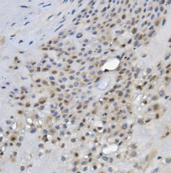
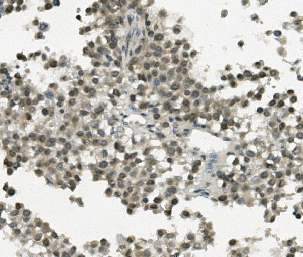
A04915-2 has IHC images from paraffin sections of human placenta, human testicular cancer, and rat lung; no IF/ICC data are supplied (catalog: A04915-2 applications and image captions).

Real IHC data IHC analysis of REC8 using anti-REC8 antibody (A04915-2). REC8 was detected in paraffin-embedded section of human placenta tissue. Heat mediated antigen retrieval was performed in EDTA buffer (pH8.0, epitope retrieval solution). The tissue section was blocked with 10% goat serum. The tissue section was then incubated with 1μg/ml rabbit anti-REC8 Antibody (A04915-2) overnight at 4°C. Biotinylated goat anti-rabbit IgG was used as secondary antibody and incubated for 30 minutes at 37°C. The tissue section was developed using Strepavidin-Biotin-Complex (SABC) (Catalog # SA1022) with DAB as the chromogen.
Anti-REC8 Antibody ®
Cat # A04915-2

A04915-2 is the only SKU listed and is validated for IHC, with images from human placenta and human testicular cancer paraffin sections (catalog: A04915-2 applications and image captions). Its IHC images also include rat lung paraffin sections; the catalog lists human and rat reactivity (catalog: A04915-2 image captions and reactivity).

Which to pick: Choose A04915-2 for tissue IHC on paraffin sections, starting at 0.5–1 μg/ml; its captions describe EDTA retrieval at pH 8.0, but do not report the fixative (datasheet: A04915-2 IHC dilution; A04915-2 image captions). No listed SKU has IF/ICC validation or an IF figure, so this payload does not support an IF/ICC recommendation (catalog: A04915-2 applications and image captions). For human and rat IHC, A04915-2 is the available choice; it is a rabbit antibody with no clone specified (catalog: A04915-2 reactivity, host, and clone).

Each figure is that product's own IHC / IF validation image from its datasheet.

References

  1. UniProt Consortium. UniProt entry O95072 (REC8_HUMAN, Meiotic recombination protein REC8 homolog).
  2. Human Protein Atlas. REC8 tissue IHC expression (reliability: Approved).
  3. Human Protein Atlas. REC8 subcellular location (ICC-IF): Localized to the nucleoplasm..
  4. Human Protein Atlas. REC8 antibody validation summary (2 antibodies).
  5. REC8 suppresses tumor angiogenesis by inhibition of NF-κB-mediated vascular endothelial growth factor expression in gastric cancer cells. Biological research 2020 — PMC7507279.
  6. Mapping separase-mediated cleavage in situ. NAR genomics and bioinformatics 2022 — PMC9673495.
  7. REC8 functions as a tumor suppressor and is epigenetically downregulated in gastric cancer, especially in EBV-positive subtype. Oncogene 2017 — PMC5241426.
  8. Human umbilical cord mesenchymal stem cells (hUC-MSCs) alleviate paclitaxel-induced spermatogenesis defects and maintain male fertility. Biological research 2023 — PMC10424423.
  9. PubMed PMID:10207075 — UniProt-cited evidence.
  10. PubMed PMID:14702039 — UniProt-cited evidence.
  11. PubMed PMID:17974005 — UniProt-cited evidence.