This website uses cookies to ensure you get the best experience on our website.


- Table of Contents
Plan RECK paraffin IHC around distinct extracellular matrix staining and additional cytoplasmic staining in several tissues (HPA tissue IHC). Breast myoepithelial cells provide a high-staining reference (HPA tissue IHC); the IHC-validated antibody has a starting dilution range of 1:100–1:300 (datasheet).

Expected localisation, validated protocols, controls and antibodies — the at-a-glance facts below, then the full design guide.
| Expected localisation | ECM and cytoplasm (HPA tissue IHC); membrane (UniProt) | |
| Staining pattern | Distinct ECM; cytoplasm in several tissues; high in breast myoepithelial cells (HPA tissue IHC) | |
| Antigen retrieval | Citrate pH 6.0 HIER, 95–98 °C, 20 min (rule: cytoplasmic / membrane antigen) | |
| Positive control | Breast+4 more · see all | |
| Negative control | Adipose tissue+4 more · see all |
| Fixation | Keep fixation conditions consistent across sections. (standard IHC practice; not target-specific) | |
| Caveat | ECM positivity can complicate cell-level scoring (HPA tissue IHC) | |
| Regulation | Undetectable in transformed cell lines (UniProt) | |
| Isoform / epitope | No isoforms listed; map epitopes to mature chain 23–942 (UniProt) |
The catalog antibody’s IHC-P protocol is accompanied by published chromogenic RECK IHC methods for salivary adenoid cystic carcinoma (PMC3997680) and paired leiomyoma and myometrium (PMC4988044).
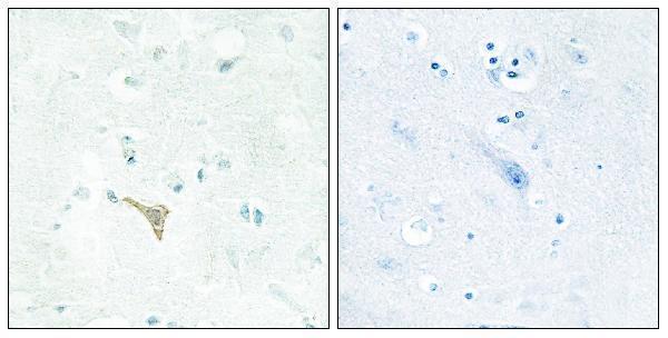
| Sample | Paraffin-embedded human brain tissue; fixative not specified (datasheet A06439) |
| Fixation | Image fixative and duration unreported (datasheet A06439); verify before use. |
| Sectioning | 4–5 µm sections on charged slides (standard) |
| Deparaffinisation | Xylene, graded ethanol series to water (standard) |
| Antigen retrieval | Heat-induced epitope retrieval in citrate buffer, pH 6.0, 20 min at 95–98 °C (standard rule: cytoplasmic / membrane antigen) |
| Peroxidase block | 3% H2O2, 10 min, room temperature (standard) |
| Blocking | 10% normal serum of the secondary host, 30 min, room temperature (standard) |
| Primary antibody | Rabbit anti-RECK, 1:100 - 1:300 (datasheet A06439) |
| Primary incubation | Overnight at 4 °C (standard) |
| Detection | HRP-polymer secondary, DAB chromogen 5–10 min (standard) |
| Counterstain | Hematoxylin, blue, dehydrate and mount (standard) |
| Expected result | RECK-positive staining in myoepithelial cells of breast (HPA tissue IHC: High). HPA tissue profile: Distinct positivity in extracellular matrix with additional cytoplasmic expression in several tissues. No signal in the no-primary control. |
RECK is a cell membrane protein with no transmembrane segment (UniProt O95980 topology). In tissue IHC, expect distinct extracellular matrix positivity and some cytoplasmic staining (HPA: tissue IHC profile; reliability Supported). Breast myoepithelial cells are a strong reference population (HPA: High); several other annotated cell populations show medium staining (HPA: tissue IHC). Interpret signal by its location and the specific cells stained, rather than by whole-tissue positivity.
| Distinct extracellular matrix staining, with signal along cell borders in an annotated positive population. | This fits the reported extracellular matrix pattern (HPA: tissue IHC profile) and cell membrane localization (UniProt O95980). Record which cells and structures carry signal; extracellular staining alone does not identify its cellular source. |
| Strong staining in breast myoepithelial cells, with lighter staining in other annotated positive populations. | This is consistent with breast myoepithelial cells at High and, for example, colon goblet cells and kidney glomerular cells at Medium (HPA: tissue IHC). These are relative HPA observations, not universal intensity thresholds for every section. |
| Dominant nuclear staining without the expected extracellular, border, or reported cytoplasmic pattern. | Nuclear localization is absent from the supplied localization records (UniProt O95980: cell membrane; HPA: tissue IHC profile). Treat a nuclear-only result as suspect and review controls and detection background before assigning it to RECK. |
| Strong signal in an HPA-annotated undetected cell population, such as adipocytes or bronchial respiratory epithelial cells. | Those specific populations are Not detected (HPA: tissue IHC). Check the stained cell identity and controls; cross-reactivity or endogenous detection activity may explain discordance. An undetected cell population does not make its entire tissue a negative control. |
| No convincing signal in breast myoepithelial cells on a section where those cells can be identified. | That conflicts with the High reference observation (HPA: tissue IHC). Review section quality, antibody and detection controls, and whether the intended cells are present before interpreting other negative populations as true absence. |
| Compartment and topology | RECK is assigned to the cell membrane and has no transmembrane segment (UniProt O95980 topology); HPA reports extracellular matrix positivity plus some cytoplasmic expression in tissue (HPA: tissue IHC profile). Interpret these reported patterns together instead of requiring every positive cell to show only a crisp membrane rim. |
| Cell-level reference pattern | Breast myoepithelial cells are High; Purkinje cells, cortical neuropil, colon goblet cells, endometrial stromal cells, glomerular cells, cholangiocytes, and alveolar cells are Medium (HPA: tissue IHC). HPA also lists some populations as Low or Not detected; specify the cell population when comparing sections. |
| Antibody validation | The catalog antibody CAB025109 has Supported IHC status (HPA: antibodies). The tissue profile has Supported reliability, meaning external characterization supports staining (HPA: tissue IHC); neither label establishes that every stained structure is annotated or that every preparation yields the same intensity. |
| Processing and epitope interpretation | UniProt annotates a signal peptide at residues 1–22 and a mature chain at 23–942 (UniProt O95980 processing). Without an epitope position in the supplied record, these annotations cannot predict whether a particular antibody will detect each processed form or explain a missing IHC signal. |
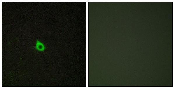
| IF/ICC Q&A: What localization should I expect? | Plasma membrane localization is supported in ICC-IF (HPA: subcellular). That observation helps interpret localization, but ICC-IF evidence is separate from the tissue IHC pattern and does not supply an IF/ICC protocol or establish identical staining intensity in paraffin sections. |
| Situation | Likely cause | Next action |
|---|---|---|
| Breast myoepithelial cells show no signal. | An expected High population is negative (HPA: tissue IHC); the supplied sources do not identify a RECK-specific fixation or retrieval failure. | General IHC practice: confirm the cells are present, check section and detection controls, then review the catalog antibody's IHC-P instructions before changing retrieval or dilution. |
| Staining is predominantly nuclear. | The dominant location conflicts with cell membrane localization and the reported tissue pattern (UniProt O95980; HPA: tissue IHC). Background or cross-reactivity is possible. | General IHC practice: compare with a no-primary control, inspect the counterstain separately, and reassess compartment calls in an annotated positive cell population. |
| Adipocytes or bronchial respiratory epithelial cells stain strongly. | Those cell populations are Not detected in HPA tissue IHC (HPA: tissue IHC); unexpected signal may reflect cell misidentification, cross-reactivity, or endogenous detection activity. | General IHC practice: verify cell morphology and review no-primary and detection-system controls. Do not label the whole adipose or bronchial section negative from one annotated cell type. |
| Nearly every compartment shows diffuse chromogen. | The distribution does not resemble distinct extracellular matrix positivity with limited additional cytoplasmic expression (HPA: tissue IHC profile). Nonspecific binding or detection background is possible. | General IHC practice: examine no-primary and detection controls, review blocking and wash steps, and score only interpretable cell and compartment patterns. |
| Only faint staining appears in a Low HPA population. | HPA lists hippocampal and caudate neuronal cells, among others, as Low (HPA: tissue IHC); a faint result there is a weak performance check. | Use an annotated High population, such as breast myoepithelial cells (HPA: tissue IHC), to judge whether the IHC run detected an expected signal. |
| An ICC-IF image looks more membrane-restricted than the tissue IHC section. | ICC-IF supports plasma membrane localization (HPA: subcellular), while tissue IHC reports extracellular matrix positivity and some cytoplasmic expression (HPA: tissue IHC profile). | Interpret each application against its own reported pattern. For this paraffin-section result, assess tissue cell identity and extracellular staining; consult the separate IF/ICC guide for that application. |


Comprehensive Human Protein Atlas IHC scoring per tissue (reliability: Supported — External characterization data supports antibody staining. Antibody staining in cells/structures not annotated, view images.). Rows are taken directly from the HPA tissue chart — click any row's HPA link to view the source.
| Tissue | Cell type | Level | Evidence | Source |
|---|---|---|---|---|
| Breast | Myoepithelial cells | High | Protein (IHC) | HPA → |
| Cerebellum | Purkinje cells | Medium | Protein (IHC) | HPA → |
| Cerebral cortex | Neuropil | Medium | Protein (IHC) | HPA → |
| Colon | Goblet cells | Medium | Protein (IHC) | HPA → |
| Endometrium | Cells in endometrial stroma | Medium | Protein (IHC) | HPA → |
| Tissue | Cell type | Level | Evidence | Source |
|---|---|---|---|---|
| Adipose tissue | Adipocytes | Not detected | Protein (IHC) | HPA → |
| Adrenal gland | Glandular cells | Not detected | Protein (IHC) | HPA → |
| Appendix | Endocrine cells | Not detected | Protein (IHC) | HPA → |
| Bronchus | Respiratory epithelial cells | Not detected | Protein (IHC) | HPA → |
| Duodenum | Endocrine cells | Not detected | Protein (IHC) | HPA → |
Troubleshoot RECK staining in paraffin-section chromogenic IHC using the page retrieval conditions, compartment-aware controls, and separately interpreted IF evidence.
Anti-RECK A06439 has IHC data from paraffin-embedded human brain and IF data from HepG2 cells (catalog image captions); listed reactivity is human and mouse (catalog reactivity).
A06439 will render with its IHC image of paraffin-embedded human brain tissue (catalog IHC image caption). The same SKU has an IF image of HepG2 cells and lists IHC, IF and ICC applications with human and mouse reactivity (catalog IF image caption; catalog applications; catalog reactivity).
Which to pick: Choose A06439 for tissue IHC in paraffin sections, as shown in its own human brain image; the fixative is unreported (catalog IHC image caption). Choose A06439 for IF/ICC based on its listed applications and HepG2 IF image (catalog applications; catalog IF image caption). For human or mouse samples, A06439 is the listed rabbit polyclonal option, although the supplied images show human brain tissue and HepG2 cells only (catalog reactivity; catalog host and clonality; catalog image captions).