REG3A / Regenerating islet-derived protein 3-alpha · IHC design guide

Design Immunohistochemistry for REG3A

Use duodenal or small-intestinal Paneth cells as high-staining references for REG3A paraffin IHC (HPA tissue IHC). This guide covers fixation consistency, titration of the IHC-validated antibody at 1:50–1:200 (datasheet), and interpretation of staining from a secreted protein (UniProt).

Evidence assembled Oct 2026 · For research use; verify linked source records and product datasheet before use
Immunohistochemistry protocol sheet for REG3A (IHC for REG3A): expected localisation Expected to be secreted; found apically in pancreatic acinar cells (UniProt), antibody A05686-1, validated IHC image, and IHC protocol steps
Printable REG3A IHC protocol sheet — expected localisation Expected to be secreted; found apically in pancreatic acinar cells (UniProt), antibody A05686-1, controls and protocol steps. Open the full REG3A IHC guide →

REG3A Immunohistochemistry Experimental Design Guide

Expected localisation, validated protocols, controls and antibodies — the at-a-glance facts below, then the full design guide.

Must know before staining
Expected localisation Expected to be secreted; found apically in pancreatic acinar cells (UniProt)
Staining pattern High in duodenal and small-intestinal Paneth cells; subcellular pattern unreported (HPA tissue IHC)
Antigen retrieval Citrate pH 6.0 HIER, 95–98 °C, 20 min (rule: cytoplasmic / membrane antigen)
Positive control ⓘ Duodenum+2 more · see all
Negative control ⓘ Adipose tissue+4 more · see all
Important caveats
Reasons your staining may differ from the expected pattern.
Fixation Keep fixation consistent across sections (standard IHC practice; not target-specific)
Caveat Secretion can separate protein staining from its RNA source (HPA tissue IHC)
Regulation Up in psoriasis and pancreatitis (UniProt)
Isoform / epitope No isoforms; processing may remove N-terminal epitopes (UniProt)
Section 1

Recommended REG3A IHC & IF Protocols

The catalog antibody’s IHC-P protocol is paired with one published REG3A protocol for FFPE colon biopsies (PMC13296221 methods).

Recommended immunohistochemistry (IHC-P) protocol parameters
SampleParaffin-embedded mouse brain tissue; fixative not specified (datasheet A05686-1)
FixationImage fixative and duration unreported (datasheet A05686-1); verify before use.
Sectioning4–5 µm sections on charged slides (standard)
DeparaffinisationXylene, graded ethanol series to water (standard)
Antigen retrievalHeat-induced epitope retrieval in citrate buffer, pH 6.0, 20 min at 95–98 °C (standard rule: cytoplasmic / membrane antigen)
Peroxidase block3% H2O2, 10 min, room temperature (standard)
Blocking10% normal serum of the secondary host, 30 min, room temperature (standard)
Primary antibodyRabbit anti-REG3A, 1:50-1:200 (datasheet A05686-1)
Primary incubationOvernight at 4 °C (standard)
DetectionHRP-polymer secondary, DAB chromogen 5–10 min (standard)
CounterstainHematoxylin, blue, dehydrate and mount (standard)
Expected resultREG3A-positive staining in paneth cells of duodenum (HPA tissue IHC: High). HPA tissue profile: Expression in a subset of cells in gastrointestinal tract. No signal in the no-primary control.
💡Decision noteStart with citrate pH 6.0 HIER at 95–98 °C for 20 min (page retrieval rule); the published colon protocol does not specify retrieval (PMC13296221 methods).
Section 2

What Is the Expected REG3A Staining Pattern?

REG3A is secreted and has no transmembrane segment (UniProt Q06141 topology). In paraffin sections, expect cell-associated staining chiefly in duodenal and small-intestinal Paneth cells, with weaker staining in appendix enterocytes (HPA tissue IHC: High in Paneth cells; Medium in appendix enterocytes). HPA rates its tissue pattern Supported but cautions that the antibodies may detect proteins from more than one gene and that secreted protein need not remain where its RNA is made (HPA tissue IHC).

What am I looking at on my slide?
Strong staining in a subset of duodenal or small-intestinal Paneth cells.This matches the reported High Paneth-cell pattern (HPA tissue IHC). Cytoplasmic or secretory-region staining is plausible for a secreted protein, but HPA does not assign a precise subcellular compartment in tissue IHC (UniProt Q06141 subcellular location; HPA tissue IHC).
Predominantly nuclear staining or a sharp continuous membrane outline.Treat this as a localization mismatch and investigate artefact before scoring it as REG3A: UniProt describes secretion and no transmembrane segment, while HPA's ICC-IF summary places the signal in the cytosol (UniProt Q06141 topology; HPA subcellular ICC-IF).
Strong staining in an unexpected cell population, including cells reported as negative.Check antibody cross-reactivity and chromogenic detection controls. HPA warns that its antibodies may target proteins from more than one gene; endogenous detection activity can also produce misleading color in IHC (HPA tissue IHC caution; standard IHC practice).
Diffuse color across tissue, luminal material, and the negative control.This does not establish cellular REG3A expression. A secreted protein may be away from its producing cell, but widespread color in a negative control points to background from detection chemistry or inadequate blocking (HPA tissue IHC caution; standard IHC practice).
No signal in duodenal or small-intestinal Paneth cells.A negative result in these reported High-staining cells is inconclusive until antibody dilution, retrieval, detection, and tissue integrity are checked using the IHC-validated antibody's instructions (HPA tissue IHC; standard IHC practice).
💡Expected REG3A appearanceCall a result consistent with REG3A when a subset of duodenal or small-intestinal Paneth cells stains strongly; regard dominant nuclear staining or broad color in negative controls as suspect (HPA tissue IHC: High in Paneth cells; UniProt Q06141 topology; standard IHC practice).
How each factor affects the staining
Secretion and tissue locationREG3A is secreted, and HPA cautions that protein and RNA locations can differ; extracellular or luminal signal should therefore be interpreted alongside identifiable positive cells and controls, rather than used alone to assign a producer cell (UniProt Q06141 subcellular location; HPA tissue IHC caution).
Protein processingUniProt lists a signal peptide at residues 1–26, a propeptide at 27–37, and chains spanning 27–175 or 38–175. Epitope position can matter when comparing antibodies; the payload does not identify their epitopes (UniProt Q06141 processing).
Antibody specificityHPA rates the tissue pattern Supported and lists IHC support for HPA047894 and HPA048334, while warning about recognition of proteins from more than one gene. Treat an unexpected positive population as a specificity question, even when staining is strong (HPA tissue IHC; HPA antibodies).
Tissue-dependent signalPaneth cells are reported High in duodenum and small intestine; appendix enterocytes are Medium, and stomach glandular cells are listed as low. Compare intensity within the appropriate cell type rather than applying one cutoff to every tissue (HPA tissue IHC).
IF/ICC question: should its cytosolic image define the IHC pattern?No. HPA calls the ICC-IF cytosolic location Approved but cautions that it is based on antibodies targeting proteins from multiple genes. Use the tissue IHC cell pattern to interpret paraffin sections; consult the separate IF/ICC guide for that application (HPA subcellular ICC-IF; HPA tissue IHC).
Why is my staining missing, weak or wrong?
SituationLikely causeNext action
Paneth cells are unstained in an expected positive section.Target-specific fixation effects are not established by the supplied assay evidence. Verify with a matched IHC source before attributing a result to fixation.Check tissue integrity and the IHC-validated antibody's retrieval and dilution instructions; verify that the chromogenic detection system worked on a suitable control (standard IHC practice).
The entire section has pale, even color.Non-specific reagent binding, inadequate blocking, or excessive antibody concentration can raise background in chromogenic IHC (standard IHC practice).Compare a negative control, review blocking and washes, and adjust the catalog antibody's dilution within its documented IHC conditions (standard IHC practice).
Unexpected cells stain as strongly as Paneth cells.Antibody cross-reactivity is plausible because HPA cautions that the antibodies may recognize proteins from multiple genes; endogenous detection activity is another IHC possibility (HPA tissue IHC caution; standard IHC practice).Review cell identity and controls; check endogenous enzyme blocking for the chosen chromogen and compare with another appropriately validated antibody if available (standard IHC practice).
Nuclei or continuous plasma-membrane outlines dominate.That pattern conflicts with secretion, the absence of a transmembrane segment, and HPA's cytosolic ICC-IF summary (UniProt Q06141 topology; HPA subcellular ICC-IF).Check counterstain and negative controls, then reassess antibody specificity before scoring the signal as REG3A (standard IHC practice; HPA tissue IHC caution).
Color appears mainly in lumen or outside recognizable cells.Extracellular signal is compatible with secretion, but the deposited protein's location may differ from its producing cell's location (UniProt Q06141 subcellular location; HPA tissue IHC caution).Record the extracellular pattern separately; use adjacent identifiable Paneth cells and negative controls before assigning cell-specific positivity (HPA tissue IHC: High in Paneth cells; standard IHC practice).
Appendix staining is weaker than duodenal staining.This may reflect the reported tissue pattern: appendix enterocytes are Medium, whereas duodenal Paneth cells are High (HPA tissue IHC).Score each tissue's reported cell population separately and confirm that controls are acceptable before treating the intensity difference as assay failure (HPA tissue IHC; standard IHC practice).

Sample controls for REG3A IHC & IF

🧪Run duodenum first and expect Paneth cells to stain (High; HPA: duodenum, Paneth cells). Use adipose tissue as the negative tissue (Not detected in adipocytes; HPA: adipose tissue), and compare adjacent non-Paneth cells on the duodenal slide for background rather than assuming every other cell type is negative (HPA: duodenum, Paneth cells High).
Positive control tissue: Duodenum (Paneth cells, HPA High)
Negative control tissue: Adipose tissue (HPA Not detected)
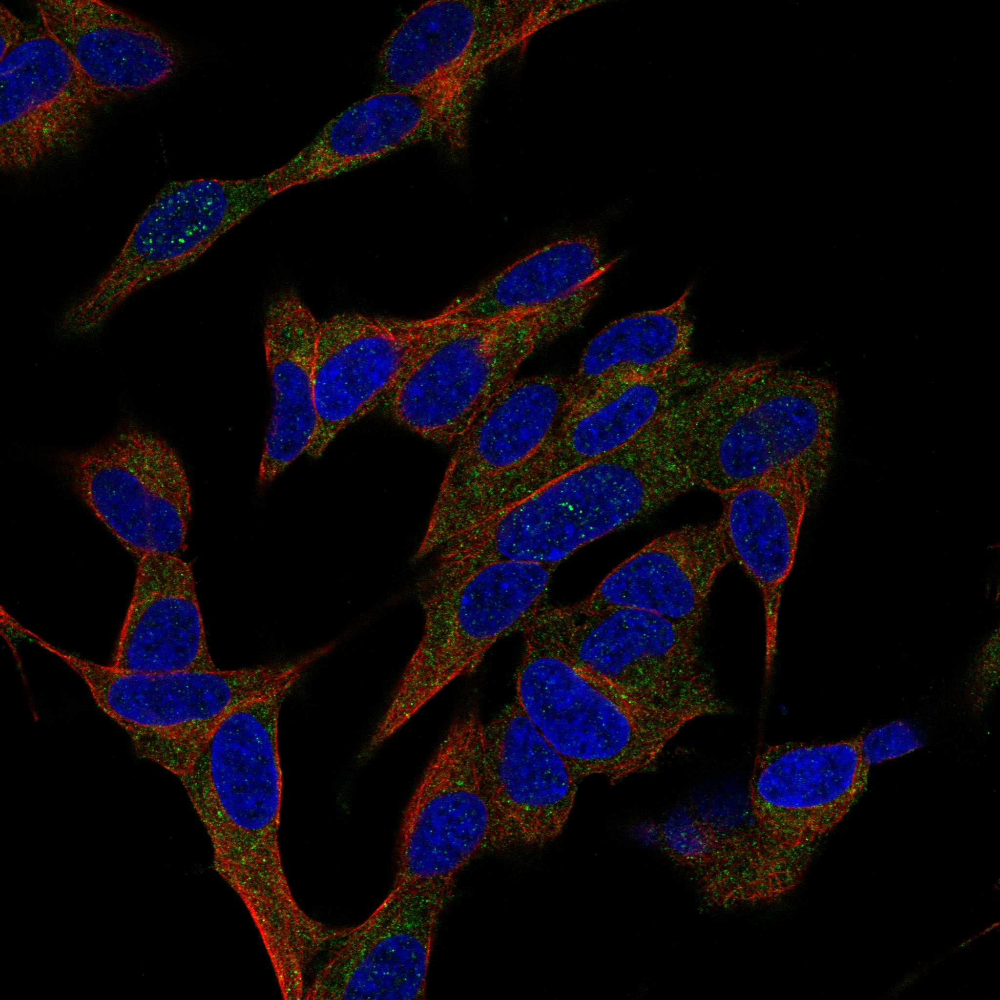
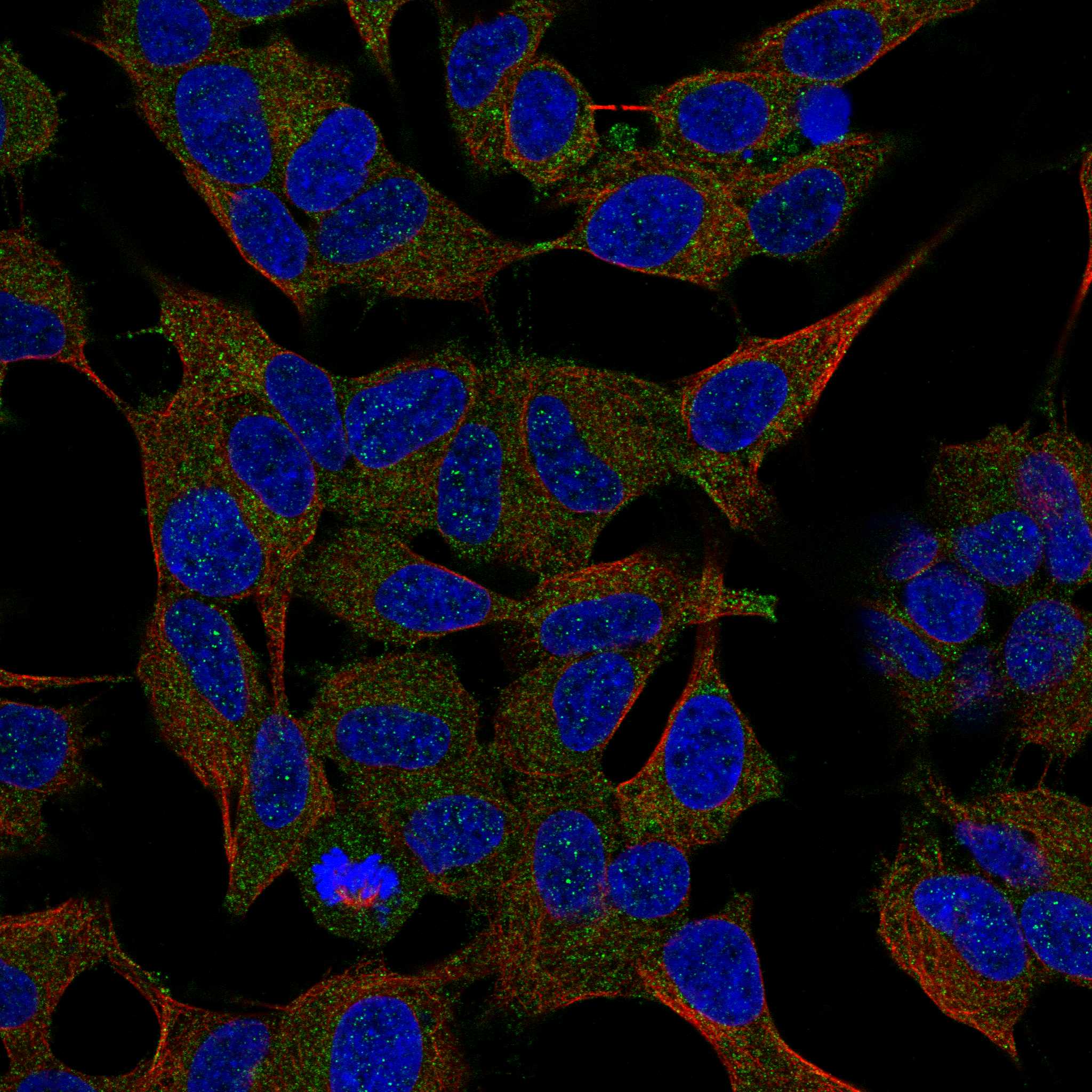
ICC-IF cell lines (HPA subcellular resource): HPA ICC-IF images show REG3A in SH-SY5Y, U-251MG, with annotated localisation: Cytosol (approved) (HPA subcellular).
Technical controls: Include a no-primary (secondary-only) control, an isotype control matched to the primary antibody’s host species and clonality, and REG3A knockout tissue as a biological negative if available (standard IHC practice). In duodenum, block endogenous peroxidase for HRP detection, check endogenous biotin if using avidin-biotin detection, and assess autofluorescence if evaluating IF (standard IHC/IF practice).
⚠️Feasibility: A target-specific fixation window or fixation effect is unreported, and the exact A05686-1 paraffin-section caption does not state a fixative (selected A05686-1 tissue-IHC caption). That caption specifies microwave retrieval in 10 mM PBS, pH 7.2, for mouse brain; whether duodenal REG3A staining depends on that retrieval is unreported (selected A05686-1 tissue-IHC caption). The supplied evidence does not establish frozen sections or IF/ICC as easier; because REG3A is secreted, distinguish Paneth-cell staining from luminal or extracellular signal when scoring duodenum (UniProt Q06141 subcellular; HPA: duodenum, Paneth cells High).

HPA tissue IHC evidence for REG3A

Comprehensive Human Protein Atlas IHC scoring per tissue (reliability: Supported — Caution, targets protein from more than one gene. Secreted protein, tissue location of RNA and protein is expected to differ.). Rows are taken directly from the HPA tissue chart — click any row's HPA link to view the source.

Positive expression · recommended positive controls

TissueCell typeLevelEvidenceSource
Duodenum Paneth cells High Protein (IHC) HPA →
Small intestine Paneth cells High Protein (IHC) HPA →
Appendix Enterocytes Medium Protein (IHC) HPA →

Undetected expression · recommended negative controls

TissueCell typeLevelEvidenceSource
Adipose tissue Adipocytes Not detected Protein (IHC) HPA →
Adrenal gland Glandular cells Not detected Protein (IHC) HPA →
Bone marrow Hematopoietic cells Not detected Protein (IHC) HPA →
Breast Adipocytes Not detected Protein (IHC) HPA →
Bronchus Respiratory epithelial cells Not detected Protein (IHC) HPA →
Section 3

Advanced REG3A IHC Tips

These questions address REG3A staining in paraffin sections by chromogenic IHC, with one entry for planning a separate IF experiment.

How should I retrieve REG3A in paraffin sections when staining is weak?
Start with citrate buffer at pH 6.0, heated to 95–98 °C for 20 minutes (page retrieval setting). Cool sections gradually, then compare signal and tissue integrity with a known positive control on the same run (standard IHC practice; HPA: high in duodenal Paneth cells). If staining remains weak, test microwave retrieval in 10 mM PBS at pH 7.2 on a parallel section; that method appears in the selected antibody’s paraffin embedded mouse brain caption (A05686-1 tissue IHC caption). Keep detection and imaging conditions constant across retrieval trials so a change in intensity can be attributed to retrieval (standard IHC practice).
Could fixation explain weak or uneven REG3A staining?
REG3A specific sensitivity to fixation is unknown from the supplied evidence; the selected paraffin section caption does not state a fixative (A05686-1 tissue IHC caption). Record the fixative and processing history for each specimen, and compare sections with matched handling when troubleshooting intensity (standard IHC practice). For routinely fixed paraffin tissue, assess staining after the stated citrate retrieval at pH 6.0 for 20 minutes, using the same detection run for controls (page retrieval setting; standard IHC practice). If staining varies with processing, repeat on comparably handled sections before attributing the difference to REG3A biology; tissue patterns and protein features do not establish fixation sensitivity (standard IHC practice).
Where should convincing REG3A staining appear within a tissue section?
Look for REG3A signal in appropriate cells and their secretory regions: REG3A is secreted and has been found apically in pancreatic acinar cells (UniProt Q06141 subcellular location). High staining in duodenal and small intestinal Paneth cells provides a useful tissue comparison (HPA: high in Paneth cells). A cytosolic pattern can occur, but the reported cytosolic localisation carries a warning that the antibodies target proteins from multiple genes (HPA subcellular: approved cytosol; multiple gene caution). Because REG3A has no transmembrane segment, treat an exclusively sharp membrane outline as a reason to check antibody specificity and section background (UniProt Q06141 topology; standard IHC practice).
Could REG3A processing change which epitope the antibody detects?
Check the antibody’s stated immunogen or mapped epitope before comparing staining across specimens, since the supplied material does not identify its binding site (standard IHC practice; supplied antibody evidence). REG3A has a signal peptide at residues 1–26, a propeptide at 27–37, and processed chains spanning 27–175 or 38–175 (UniProt Q06141 processing). An epitope within the propeptide could distinguish those processed chains, whereas an epitope in their shared sequence would not distinguish them by sequence alone (UniProt Q06141 processing). REG3A has no listed alternative isoforms or glycosylation sites, so investigate processing and epitope access before assigning a staining difference to either mechanism (UniProt Q06141 isoforms and glycosylation; standard IHC practice).
How can I design a separate REG3A immunofluorescence check?
For a separate IF experiment, pair REG3A with a Paneth cell marker such as lysozyme in intestinal tissue, and inspect single channels before interpreting overlap (HPA: high in intestinal Paneth cells; standard IF practice). Choose spectrally separated fluorophores after checking tissue autofluorescence in an unstained section, especially if a channel shows diffuse signal (standard IF practice). REG3A is secreted and lacks a transmembrane segment, so select permeabilisation according to the antibody’s mapped epitope and whether intracellular protein or deposited extracellular protein is being assessed (UniProt Q06141 subcellular location and topology; standard IF practice). Document fixation, permeabilisation and antibody controls within the IF experiment; the selected paraffin section caption does not supply IF conditions (A05686-1 tissue IHC caption; standard IF practice).
How do I reduce diffuse brown staining without losing REG3A signal?
First compare a no primary control with the stained section to separate detection background from antibody dependent signal (standard IHC practice). Block endogenous peroxidase before chromogenic detection, for example with 3% hydrogen peroxide for 10 minutes, and rinse thoroughly before applying antibody (standard IHC practice). Titrate the primary antibody and detection reagent using matched sections while preserving signal in a positive tissue control such as duodenal Paneth cells (standard IHC practice; HPA: high in duodenal Paneth cells). Because REG3A is secreted, diffuse extracellular staining deserves evaluation against section edges and control staining before it is counted as cellular expression (UniProt Q06141 subcellular location; standard IHC practice).
What should I score when REG3A staining is patchy? ⚠ ANSWER MARKED FOR VERIFICATION
Define the anatomical compartment and eligible cell population before scoring, then apply the same rule to every section (standard IHC practice). For cellular chromogenic staining, report the percentage of positive cells and an intensity weighted H score from 0–300, with thresholds set from controls (standard IHC practice). Normalise positive cell counts to the total eligible cells, or report positive cell density per mm² of viable sampled tissue when cell boundaries are difficult to count (standard IHC practice). Score extracellular deposits separately from cellular signal because REG3A is secreted, and document whether Paneth cells or another defined population was sampled (UniProt Q06141 subcellular location; HPA: high in intestinal Paneth cells; standard IHC practice).
How can I distinguish real REG3A staining from artefact?
A convincing positive combines reproducible staining in an expected cell population with little signal in matched negative controls (HPA: high in intestinal Paneth cells; standard IHC practice). Compare suspect staining with the HPA pattern: duodenal and small intestinal Paneth cells are high, whereas adipose adipocytes are reported as not detected (HPA tissue IHC). Exclude section edge staining, necrotic areas and residual endogenous peroxidase signal by reviewing morphology alongside no primary and peroxidase block controls (standard IHC practice). Interpret an exclusively nuclear or crisp membrane pattern cautiously because REG3A is secreted and has no transmembrane segment; HPA also warns that its tissue antibodies can target proteins from multiple genes (UniProt Q06141 subcellular location and topology; HPA tissue IHC caution).
Boster reagents

Best REG3A / Regenerating islet-derived protein 3-alpha IHC Antibodies

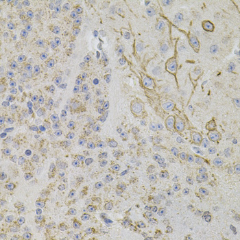
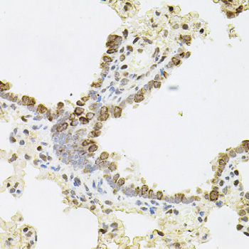
A05686-1 has real REG3A IHC images from paraffin-embedded mouse brain and rat lung and heart (catalog image captions). Human, Mouse and Rat reactivity is listed; IF data are absent (catalog: A05686-1).

Real IHC data Immunohistochemistry of paraffin-embedded mouse brain using REG3A antibody .Perform microwave antigen retrieval with 10 mM PBS buffer pH 7.2 before commencing with IHC staining protocol.
Anti-REG3A Antibody
Cat # A05686-1

The card for A05686-1 will render with an IHC image of paraffin-embedded mouse brain (catalog image caption). Additional IHC images show paraffin-embedded rat lung and heart; the application list includes IHC, and the reactivity list includes Human, Mouse and Rat (catalog: A05686-1).

Which to pick: For tissue IHC, choose A05686-1: it is a rabbit polyclonal antibody with an IHC dilution of 1:50–1:200, and its images document paraffin-embedded mouse brain and rat lung and heart (catalog: A05686-1; catalog image captions). No listed SKU has IF/ICC validation, an IF figure or an IF dilution (catalog: A05686-1). For cross-species IHC, A05686-1 lists Human, Mouse and Rat reactivity, though its IHC images show only mouse and rat tissue; the paraffin-section captions do not report a fixative (catalog: A05686-1; catalog image captions).

Each figure is that product's own IHC / IF validation image from its datasheet.

References

  1. UniProt Consortium. UniProt entry Q06141 (REG3A_HUMAN, Regenerating islet-derived protein 3-alpha).
  2. Human Protein Atlas. REG3A tissue IHC expression (reliability: Supported).
  3. Human Protein Atlas. REG3A subcellular location (ICC-IF): Localized to the cytosol. Caution: Based on antibodies targeting proteins from multiple genes..
  4. Human Protein Atlas. REG3A antibody validation summary (3 antibodies).
  5. REG3A: A Potential Fecal Biomarker for Ulcerative Colitis. Mediators of inflammation 2026 — PMC13296221.
  6. P. gingivalis Lipopolysaccharide Stimulates the Upregulated Expression of the Pancreatic Cancer-Related Genes Regenerating Islet-Derived 3 A/G in Mouse Pancreas. International journal of molecular sciences 2020 — PMC7583020.
  7. Deciphering the roadmap of in vivo reprogramming toward pluripotency. Stem cell reports 2022 — PMC9669407.
  8. A Single-Cell Transcriptome Atlas of the Human Pancreas. Cell systems 2016 — PMC5092539.
  9. PubMed PMID:7679928 — UniProt-cited evidence.
  10. PubMed PMID:1469087 — UniProt-cited evidence.
  11. PubMed PMID:1325291 — UniProt-cited evidence.