This website uses cookies to ensure you get the best experience on our website.


- Table of Contents
Plan REG3G IHC-P using the catalog antibody at 1:25 (datasheet M05473). Compare staining with the reported high Paneth-cell signal in duodenum and small intestine (HPA tissue IHC), while accounting for REG3G secretion (UniProt).

Expected localisation, validated protocols, controls and antibodies — the at-a-glance facts below, then the full design guide.
| Expected localisation | Secreted; cytoplasmic location also annotated (UniProt) | |
| Staining pattern | High Paneth-cell staining; compartment unreported (HPA tissue IHC) | |
| Antigen retrieval | Citrate pH 6 HIER, heat-mediated (datasheet M05473) | |
| Positive control | Duodenum+2 more · see all | |
| Negative control | Adipose tissue+4 more · see all |
| Fixation | Use consistent formaldehyde fixation for paraffin sections (datasheet M05473 IHC-P) (selected-SKU IHC image M05473) | |
| Caveat | Secreted protein may appear away from its source cells (HPA tissue IHC) | |
| Regulation | Acute-phase associated (UniProt) | |
| Isoform / epitope | 2 isoforms; processing may remove residues 27–37 (UniProt) |
Compare the catalog antibody’s IHC-P protocol with three published REG3G IHC workflows (PMC7144519; PMC5636971; PMC7583020).
| Sample | Formaldehyde-fixed, paraffin-embedded human pancreas tissue (datasheet M05473) |
| Fixation | Image formalin-fixed; duration unreported (datasheet M05473); verify before use. |
| Sectioning | 4–5 µm sections on charged slides (standard) |
| Deparaffinisation | Xylene, graded ethanol series to water (standard) |
| Antigen retrieval | Heat retrieval: Citrate pH 6 (datasheet M05473); 20 min, 95–100 °C (standard) |
| Peroxidase block | 3% H2O2, 10 min, room temperature (standard) |
| Blocking | 10% normal serum of the secondary host, 30 min, room temperature (standard) |
| Primary antibody | Mouse monoclonal (clone 1790CT313.66.2) anti-REG3G, 1:25 (datasheet M05473) |
| Primary incubation | 1 hours at 37°C (datasheet M05473) |
| Detection | HRP-polymer secondary, DAB chromogen 5–10 min (standard) |
| Counterstain | Hematoxylin, blue, dehydrate and mount (standard) |
| Expected result | REG3G-positive staining in paneth cells of duodenum (HPA tissue IHC: High). HPA tissue profile: Plasma protein. No signal in the no-primary control. |
For paraffin-section IHC, expect REG3G-associated staining in Paneth cells of the duodenum and small intestine, where HPA reports high staining; appendix enterocytes show medium staining (HPA tissue IHC). REG3G is secreted and also annotated in the cytoplasm, with no transmembrane segment (UniProt Q6UW15). Interpret cellular staining cautiously: HPA rates tissue IHC Uncertain because the antibodies may recognize proteins from more than one gene (HPA tissue IHC).
| Distinct staining in duodenal or small-intestinal Paneth cells, stronger than adjacent cells. | This matches the reported high Paneth-cell pattern (HPA tissue IHC). Record cell identity and distribution as well as intensity; staining alone does not resolve REG3G from another protein recognized by the antibody because the IHC evidence remains Uncertain (HPA tissue IHC). |
| Predominantly nuclear staining or a crisp, continuous membrane outline, with little cellular signal. | That compartment pattern is unsupported: REG3G is annotated as secreted and cytoplasmic and has no transmembrane segment (UniProt Q6UW15). Check section morphology and detection controls before interpreting it as specific; an isolated membrane outline is especially weak evidence for REG3G (general IHC practice). |
| Strong staining in cells outside the reported positive populations, including a listed negative cell type. | Compare the exact tissue and cell type with HPA, rather than assigning a whole tissue a single status (HPA tissue IHC). Unexpected staining may reflect antibody cross-reactivity, which HPA flags, or endogenous detection activity; use appropriate antibody and detection controls to distinguish them (HPA tissue IHC; general IHC practice). |
| Diffuse color across stroma, lumina, and unrelated cells, without a clear Paneth-cell pattern. | REG3G is secreted, so extracellular material cannot be dismissed solely by location (UniProt Q6UW15). Broad, uniform color that persists outside the expected cellular pattern is less persuasive; compare a no-primary control and assess nonspecific binding or detection background (HPA tissue IHC; general IHC practice). |
| No discernible staining in well-preserved duodenal or small-intestinal Paneth cells. | This conflicts with HPA's reported high staining in those cells, but does not by itself prove biological absence (HPA tissue IHC). Check cell identification, run controls, and review the antibody and detection workflow; HPA's uncertain IHC reliability limits conclusions from either a positive or negative result (HPA tissue IHC; general IHC practice). |
| Tissue and cell selection | Use Paneth cells in duodenum or small intestine as reported high IHC references; appendix enterocytes are reported medium (HPA tissue IHC). UniProt calls pancreas the predominant expression site, but that statement does not establish a pancreatic IHC pattern (UniProt Q6UW15). |
| Secretion and tissue location | REG3G is annotated as secreted and cytoplasmic (UniProt Q6UW15). HPA warns that RNA and protein locations can differ for a secreted protein; do not treat pancreatic RNA enrichment as a substitute for cell-level protein staining (HPA tissue IHC). |
| Antibody evidence | HPA047894 and HPA048334 each have Uncertain IHC status, and HPA warns of recognition of proteins from more than one gene (HPA antibodies; HPA tissue IHC). Match the staining pattern to morphology and controls before assigning REG3G specificity (general IHC practice). |
| Processing and epitope coverage | UniProt lists a signal peptide at residues 1–26, a propeptide at 27–37, and chains spanning 27–175 or 38–175 (UniProt Q6UW15). Antibody epitope information is absent here, so the record cannot predict which processed form an IHC stain detects. |
| Isoforms and retrieval | Target-specific fixation effects are not established by the supplied assay evidence. Verify with a matched IHC source before attributing a result to fixation. |
| IF/ICC Q&A: What localisation should an IF image show? | HPA reports approved cytosolic ICC-IF localisation, while cautioning that the antibody targets proteins from multiple genes (HPA subcellular). That IF observation supports a cytosolic comparison; it does not validate the paraffin-IHC antibody or establish an IF protocol (HPA subcellular; HPA antibodies). |
| Situation | Likely cause | Next action |
|---|---|---|
| Paneth cells are negative in a duodenal or small-intestinal section. | Possible cell-identification, staining-workflow, or detection failure; HPA reports high staining there but rates IHC Uncertain (HPA tissue IHC; general IHC practice). | Confirm Paneth-cell morphology and section quality, then review retrieval, primary-antibody use, detection, and run controls together; avoid calling biological absence from this slide alone (general IHC practice). |
| A listed negative population stains strongly. | Cross-reactivity is plausible given HPA's multi-gene warning; endogenous detection activity is another general IHC possibility (HPA tissue IHC; general IHC practice). | Identify the stained cell type, compare its HPA entry, and inspect no-primary and relevant detection controls before attributing the signal to REG3G (HPA tissue IHC; general IHC practice). |
| Color covers most of the section without a readable cellular pattern. | Nonspecific binding or detection background may obscure interpretation; secretion alone cannot establish that every extracellular deposit is specific (UniProt Q6UW15; general IHC practice). | Compare the no-primary control, reassess blocking and washes, and score only interpretable structures against the reported Paneth-cell pattern (HPA tissue IHC; general IHC practice). |
| Signal appears nuclear or confined to sharply outlined cell membranes. | Those dominant compartments are unsupported by the secreted/cytoplasmic annotation and lack of a transmembrane segment (UniProt Q6UW15). | Check morphology and detection controls, then compare the same run with a reported positive tissue; treat the discordant compartment as unresolved until specificity is supported (HPA tissue IHC; general IHC practice). |
| Pancreatic staining differs from what its high RNA expression suggests. | UniProt describes predominant pancreatic expression, whereas HPA warns that secreted protein and RNA locations may differ (UniProt Q6UW15; HPA tissue IHC). | Document the pancreatic cell-level pattern without assigning an expected IHC intensity from RNA; compare staining with HPA's reported positive cell populations (HPA tissue IHC; general IHC practice). |
| A positive stain varies between antibody preparations or cannot be assigned to one processed form. | HPA IHC antibody status is Uncertain; UniProt lists two chains, while antibody epitopes are unspecified in this payload (HPA antibodies; UniProt Q6UW15). | Report the antibody identifier, cellular pattern, and controls; keep the interpretation at REG3G-associated staining unless specificity and epitope coverage are independently established (HPA tissue IHC; general IHC practice). |


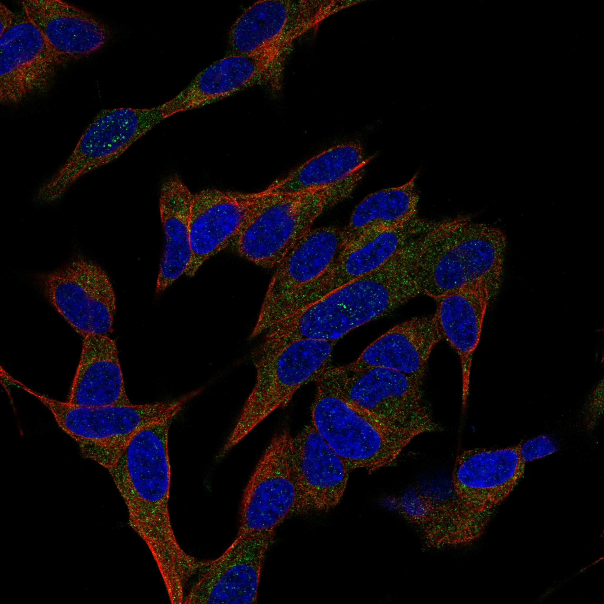
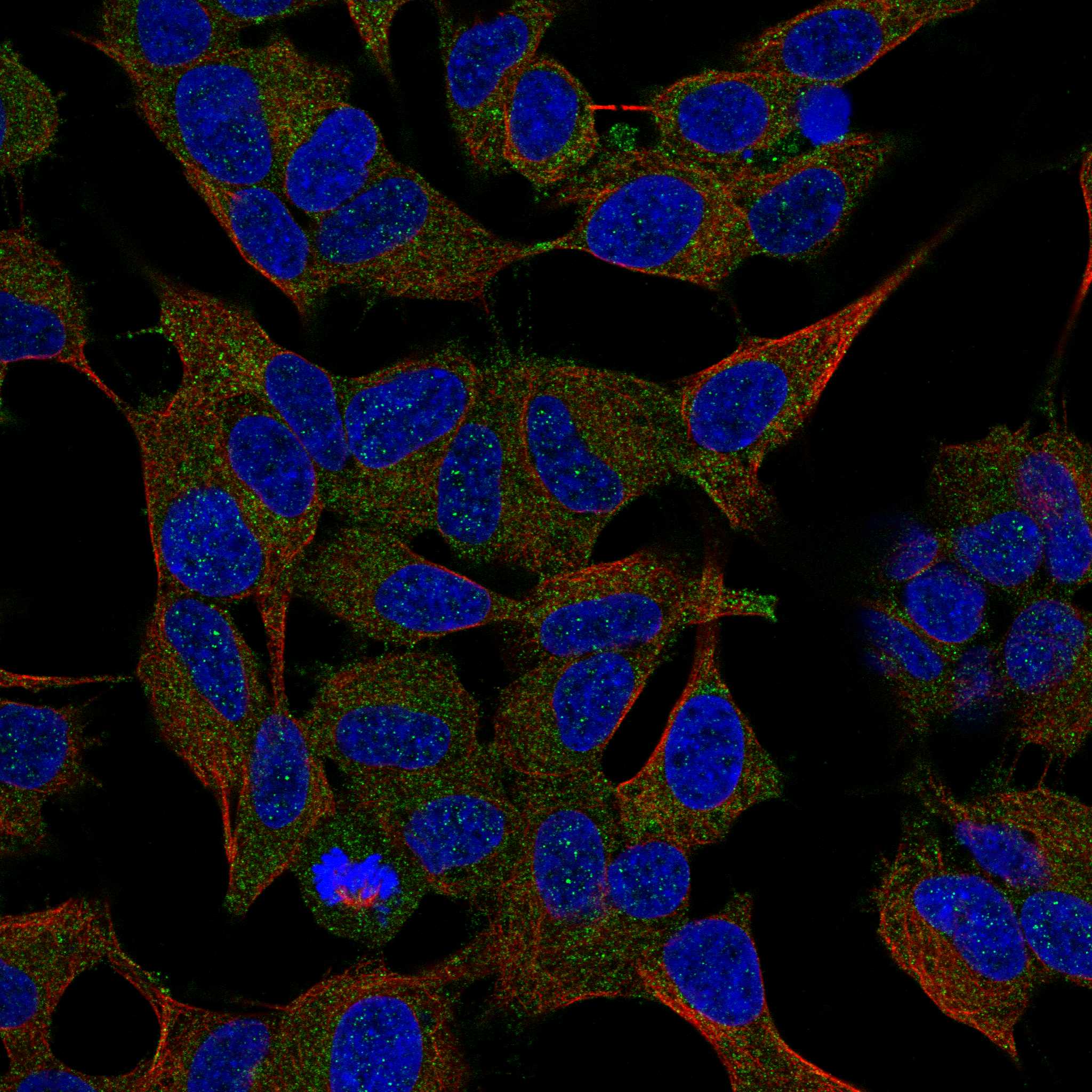
Comprehensive Human Protein Atlas IHC scoring per tissue (reliability: Uncertain — Caution, targets protein from more than one gene. Secreted protein, tissue location of RNA and protein is expected to differ. Pending external verification.). Rows are taken directly from the HPA tissue chart — click any row's HPA link to view the source.
| Tissue | Cell type | Level | Evidence | Source |
|---|---|---|---|---|
| Duodenum | Paneth cells | High | Protein (IHC) | HPA → |
| Small intestine | Paneth cells | High | Protein (IHC) | HPA → |
| Appendix | Enterocytes | Medium | Protein (IHC) | HPA → |
| Tissue | Cell type | Level | Evidence | Source |
|---|---|---|---|---|
| Adipose tissue | Adipocytes | Not detected | Protein (IHC) | HPA → |
| Adrenal gland | Glandular cells | Not detected | Protein (IHC) | HPA → |
| Bone marrow | Hematopoietic cells | Not detected | Protein (IHC) | HPA → |
| Breast | Adipocytes | Not detected | Protein (IHC) | HPA → |
| Bronchus | Respiratory epithelial cells | Not detected | Protein (IHC) | HPA → |
Troubleshoot REG3G chromogenic IHC by checking retrieval, tissue context, and secreted protein distribution before interpreting staining intensity.
M05473 is a human-reactive REG3G antibody with real IHC-P staining data from paraffin-embedded human pancreas sections (M05473 catalog reactivity; M05473 IHC image caption).
M05473 is listed for human IHC-P (M05473 catalog applications and reactivity). Its IHC image caption reports REG3G staining in formaldehyde-fixed, paraffin-embedded human pancreas sections (M05473 IHC image caption).
Which to pick: For tissue IHC, choose M05473, a mouse monoclonal listed for human IHC-P at 1:25 (M05473 catalog host, clone, applications, reactivity, and dilution). Its own image caption documents staining in formaldehyde-fixed, paraffin-embedded human pancreas sections (M05473 IHC image caption). There is no supported IF/ICC or cross-species pick in this payload (M05473 catalog applications, reactivity, and IF image data).