REV3L / DNA polymerase zeta catalytic subunit · IHC design guide

Design Immunohistochemistry for REV3L

Use this guide to plan chromogenic paraffin IHC for REV3L with the catalog antibody at 1:100–1:300 (datasheet). Score the mainly cytoplasmic tissue staining (HPA tissue IHC) with the nuclear annotation in mind (UniProt).

Evidence assembled Oct 2026 · For research use; verify linked source records and product datasheet before use
Immunohistochemistry protocol sheet for REV3L (IHC for REV3L): expected localisation Mainly cytoplasmic in tissue (HPA tissue IHC); nuclear annotation (UniProt), antibody A04785, validated IHC image, and IHC protocol steps
Printable REV3L IHC protocol sheet — expected localisation Mainly cytoplasmic in tissue (HPA tissue IHC); nuclear annotation (UniProt), antibody A04785, controls and protocol steps. Open the full REV3L IHC guide →

REV3L Immunohistochemistry Experimental Design Guide

Expected localisation, validated protocols, controls and antibodies — the at-a-glance facts below, then the full design guide.

Must know before staining
Expected localisation Mainly cytoplasmic in tissue (HPA tissue IHC); nuclear annotation (UniProt)
Staining pattern Cytoplasmic staining in glandular cells and kidney tubules (HPA tissue IHC)
Antigen retrieval Tris-EDTA pH 9.0 HIER, 95–98 °C, 20 min (rule: nuclear antigen)
Positive control ⓘ Adrenal gland+4 more · see all
Negative control ⓘ Adipose tissue+4 more · see all
Important caveats
Reasons your staining may differ from the expected pattern.
Fixation Keep formalin fixation consistent across paraffin sections. (standard IHC practice; not target-specific)
Caveat IHC localisation is uncertain and conflicts with nuclear annotation (HPA tissue IHC; UniProt)
Regulation Ubiquitous expression; no regulator given (UniProt)
Isoform / epitope 2 isoforms; epitope coverage is unreported (UniProt)
Section 1

Recommended REV3L IHC & IF Protocols

The catalog antibody’s IHC-P protocol is paired with a published mouse endometrial and EM lesion IHC protocol (PMC10034389).

Recommended immunohistochemistry (IHC-P) protocol parameters
SampleParaffin-embedded human brain tissue; fixative not specified (datasheet A04785)
FixationImage fixative and duration unreported (datasheet A04785); verify before use.
Sectioning4–5 µm sections on charged slides (standard)
DeparaffinisationXylene, graded ethanol series to water (standard)
Antigen retrievalHeat-induced epitope retrieval in Tris-EDTA buffer, pH 9.0, 20 min at 95–98 °C (standard rule: nuclear antigen)
Peroxidase block3% H2O2, 10 min, room temperature (standard)
Blocking10% normal serum of the secondary host, 30 min, room temperature (standard)
Primary antibodyRabbit anti-REV3L, 1:100-1:300 (datasheet A04785)
Primary incubationOvernight at 4 °C (standard)
DetectionHRP-polymer secondary, DAB chromogen 5–10 min (standard)
CounterstainHematoxylin, blue, dehydrate and mount (standard)
Expected resultREV3L-positive staining in glandular cells of adrenal gland (HPA tissue IHC: Medium). HPA tissue profile: Mainly cytoplasmic expression in several different tissue types. No signal in the no-primary control.
💡Decision noteStart with Tris-EDTA pH 9.0 HIER at 95–98 °C for 20 min for nuclear REV3L (page retrieval rule; UniProt O60673: nucleus).
Section 2

What Is the Expected REV3L Staining Pattern?

REV3L is a nuclear protein with no transmembrane segment (UniProt O60673: location, topology). In paraffin tissue IHC, HPA instead reports mainly cytoplasmic staining, including medium staining in glandular cells, renal tubule cells, hepatocytes and lung macrophages (HPA: tissue IHC). Treat that observed pattern as provisional: HPA rates tissue IHC reliability Uncertain because its subcellular location conflicts with external data (HPA: tissue IHC reliability).

What am I looking at on my slide?
Medium cytoplasmic staining in colon glandular cells, kidney tubule cells or liver hepatocytes.This matches examples of observed tissue staining (HPA: tissue IHC, Medium), but does not independently establish REV3L specificity: the tissue IHC assessment is Uncertain (HPA: tissue IHC reliability). Compare the relevant cell population and compartment before scoring a section positive.
Predominantly nuclear staining, or both nuclear and cytoplasmic staining, in an otherwise plausible tissue.A nuclear component fits UniProt's nuclear location and HPA's approved nucleoplasmic ICC-IF result (UniProt O60673: location; HPA: subcellular). Tissue IHC is reported mainly cytoplasmic and Uncertain (HPA: tissue IHC). Record the compartments separately and seek antibody-specific confirmation before calling either pattern definitive.
Strong membrane, luminal or extracellular staining dominates the section.That compartment is unsupported by the reported nuclear location and absence of a transmembrane segment (UniProt O60673: location, topology). Treat it as a possible artefact; inspect section morphology, the no-primary control and the detection reagents (general IHC practice).
A cell population reported as unstained, such as adipocytes or skin keratinocytes, stains strongly.HPA reports these populations as Not detected in tissue IHC (HPA: adipose tissue, skin). The mismatch raises cross-reactivity or endogenous detection activity as possibilities, rather than proving either cause. Compare matched negative controls and the distribution within the section (general IHC practice).
Color is spread evenly across cells and surrounding tissue, obscuring cell boundaries.This is diffuse background, not a scorable cell-specific pattern (general IHC practice). Evaluate a no-primary control, wash adequacy and detection background before interpreting the signal; HPA's Uncertain tissue IHC rating makes compartment-based claims especially tentative (HPA: tissue IHC reliability).
💡Expected REV3L appearanceA provisional positive IHC result is medium cytoplasmic signal in an HPA-reported cell population, such as colon glandular cells or kidney tubule cells; strong staining in reported negative populations or diffuse acellular color is suspect (HPA: tissue IHC, Uncertain; general IHC practice).
How each factor affects the staining
Compartment evidenceUniProt assigns REV3L to the nucleus, while HPA reports mainly cytoplasmic tissue IHC and approved nucleoplasmic ICC-IF (UniProt O60673: location; HPA: tissue IHC, subcellular). Score nuclear and cytoplasmic signals separately; the IHC discrepancy remains unresolved.
Cell-population selectionHPA reports Medium staining in bronchial respiratory epithelium and lung macrophages, but Not detected in adipocytes and skin keratinocytes (HPA: tissue IHC). Those are practical comparison populations, with no guarantee that every section will reproduce the reported level.
Antibody validationThe tissue IHC antibody HPA064853 is rated Uncertain, whereas ICC antibody HPA069382 is Approved for ICC (HPA: antibodies). These labels apply to different antibodies and applications; the ICC rating cannot validate a tissue IHC result.
Isoforms and epitope coverageUniProt lists two REV3L isoforms (UniProt O60673: isoforms). The supplied evidence gives no antibody epitope or isoform coverage, so a negative section cannot be explained as isoform exclusion on this record.
IF/ICC Q&AQ: Should nuclear IF settle a cytoplasmic IHC result? A: No. HPA reports approved nucleoplasmic ICC-IF, but tissue IHC remains Uncertain and mainly cytoplasmic (HPA: subcellular, tissue IHC). Interpret each application with its own validation evidence.
Why is my staining missing, weak or wrong?
SituationLikely causeNext action
No visible signal in a colon or kidney section.These contain HPA-reported Medium populations, but staining may vary and the tissue IHC assessment is Uncertain (HPA: colon, kidney; HPA: tissue IHC reliability).Confirm that the expected glandular or tubule cells are present; then check the IHC-validated antibody's supplied paraffin protocol, detection reagents and a run control (general IHC practice).
Only a strong nuclear signal appears in an HPA-reported positive tissue.Nuclear localization is plausible from UniProt and ICC-IF, while the reported tissue IHC pattern is mainly cytoplasmic (UniProt O60673: location; HPA: subcellular, tissue IHC).Document nuclear and cytoplasmic scores separately. Confirm the antibody identity and assess specificity with suitable controls before treating the discrepancy as a biological result (general IHC practice).
Reported negative populations stain as strongly as positive populations.Specificity is uncertain: adipocytes and skin keratinocytes are reported Not detected, while several epithelial populations are Medium (HPA: tissue IHC). Cross-reactivity or endogenous detection activity may contribute.Compare a no-primary control and the same detection system across sections; block endogenous detection activity where relevant to that system (general IHC practice).
Diffuse color persists across cells, stroma and blank areas.Non-cell-specific background can arise from the staining workflow (general IHC practice); it cannot be assigned to REV3L from the supplied HPA or UniProt evidence.Check the no-primary control, washing, blocking and detection development; repeat scoring only where cell boundaries and compartment are discernible (general IHC practice).
Membrane or extracellular signal is the dominant pattern.Neither compartment matches REV3L's nuclear assignment or lack of a transmembrane segment (UniProt O60673: location, topology).Check morphology and no-primary staining, and compare with a tissue containing an HPA-reported Medium cell population before interpreting the signal (general IHC practice; HPA: tissue IHC).
A weak or negative slide is attributed to fixation or an isoform.Target-specific fixation sensitivity is unreported in the supplied evidence; two isoforms are listed, but antibody epitope coverage is unspecified (UniProt O60673: isoforms).Record processing and antibody details, verify routine IHC run controls, and avoid assigning a REV3L-specific mechanism without additional evidence (general IHC practice).

Sample controls for REV3L IHC & IF

🧪Run colon first and look for nuclear staining in glandular cells (HPA: colon glandular cells Medium; UniProt O60673: nucleus); use adipose tissue as a negative comparator, scoring adipocytes as not detected (HPA: adipocytes Not detected). On the colon slide, cells without nuclear chromogen can show background staining, but no specific cell type there is established as REV3L-negative (HPA: colon glandular cells Medium; UniProt O60673: ubiquitously expressed).
Positive control tissue: Adrenal gland (Glandular cells, HPA Medium)
Negative control tissue: Adipose tissue (HPA Not detected)
ICC-IF cell lines (HPA subcellular resource): HPA ICC-IF images show REV3L in Hep-G2, SK-MEL-30, U2OS, with annotated localisation: Nucleoplasm (approved) (HPA subcellular).
Technical controls: Include a no-primary, secondary-only control and an isotype control matched to the primary antibody’s host species, immunoglobulin class and clonality where applicable (standard IHC practice). Use a REV3L knockout specimen or cognate-peptide block as a biological specificity control (caption: A04785 peptide block); for colon chromogenic IHC, quench endogenous peroxidase and check endogenous biotin if using avidin-biotin detection (standard IHC practice).
⚠️Feasibility: No target-specific fixation window or fixation effect is reported, and the selected A04785 paraffin-section caption does not state a fixative (caption: fixative not stated). Antigen-retrieval dependence is unreported, so assess retrieval conditions with the IHC-validated antibody (standard paraffin IHC practice). The supplied evidence does not establish whether frozen sections or IF/ICC are easier, or document a colon-specific artefact; HPA reports nucleoplasmic ICC-IF localization (HPA: nucleoplasm approved).

HPA tissue IHC evidence for REV3L

Comprehensive Human Protein Atlas IHC scoring per tissue (reliability: Uncertain — Subcellular location is contradicted by external data.). Rows are taken directly from the HPA tissue chart — click any row's HPA link to view the source.

Positive expression · recommended positive controls

TissueCell typeLevelEvidenceSource
Adrenal gland Glandular cells Medium Protein (IHC) HPA →
Appendix Glandular cells Medium Protein (IHC) HPA →
Bronchus Respiratory epithelial cells Medium Protein (IHC) HPA →
Colon Glandular cells Medium Protein (IHC) HPA →
Duodenum Glandular cells Medium Protein (IHC) HPA →

Undetected expression · recommended negative controls

TissueCell typeLevelEvidenceSource
Adipose tissue Adipocytes Not detected Protein (IHC) HPA →
Caudate Glial cells Not detected Protein (IHC) HPA →
Cervix Glandular cells Not detected Protein (IHC) HPA →
Endometrium Cells in endometrial stroma Not detected Protein (IHC) HPA →
Lymph node Germinal center cells Not detected Protein (IHC) HPA →
Section 3

Advanced REV3L IHC Tips

Troubleshoot REV3L chromogenic IHC by checking retrieval, nuclear localisation and cell level controls before interpreting staining intensity (UniProt O60673; HPA tissue IHC).

How should I retrieve REV3L in paraffin sections when nuclear staining is weak?
Start with Tris-EDTA pH 9.0 heat-induced retrieval at 95–98 °C for 20 min (page retrieval rule: nuclear antigen). Keep section thickness, heating time and cooling conditions consistent across the run so that changes in nuclear signal can be attributed to the retrieval comparison (standard IHC practice). If staining remains weak, compare a separately optimised retrieval condition on adjacent sections, using the same detection and counterstain settings; treat it as a fallback, not an established REV3L method (standard IHC practice). Score nuclear staining separately from cytoplasmic staining because REV3L is assigned to the nucleus, while HPA tissue IHC mainly reports cytoplasmic signal with uncertain reliability (UniProt O60673; HPA tissue IHC).
Could fixation explain weak or variable REV3L staining across paraffin blocks?
REV3L-specific fixation sensitivity is unknown from the supplied evidence; the catalog image shows paraffin-embedded human brain tissue but does not state its fixative (A04785 tissue-IHC caption). Compare blocks with documented fixation histories using matched 5 µm sections, identical Tris-EDTA pH 9.0 retrieval and one detection run (standard IHC practice; page retrieval rule: nuclear antigen). If fixation history differs, assess nuclear signal and tissue preservation together before assigning a weak result to antigen loss (standard IHC practice; UniProt O60673 localisation). Do not infer a REV3L fixation effect from tissue staining patterns or from its sequence features; neither establishes fixation sensitivity (HPA tissue IHC; UniProt O60673).
What should I do if REV3L staining looks cytoplasmic rather than nuclear?
Prioritise a nuclear readout because UniProt places REV3L in the nucleus and HPA subcellular imaging identifies the nucleoplasm as its approved location (UniProt O60673; HPA subcellular). HPA tissue IHC instead reports mainly cytoplasmic staining and labels its reliability uncertain because that location conflicts with external data (HPA tissue IHC). On matched 5 µm sections, compare nuclear and cytoplasmic signal after the page’s Tris-EDTA pH 9.0 retrieval, keeping detection and counterstaining identical (page retrieval rule; standard IHC practice). Record cytoplasmic signal as a separate observation requiring validation, rather than combining it with nuclear positivity in a single score (UniProt O60673; HPA tissue IHC).
Could REV3L isoforms or epitope accessibility change my IHC result?
REV3L has 2 annotated isoforms and is a 3,130-residue protein, so check the catalog antibody’s stated immunogen or epitope before interpreting discordant staining (UniProt O60673). The supplied product caption does not identify its epitope, and peptide blocking in the pictured paraffin brain section alone does not establish which isoform is detected (A04785 tissue-IHC caption; UniProt O60673). Compare matched sections under the page’s Tris-EDTA pH 9.0, 20 min retrieval while holding antibody and detection settings constant (page retrieval rule; standard IHC practice). Interpret an epitope-dependent difference only after checking available sequence coverage and an independent specificity control; REV3L’s annotated phosphorylation sites include residues 1030 and 1041 (UniProt O60673; standard IHC practice).
How can IF help assess a disputed REV3L IHC staining pattern?
Use IF as an orthogonal localisation check: REV3L is assigned to the nucleus and HPA reports approved nucleoplasmic localisation in its subcellular data (UniProt O60673; HPA subcellular). For a lung macrophage comparison, multiplex with a separately validated macrophage marker and a nuclear counterstain, then inspect whether REV3L signal falls within the expected cells and nuclei (HPA tissue IHC: lung macrophages; standard IF practice). Choose spectrally separated fluorophores after checking tissue autofluorescence with an unstained control (standard IF practice). Because REV3L has no annotated transmembrane segment and the expected signal is nuclear, optimise permeabilisation for nuclear access without assuming that the catalog antibody’s epitope is known (UniProt O60673; standard IF practice).
How do I distinguish REV3L signal from chromogenic background?
Run a no-primary control through the same retrieval, secondary detection and chromogen steps, and inspect whether the suspected staining persists (standard IHC practice). Include a peroxidase block before DAB detection and compare staining with tissue pigment, section edges and damaged areas; these are general chromogenic workflow checks, not REV3L-specific evidence (standard IHC practice). The A04785 caption describes peptide blocking in a paraffin-embedded human brain image, which supports a blocking comparison for that pictured preparation but does not identify its fixative (A04785 tissue-IHC caption). Give greater weight to reproducible nuclear signal than diffuse cytoplasmic colour because the reported tissue IHC pattern conflicts with approved nucleoplasmic localisation (UniProt O60673; HPA tissue IHC; HPA subcellular).
What is a defensible way to quantify REV3L chromogenic IHC? ⚠ ANSWER MARKED FOR VERIFICATION
Define the cell population and compartment before scoring, and report nuclear REV3L separately from cytoplasmic staining because the localisation sources disagree (UniProt O60673; HPA subcellular; HPA tissue IHC). For a nuclear H-score, multiply the percentage of cells at each intensity category 0–3 by that category and sum to a 0–300 score; also report percent positive nuclei (standard IHC scoring practice). Normalise positive counts to all evaluable nuclei in the same defined cell population, or report positive-cell density per mm² of viable tissue (standard IHC scoring practice). Use matched retrieval, detection and counterstain conditions across specimens, and exclude damaged or necrotic areas before comparison (standard IHC practice).
When should a positive REV3L stain be treated as uncertain?
Treat nuclear staining in intact, morphologically identifiable cells as the localisation-consistent finding: UniProt assigns REV3L to the nucleus and HPA subcellular data approve nucleoplasmic localisation (UniProt O60673; HPA subcellular). Check the cell identity against the sampled tissue; HPA reports medium staining in lung macrophages, while its tissue IHC profile is mainly cytoplasmic and rated uncertain (HPA tissue IHC). Discount staining confined to section edges, necrotic regions or no-primary controls, and check for endogenous peroxidase before interpreting DAB colour as antigen signal (standard IHC practice). Peptide blocking in the A04785 brain image is useful supporting evidence for that preparation, but it does not resolve the nuclear-versus-cytoplasmic discrepancy across assays (A04785 tissue-IHC caption; HPA tissue IHC; HPA subcellular).
Boster reagents

Best REV3L / DNA polymerase zeta catalytic subunit IHC Antibodies

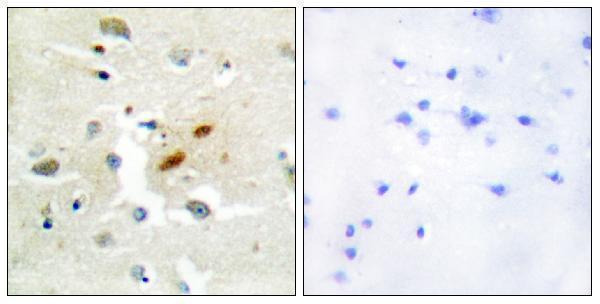
A04785 lists IHC and IF applications with human and mouse reactivity (catalog applications/reactivity). Its IHC figure shows paraffin-embedded human brain tissue; no IF figure is supplied (IHC image caption; IF image alts).

Real IHC data Immunohistochemistry analysis of paraffin-embedded human brain tissue, using DNA Polymerase zeta Antibody. The picture on the right is blocked with the synthesized peptide.
Anti-DNA pol zeta REV3L Antibody
Cat # A04785

A04785 is listed for IHC and IF with human and mouse reactivity (catalog applications/reactivity). Its IHC figure shows paraffin-embedded human brain tissue alongside a peptide-blocked comparison; no IF image is supplied (IHC image caption; IF image alts).

Which to pick: For tissue IHC, choose A04785: its own figure shows paraffin-embedded human brain tissue, with the fixative unreported (A04785 IHC image caption). For IF/ICC, A04785 lists IF at 1:50; ICC is not listed, and no IF figure is supplied (catalog applications/dilution; IF image alts). For cross-species work, A04785 lists human and mouse reactivity, although its IHC figure shows only human tissue; clonality is unspecified (catalog reactivity/clone; A04785 IHC image caption).

Each figure is that product's own IHC / IF validation image from its datasheet.

References

  1. UniProt Consortium. UniProt entry O60673 (REV3L_HUMAN, DNA polymerase zeta catalytic subunit).
  2. Human Protein Atlas. REV3L tissue IHC expression (reliability: Uncertain).
  3. Human Protein Atlas. REV3L subcellular location (ICC-IF): Mainly localized to the nucleoplasm..
  4. Human Protein Atlas. REV3L antibody validation summary (2 antibodies).
  5. DNA polymerase ζ deficiency causes impaired wound healing and stress-induced skin pigmentation. Life science alliance 2018 — PMC6055517.
  6. Telomere-related genes as potential biomarkers to predict endometriosis and immune response: Development of a machine learning-based risk model. Frontiers in medicine 2023 — PMC10034389.
  7. Epigenetics and DNA Base Substitutions of Epstein-Barr Virus (EBV)-Related Gastric Cancers: Implications for Targeted Therapies. Genes 2026 — PMC13408365.
  8. REV3L, a promising target in regulating the chemosensitivity of cervical cancer cells. PloS one 2015 — PMC4364373.
  9. PubMed PMID:9618506 — UniProt-cited evidence.
  10. PubMed PMID:10102035 — UniProt-cited evidence.
  11. PubMed PMID:9925914 — UniProt-cited evidence.