This website uses cookies to ensure you get the best experience on our website.


- Table of Contents
Plan chromogenic RGS7 IHC around high cerebellar GLUC-cell staining and medium neuronal staining in caudate and cortex (HPA tissue IHC). This guide covers FFPE handling, brain-region controls and interpretation of the cytoplasmic/membrane pattern (HPA tissue IHC).

Expected localisation, validated protocols, controls and antibodies — the at-a-glance facts below, then the full design guide.
| Expected localisation | Cytoplasm/membrane in cerebellar GLUC cells (HPA tissue IHC) | |
| Staining pattern | High in cerebellar GLUC cells; medium in caudate and cortical neurons (HPA tissue IHC) | |
| Antigen retrieval | Tris-EDTA pH 9.0 HIER, heat-mediated (datasheet A04305) | |
| Positive control | Cerebellum+2 more · see all | |
| Negative control | Adipose tissue+4 more · see all |
| Fixation | Matched tissue-IHC evidence does not establish the fixation claim. Validate the specimen-specific method before use. (selected-SKU IHC image A04305) | |
| Caveat | Low hippocampal staining may complicate interpretation (HPA tissue IHC) | |
| Regulation | Brain-specific expression (HPA tissue IHC) | |
| Isoform / epitope | Five isoforms; check antibody epitope coverage (UniProt) |
The catalog antibody’s paraffin IHC protocol is complemented by three published chromogenic RGS7 IHC protocols using tissue microarrays or brain sections (PMC5766496; PMC5127842; PMC4060866).
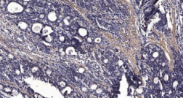
| Sample | Paraffin-embedded human Gastric adenocarcinoma tissue; fixative not specified (datasheet A04305) |
| Fixation | Image fixative and duration unreported (datasheet A04305); verify before use. |
| Sectioning | 4–5 µm sections on charged slides (standard) |
| Deparaffinisation | Xylene, graded ethanol series to water (standard) |
| Antigen retrieval | Heat retrieval: Tris-EDTA pH 9.0 (datasheet A04305); 20 min, 95–100 °C (standard) |
| Peroxidase block | 3% H2O2, 10 min, room temperature (standard) |
| Blocking | 10% normal serum of the secondary host, 30 min, room temperature (standard) |
| Primary antibody | Rabbit anti-RGS7, 1:50 recommended; image 1:200 (datasheet A04305) |
| Primary incubation | Overnight at 4 °C (standard) |
| Detection | HRP-polymer secondary, DAB chromogen 5–10 min (standard) |
| Counterstain | Hematoxylin, blue, dehydrate and mount (standard) |
| Expected result | RGS7-positive staining in gLUC cells - cytoplasm/membrane of cerebellum (HPA tissue IHC: High). HPA tissue profile: Brain-specific expression. No signal in the no-primary control. |
RGS7 should stain mainly neuronal cytoplasm, with membrane staining possible: HPA reports High cytoplasmic/membranous staining in cerebellar GLUC cells and Medium staining in caudate and cerebral cortex neuronal cells (HPA tissue IHC). HPA rates the tissue pattern Enhanced for consistency with RNA expression (HPA tissue IHC). RGS7 has no transmembrane segment, but interacting proteins can promote membrane localization (UniProt P49802 topology and subunit).
| Cerebellar GLUC cells show strong cytoplasmic staining, with some membrane accentuation; neuronal cells stain in caudate or cortex. | This fits the reported tissue pattern: cerebellar GLUC cells are High in cytoplasm/membrane, while caudate and cortical neuronal cells are Medium (HPA tissue IHC). Membrane staining is plausible because PKD1 or RGS7BP interaction promotes RGS7 membrane localization (UniProt P49802 subcellular location). |
| Staining is predominantly nuclear, with little cytoplasmic or membrane signal in expected positive cells. | A predominantly nuclear pattern conflicts with UniProt's cytoplasmic and membrane locations and HPA's cerebellar cytoplasm/membrane observation (UniProt P49802 subcellular location; HPA tissue IHC). Review the control pattern and antibody-dependent signal before calling nuclei RGS7-positive (general IHC practice). |
| Strong chromogen appears in adipocytes, glandular cells, or respiratory epithelium. | These cells were Not detected in the corresponding HPA adipose, adrenal or appendix, and bronchus tissue examples (HPA tissue IHC). Treat staining as unexpected; antibody cross-reactivity or endogenous detection activity are possibilities to investigate, not established explanations (general IHC practice). |
| Color spreads across multiple compartments or tissue regions without a clear cell-associated pattern. | Diffuse background cannot establish RGS7 localization or cell-specific expression (general IHC practice). Compare a no-primary control with the expected neuronal pattern; nonspecific antibody binding, incomplete blocking, or detection background may contribute (general IHC practice). |
| No convincing staining appears in a cerebellar section expected to contain GLUC cells. | HPA reports High staining in cerebellar GLUC cells, so this is a failed or discrepant positive-control result rather than evidence that RGS7 is universally absent (HPA tissue IHC). Check that the relevant cells are present, then review the IHC detection workflow (general IHC practice). |
| Tissue and cell selection | Cerebellar GLUC cells provide the strongest listed positive example; caudate and cortical neuronal cells are Medium, while hippocampal neuronal cells are Low (HPA tissue IHC). Compare the same cell types when judging intensity, because these reported levels differ (HPA tissue IHC). |
| Intracellular distribution | UniProt lists cytosol/cytoplasm and membrane, and reports that PKD1 or RGS7BP interaction promotes membrane location (UniProt P49802 subcellular location). RGS7 has no transmembrane segment, so membrane accentuation does not imply a membrane-spanning protein (UniProt P49802 topology). |
| Antibody validation | CAB017561 is listed as IHC Enhanced, and HPA describes the tissue result as highly consistent with RNA expression (HPA antibodies; HPA tissue IHC). These assessments support the reported pattern; evaluate unexpected staining with controls rather than assuming every colored structure identifies RGS7 (general IHC practice). |
| Isoforms and processing | UniProt lists 5 isoforms, no signal peptide or propeptide, and a chain spanning residues 1–495 (UniProt P49802). The supplied sources do not map the antibody epitope or establish which isoforms its IHC staining detects (HPA antibodies; UniProt P49802). |
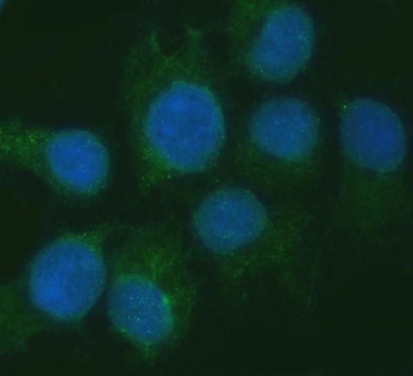
| IF/ICC Q: What localization should I expect? | A: HPA reports mainly supported cytosolic localization in ICC-IF, with mitochondrial localization marked uncertain; its ICC antibody status is Supported (HPA subcellular ICC-IF; HPA antibodies). Interpret the mitochondrial observation cautiously and do not transfer its certainty to paraffin-section IHC (HPA subcellular ICC-IF). |
| Situation | Likely cause | Next action |
|---|---|---|
| Cerebellar positive control has no detectable chromogen. | The result conflicts with HPA's High GLUC-cell observation; cell sampling or an IHC workflow failure is possible (HPA tissue IHC; general IHC practice). | Confirm GLUC cells are represented, then review primary-antibody application, detection reagents and chromogen development using routine IHC controls (general IHC practice). |
| Only nuclei stain in a neuronal-rich field. | Predominant nuclear localization conflicts with the supplied RGS7 locations and the reported cerebellar cell pattern (UniProt P49802 subcellular location; HPA tissue IHC). | Compare the cerebellar positive control and a no-primary control; reassess whether the nuclear signal follows the primary antibody or the detection system (general IHC practice). |
| Adipose or bronchial cells stain strongly. | HPA reports adipocytes in adipose tissue and respiratory epithelial cells in bronchus as Not detected (HPA tissue IHC). Cross-reactivity or endogenous detection activity is possible (general IHC practice). | Run a no-primary control and review the detection block; score those cells separately from expected neuronal staining (general IHC practice). |
| Background obscures cell borders and cytoplasm. | Nonspecific binding or background from the chromogenic detection workflow may obscure localization (general IHC practice). | Check the no-primary control, blocking and washing steps; interpret a positive result only where the cell-associated pattern remains clear (general IHC practice). |
| Cortex looks weaker than cerebellum. | That contrast can match the reported levels: cortical neuronal cells are Medium and cerebellar GLUC cells are High (HPA tissue IHC). | Compare like cell types and confirm the cortical neuronal signal is cell-associated before changing the assay based on intensity alone (HPA tissue IHC; general IHC practice). |
| Punctate signal is called mitochondrial RGS7 in a tissue section. | HPA marks mitochondrial localization uncertain in ICC-IF; it does not establish that interpretation for tissue IHC (HPA subcellular ICC-IF). | Describe the observed IHC pattern without assigning an organelle; assess any mitochondrial claim separately with an appropriate localization control (general IHC practice). |


Comprehensive Human Protein Atlas IHC scoring per tissue (reliability: Enhanced — High consistency between antibody staining and RNA expression data.). Rows are taken directly from the HPA tissue chart — click any row's HPA link to view the source.
| Tissue | Cell type | Level | Evidence | Source |
|---|---|---|---|---|
| Cerebellum | GLUC cells - cytoplasm/membrane | High | Protein (IHC) | HPA → |
| Caudate | Neuronal cells | Medium | Protein (IHC) | HPA → |
| Cerebral cortex | Neuronal cells | Medium | Protein (IHC) | HPA → |
| Tissue | Cell type | Level | Evidence | Source |
|---|---|---|---|---|
| Adipose tissue | Adipocytes | Not detected | Protein (IHC) | HPA → |
| Adrenal gland | Glandular cells | Not detected | Protein (IHC) | HPA → |
| Appendix | Glandular cells | Not detected | Protein (IHC) | HPA → |
| Bone marrow | Hematopoietic cells | Not detected | Protein (IHC) | HPA → |
| Breast | Adipocytes | Not detected | Protein (IHC) | HPA → |
Troubleshoot RGS7 staining in paraffin sections by checking retrieval, compartment, cell type and controls before comparing signal intensity.
Anti-RGS7 antibodies have IHC data from human paraffin-embedded gastric adenocarcinoma (A04305 image caption) and IF/ICC data from SiHa cells (A04305-1 image caption); both list Human, Mouse, and Rat reactivity (catalog).
A04305 will render with its IHC image of paraffin-embedded human gastric adenocarcinoma (A04305 image caption); its listed applications include IHC (catalog). A04305-1 will render with its IF image of SiHa cells (A04305-1 image caption); its listed applications include IF and ICC (catalog).
Which to pick: For paraffin-section IHC, choose A04305: it lists IHC and is polyclonal (catalog), and its image caption shows paraffin-embedded human gastric adenocarcinoma but does not report the fixative (A04305 image caption). For IF/ICC, choose A04305-1: it lists both applications (catalog) and has a SiHa cell IF image (A04305-1 image caption). For cross-species planning, both list Human, Mouse, and Rat reactivity (catalog); the A04305-1 IF/ICC dilution entry specifies Human (catalog dilution entry).