RIPK4 / Receptor-interacting serine/threonine-protein kinase 4 · IHC design guide

Design Immunohistochemistry for RIPK4

Plan RIPK4 IHC in paraffin sections with the catalog antibody at 1:100–1:200 (datasheet: IHC). Assess membranous staining in esophageal squamous epithelial cells (HPA tissue IHC), using negative controls and scoring by cell type and intensity (standard IHC practice).

Evidence assembled Oct 2026 · For research use; verify linked source records and product datasheet before use
Immunohistochemistry protocol sheet for RIPK4 (IHC for RIPK4): expected localisation Predominantly membranous in squamous epithelia (HPA tissue IHC), antibody A07084, validated IHC image, and IHC protocol steps
Printable RIPK4 IHC protocol sheet — expected localisation Predominantly membranous in squamous epithelia (HPA tissue IHC), antibody A07084, controls and protocol steps. Open the full RIPK4 IHC guide →

RIPK4 Immunohistochemistry Experimental Design Guide

Expected localisation, validated protocols, controls and antibodies — the at-a-glance facts below, then the full design guide.

Must know before staining
Expected localisation Predominantly membranous in squamous epithelia (HPA tissue IHC)
Staining pattern Membranous staining in squamous epithelial cells (HPA tissue IHC)
Antigen retrieval Citrate pH 6.0 HIER, 95–98 °C, 20 min (rule: cytoplasmic / membrane antigen)
Positive control ⓘ Esophagus+1 more · see all
Negative control ⓘ Adipose tissue+4 more · see all
Important caveats
Reasons your staining may differ from the expected pattern.
Fixation Keep paraffin-section fixation consistent. (standard IHC practice; not target-specific)
Caveat Staining and RNA show medium consistency (HPA tissue IHC)
Regulation Direct TP63 transcriptional target (UniProt)
Isoform / epitope 2 isoforms; epitope coverage needs checking (UniProt)
Section 1

Recommended RIPK4 IHC & IF Protocols

The catalog antibody’s IHC-P protocol is supplemented by 2 published RIPK4 protocols for paraffin-embedded cervical tissue (PMC7841057; PMC4493702).

Recommended immunohistochemistry (IHC-P) protocol parameters
SampleParaffin-embedded human liver damage tissue; fixative not specified (datasheet A07084)
FixationImage fixative and duration unreported (datasheet A07084); verify before use.
Sectioning4–5 µm sections on charged slides (standard)
DeparaffinisationXylene, graded ethanol series to water (standard)
Antigen retrievalHeat-induced epitope retrieval in citrate buffer, pH 6.0, 20 min at 95–98 °C (standard rule: cytoplasmic / membrane antigen)
Peroxidase block3% H2O2, 10 min, room temperature (standard)
Blocking10% normal serum of the secondary host, 30 min, room temperature (standard)
Primary antibodyRabbit anti-RIPK4, 1:100-1:200 (datasheet A07084)
Primary incubationOvernight at 4 °C (standard)
DetectionHRP-polymer secondary, DAB chromogen 5–10 min (standard)
CounterstainHematoxylin, blue, dehydrate and mount (standard)
Expected resultRIPK4-positive staining in squamous epithelial cells of esophagus (HPA tissue IHC: Medium). HPA tissue profile: Membranous expression mainly in squamous epithelia. No signal in the no-primary control.
💡Decision noteStart with citrate pH 6.0 retrieval for this page (page protocol); EDTA pH 8.0 is a published alternative (PMC4493702).
Section 2

What Is the Expected RIPK4 Staining Pattern?

RIPK4 is listed in the cytoplasm and at membranes, with no transmembrane segment (UniProt P57078 topology). In tissue IHC, expect mainly membranous staining in squamous epithelia, including medium staining of esophageal squamous epithelial cells; stomach glandular cells also show medium staining (HPA tissue IHC). HPA rates the tissue pattern Enhanced, but reports only medium agreement with RNA data and pending external verification (HPA tissue IHC).

What am I looking at on my slide?
Membranous staining in esophageal squamous epithelial cells, with an interpretable counterstain.This matches HPA’s main tissue pattern; the reported esophageal intensity is medium (HPA tissue IHC). Judge localization in the epithelial cells, since nearby unstained areas do not establish assay failure (general IHC practice).
Predominantly nuclear staining, with little discernible epithelial membrane staining.A nuclear dominant pattern does not match the reported cytoplasmic and membrane locations (UniProt P57078; HPA tissue IHC). Review antibody specificity and whether the signal follows tissue edges or other staining artifacts before assigning it to RIPK4 (general IHC practice).
Strong staining in adipocytes or bone marrow hematopoietic cells while the expected epithelial pattern is absent.HPA reports RIPK4 as not detected in those cell populations (HPA tissue IHC). Consider cross reactivity or endogenous detection activity, and compare a reagent omission control with the stained section (general IHC practice).
Color spreads across stroma, empty spaces, or many unrelated cell types, obscuring cell boundaries.That distribution cannot be scored confidently as the mainly membranous squamous epithelial pattern (HPA tissue IHC). Diffuse background can arise from inadequate blocking, washing, or chromogen control (general IHC practice).
No discernible epithelial signal in esophagus despite an intact section and counterstain.Esophageal squamous epithelial cells are an observed medium positive population, so a blank result warrants an assay check (HPA tissue IHC). Compare a concurrently stained control before interpreting the specimen as RIPK4 negative (general IHC practice).
💡Expected RIPK4 appearanceCall a positive result when esophageal squamous epithelial cells show discernible, mainly membranous medium staining (HPA tissue IHC); widespread nuclear or cell free color is suspect background (general IHC practice).
How each factor affects the staining
Tissue choice and intensityEsophageal squamous epithelial cells and stomach glandular cells show medium staining; adipocytes and bone marrow hematopoietic cells are not detected (HPA tissue IHC). Compare like cell types when selecting controls.
Antibody evidenceHPA030942 has Enhanced IHC validation, while HPA079641 has Supported ICC validation with no IHC status listed (HPA antibodies). The tissue profile has medium RNA agreement and awaits external verification (HPA tissue IHC).
Membrane interpretationRIPK4 has no transmembrane segment despite its membrane and cytoplasmic annotations (UniProt P57078 topology). Membranous IHC therefore describes the observed distribution, without establishing membrane insertion (HPA tissue IHC; UniProt P57078 topology).
Isoforms and epitope scopeUniProt lists 2 isoforms and a protein kinase domain at residues 22–286 (UniProt P57078). No supplied antibody epitope map establishes whether either isoform should stain differently; avoid isoform specific calls.
Q: What should IF/ICC show?A: HPA reports vesicles and cytosol as approved main locations, with plasma membrane as an uncertain additional location (HPA subcellular ICC-IF). Interpret this as IF/ICC evidence, separate from the tissue IHC pattern.
Why is my staining missing, weak or wrong?
SituationLikely causeNext action
Esophageal epithelium is blank.The expected medium epithelial staining is missing (HPA tissue IHC); the failed step cannot be identified from appearance alone.Check the concurrent positive control, antibody application, detection reagents, and chromogen development (general IHC practice).
The section shows nuclear dominant staining.This conflicts with the reported cytoplasmic and membrane locations (UniProt P57078; HPA tissue IHC); specificity or staining artifact is possible.Inspect an antibody omission control and reassess morphology before scoring nuclear color as RIPK4 (general IHC practice).
Adipocytes or bone marrow cells stain strongly.Those populations are reported as not detected (HPA tissue IHC); cross reactivity or endogenous detection activity is possible (general IHC practice).Compare the same cell types in a reagent omission control; review blocking and detection chemistry (general IHC practice).
Diffuse color masks epithelial borders.Background prevents assessment of the mainly membranous pattern (HPA tissue IHC); nonspecific reagent binding or excess chromogen may contribute (general IHC practice).Use the omission control, review blocking and washes, and stop chromogen development at a readable endpoint (general IHC practice).
Oral mucosa stains more faintly than esophagus.HPA lists oral mucosa squamous epithelial cells as low and esophageal cells as medium (HPA tissue IHC).Assess each tissue against its reported level; use esophageal epithelium to check whether the run can reveal the stronger expected signal (HPA tissue IHC; general IHC practice).
ICC/IF shows cytosolic or vesicular signal without crisp membrane outlines.Vesicles and cytosol are approved ICC-IF locations, whereas plasma membrane is an uncertain additional location (HPA subcellular ICC-IF).Do not reject the IF/ICC image solely for lacking membrane outlines; assess it against the ICC-IF localization evidence (HPA subcellular ICC-IF).

Sample controls for RIPK4 IHC & IF

🧪Run esophagus first: squamous epithelial cells should stain (HPA: Medium). Use adipose tissue as the negative comparison, with adipocytes near background (HPA: Not detected); on the esophagus slide, use adjacent stromal cells as an internal background reference, without treating them as a validated RIPK4-negative population (HPA: esophageal squamous epithelial cells Medium).
Positive control tissue: Esophagus (Squamous epithelial cells, HPA Medium)
Negative control tissue: Adipose tissue (HPA Not detected)
ICC-IF cell lines (HPA subcellular resource): HPA ICC-IF images show RIPK4 in CACO-2, HaCaT, RT-4, with annotated localisation: Vesicles (approved), Cytosol (approved) (HPA subcellular).
Technical controls: Include a no-primary, secondary-only control; an isotype control matched to the primary antibody’s host species and clonality; and a RIPK4 knockout biological negative if available (standard IHC practice). Quench endogenous peroxidase and assess background in the esophagus section before interpreting chromogenic signal (standard chromogenic IHC practice).
⚠️Feasibility: A target-specific fixation window and fixation effect are unreported in the supplied evidence; the selected A07084 paraffin-section caption does not state a fixative (A07084 caption: fixative unreported). That caption uses microwave retrieval in 10 mM PBS, pH 7.2, but it does not establish that retrieval is required for esophagus (A07084 caption). The supplied evidence does not establish whether frozen sections or IF are easier; inspect the esophageal surface for nonspecific staining when scoring squamous epithelium (standard IHC practice).

HPA tissue IHC evidence for RIPK4

Comprehensive Human Protein Atlas IHC scoring per tissue (reliability: Enhanced — Medium consistency between antibody staining and RNA expression data. Pending external verification.). Rows are taken directly from the HPA tissue chart — click any row's HPA link to view the source.

Positive expression · recommended positive controls

TissueCell typeLevelEvidenceSource
Esophagus Squamous epithelial cells Medium Protein (IHC) HPA →
Stomach Glandular cells Medium Protein (IHC) HPA →

Undetected expression · recommended negative controls

TissueCell typeLevelEvidenceSource
Adipose tissue Adipocytes Not detected Protein (IHC) HPA →
Appendix Glandular cells Not detected Protein (IHC) HPA →
Bone marrow Hematopoietic cells Not detected Protein (IHC) HPA →
Breast Adipocytes Not detected Protein (IHC) HPA →
Bronchus Respiratory epithelial cells Not detected Protein (IHC) HPA →
Section 3

Advanced RIPK4 IHC Tips

Troubleshoot RIPK4 chromogenic IHC in paraffin sections by checking retrieval, compartment, tissue context, and controls before interpreting staining.

How should I retrieve RIPK4 antigen in paraffin sections?
Start with citrate buffer at pH 6.0 for heat-induced retrieval at 95–98 °C for 20 min (page retrieval protocol). If staining is weak, compare an adjacent section using microwave retrieval in 10 mM PBS, pH 7.2, the condition reported for catalog antibody A07084 on paraffin-embedded human liver damage (A07084 tissue-IHC caption). Keep antibody concentration, detection chemistry, and development time matched between sections so the retrieval comparison is interpretable (standard IHC practice). Assess signal in viable cells and watch for tissue lifting or diffuse background after heating (standard IHC practice). The caption gives no microwave duration, so establish that variable locally (A07084 tissue-IHC caption).
Could fixation explain weak or patchy RIPK4 staining?
Target-specific fixation sensitivity is unknown: the selected paraffin-section caption does not state a fixative (A07084 tissue-IHC caption). Record the actual fixative, time before fixation, fixation duration, and processing history for each specimen, then compare sections with matched histories (standard IHC practice). Inconsistent processing can complicate interpretation of patchy chromogenic staining, so inspect tissue preservation and use an on-slide control processed alongside the sample (standard IHC practice). Apply the stated pH 6.0, 95–98 °C, 20 min retrieval condition consistently before adjusting antibody concentration (page retrieval protocol). Neither the tissue expression profile nor protein topology establishes a RIPK4-specific fixation effect (HPA tissue IHC; UniProt P57078 topology).
Which cellular pattern should count as plausible RIPK4 staining?
Examine epithelial cell borders and cytoplasm separately: tissue IHC reports mainly membranous expression in squamous epithelia, while the protein is annotated in cytoplasm and at membranes (HPA tissue IHC; UniProt P57078 subcellular location). Its sequence has no transmembrane segment, so a membrane-associated pattern does not require a membrane-spanning protein (UniProt P57078 topology). Cell imaging places RIPK4 mainly in vesicles and cytosol, with plasma-membrane localisation listed as uncertain (HPA subcellular). On chromogenic sections, score convincing signal only where cell outlines and tissue structure permit compartment assignment (standard IHC practice). Treat diffuse staining outside intact cells as inconclusive until section quality and background controls are checked (standard IHC practice).
Could the antibody epitope affect staining across RIPK4 isoforms?
RIPK4 has 2 listed isoforms, but the supplied evidence does not map the catalog antibody’s epitope to either one (UniProt P57078 isoforms; supplied antibody evidence). Obtain the immunogen sequence and align it with both isoforms before claiming that the IHC result represents total RIPK4 (standard antibody-validation practice). The annotated kinase domain spans residues 22–286, and the protein has no transmembrane segment; neither fact locates this antibody’s binding site (UniProt P57078 domains and topology). If staining differs between specimens, compare epitope coverage and section processing before assigning the difference to splicing (standard IHC practice). Report the antibody identity and retrieval condition with any compartment-specific score (standard IHC practice).
How can IF help check an ambiguous chromogenic RIPK4 pattern?
On an adjacent section, multiplex RIPK4 with a validated squamous epithelial marker to identify the cells being compared with the chromogenic result (HPA tissue IHC: mainly squamous epithelial expression; standard IF practice). Choose a spectrally separated, preferably far-red fluorophore after checking autofluorescence in an unstained section, and include single-stain controls for channel bleed-through (standard IF practice). For a cytosolic or vesicular epitope, use controlled permeabilisation and compare it with a minimally permeabilised condition if a membrane-associated signal is under study (HPA subcellular; standard IF practice). The epitope’s membrane-facing side is not supplied, and RIPK4 has no transmembrane segment, so do not assume surface accessibility (UniProt P57078 topology; supplied antibody evidence). Keep IF observations distinct from the antibody’s paraffin-section IHC evidence (A07084 tissue-IHC caption).
What should I check when RIPK4 chromogenic staining is diffuse?
Run a no-primary control and examine tissue edges, folds, damaged regions, and pigment before increasing the RIPK4 antibody concentration (standard IHC practice). Apply a peroxidase block before peroxidase-based detection and use matched DAB development times; these are general chromogenic IHC steps, not RIPK4-specific validation (standard IHC practice). The selected antibody image used 1:100 on a paraffin section, which is a reference condition rather than proof that the dilution suits every specimen (A07084 tissue-IHC caption). If background persists, optimise blocking, washes, primary-antibody dilution, and detection exposure one variable at a time (standard IHC practice). Judge the result against intact-cell localisation and the no-primary control (standard IHC practice).
How should I quantify RIPK4 staining across sections? ⚠ ANSWER MARKED FOR VERIFICATION
Define the cell population and compartment before scoring, because tissue IHC describes mainly membranous staining in squamous epithelia (HPA tissue IHC). For epithelial cells, report the percentage positive and an H-score from 0–300 using intensity categories 0–3; record membranous and cytoplasmic scores separately when both are evaluable (standard IHC scoring practice). Normalise each score to viable epithelial cells assessed, not to all cells or blank tissue area (standard IHC quantification practice). If counting discrete positive cells, report density per mm² of viable annotated tissue and keep the region-selection rule fixed (standard IHC quantification practice). Use matched processing and detection settings across compared sections (standard IHC practice).
How do I distinguish true RIPK4 staining from artefact?
A plausible result follows intact cellular structures and fits the reported epithelial distribution: medium staining occurs in esophageal squamous epithelial cells, while adipocytes in adipose tissue were not detected (HPA tissue IHC). Membranous epithelial staining is compatible with the tissue profile, and cytoplasmic signal is biologically plausible from the protein annotation (HPA tissue IHC; UniProt P57078 subcellular location). Recheck signal concentrated at cut edges, folds, necrotic areas, or tissue debris before calling cells positive (standard IHC practice). Compare suspicious staining with a no-primary control, especially where endogenous enzyme activity could produce chromogen (standard IHC practice). Treat staining in an unexpected cell type or compartment as provisional until supported by appropriate controls (standard IHC practice).
Boster reagents

Best RIPK4 / Receptor-interacting serine/threonine-protein kinase 4 IHC Antibodies

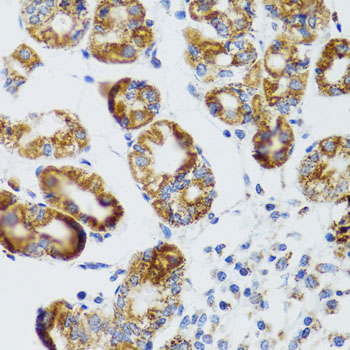
A07084 has real RIPK4 IHC data from paraffin-embedded human liver damage tissue (A07084 image caption). Human and mouse reactivity is listed; no IF image is supplied (catalog: A07084 reactivity/if_image_alts).

Real IHC data Immunohistochemistry of paraffin-embedded human liver damage using RIPK4 antibody at dilution of 1:100 .Perform microwave antigen retrieval with 10 mM PBS buffer pH 7.2 before commencing with IHC staining protocol.
Anti-RIPK4 Antibody
Cat # A07084

A07084 is listed for IHC in human and mouse (catalog: A07084 applications/reactivity). Its IHC figure shows paraffin-embedded human liver damage tissue stained at 1:100 after microwave retrieval in 10 mM PBS, pH 7.2 (A07084 image caption).

Which to pick: For tissue IHC, choose A07084, a rabbit antibody with listed human and mouse reactivity; its own figure demonstrates staining in paraffin-embedded human tissue (catalog: A07084 host/applications/reactivity; A07084 image caption). For cross-species IHC, A07084 is listed for both species, but the figure shows human tissue only (catalog: A07084 reactivity; A07084 image caption). No IF/ICC application or IF image is listed for A07084, and its IHC caption does not report the fixative (catalog: A07084 applications/if_image_alts; A07084 image caption).

Each figure is that product's own IHC / IF validation image from its datasheet.

References

  1. UniProt Consortium. UniProt entry P57078 (RIPK4_HUMAN, Receptor-interacting serine/threonine-protein kinase 4).
  2. Human Protein Atlas. RIPK4 tissue IHC expression (reliability: Enhanced).
  3. Human Protein Atlas. RIPK4 subcellular location (ICC-IF): Mainly localized to vesicles and cytosol. In addition localized to the plasma membrane..
  4. Human Protein Atlas. RIPK4 antibody validation summary (2 antibodies).
  5. High-Level Expression of RIPK4 and EZH2 Contributes to Lymph Node Metastasis and Predicts Favorable Prognosis in Patients With Cervical Cancer. Oncology research 2017 — PMC7841057.
  6. Increased RIPK4 expression is associated with progression and poor prognosis in cervical squamous cell carcinoma patients. Scientific reports 2015 — PMC4493702.
  7. RIPK4 Suppresses the Invasion and Metastasis of Hepatocellular Carcinoma by Inhibiting the Phosphorylation of STAT3. Frontiers in molecular biosciences 2021 — PMC8249771.
  8. RIPK4 function interferes with melanoma cell adhesion and metastasis. Molecular oncology 2026 — PMC13155153.
  9. PubMed PMID:10830953 — UniProt-cited evidence.
  10. PubMed PMID:12446564 — UniProt-cited evidence.
  11. PubMed PMID:21931591 — UniProt-cited evidence.