RNF149 / E3 ubiquitin-protein ligase RNF149 · IHC design guide

Design Immunohistochemistry for RNF149

This guide helps assess cytoplasmic, membrane-prominent RNF149 staining in paraffin sections (HPA tissue IHC). Use colon glandular cells as a positive reference (HPA tissue IHC) and start the catalog antibody at 1:100–1:300 (datasheet).

Evidence assembled Oct 2026 · For research use; verify linked source records and product datasheet before use
Immunohistochemistry protocol sheet for RNF149 (IHC for RNF149): expected localisation Cytoplasmic staining, most prominent at membranes (HPA tissue IHC), antibody A30667, validated IHC image, and IHC protocol steps
Printable RNF149 IHC protocol sheet — expected localisation Cytoplasmic staining, most prominent at membranes (HPA tissue IHC), antibody A30667, controls and protocol steps. Open the full RNF149 IHC guide →

RNF149 Immunohistochemistry Experimental Design Guide

Expected localisation, validated protocols, controls and antibodies — the at-a-glance facts below, then the full design guide.

Must know before staining
Expected localisation Cytoplasmic staining, most prominent at membranes (HPA tissue IHC)
Staining pattern Glandular and tubular cells: cytoplasmic, membrane prominent (HPA tissue IHC)
Antigen retrieval Citrate pH 6.0 HIER, 95–98 °C, 20 min (rule: cytoplasmic / membrane antigen)
Positive control ⓘ Appendix+4 more · see all
Negative control ⓘ Bone marrow+4 more · see all
Important caveats
Reasons your staining may differ from the expected pattern.
Fixation Keep fixation consistent across sections (standard IHC practice; not target-specific)
Caveat Antibody staining has low consistency with RNA expression (HPA tissue IHC)
Regulation Expression regulation is not annotated (UniProt)
Isoform / epitope No isoforms listed; extracellular versus cytoplasmic epitope matters (UniProt)
Section 1

Recommended RNF149 IHC & IF Protocols

The catalog antibody’s IHC-P protocol is accompanied by published RNF149 staining methods for rat tissues and an HCC tissue microarray (PMC9136010; PMC10648572).

Recommended immunohistochemistry (IHC-P) protocol parameters
SampleParaffin-embedded human brain tissue; fixative not specified (datasheet A30667)
FixationImage fixative and duration unreported (datasheet A30667); verify before use.
Sectioning4–5 µm sections on charged slides (standard)
DeparaffinisationXylene, graded ethanol series to water (standard)
Antigen retrievalHeat-induced epitope retrieval in citrate buffer, pH 6.0, 20 min at 95–98 °C (standard rule: cytoplasmic / membrane antigen)
Peroxidase block3% H2O2, 10 min, room temperature (standard)
Blocking10% normal serum of the secondary host, 30 min, room temperature (standard)
Primary antibodyRabbit anti-RNF149, 1:100 - 1:300 (datasheet A30667)
Primary incubationOvernight at 4 °C (standard)
DetectionHRP-polymer secondary, DAB chromogen 5–10 min (standard)
CounterstainHematoxylin, blue, dehydrate and mount (standard)
Expected resultRNF149-positive staining in glandular cells of appendix (HPA tissue IHC: High). HPA tissue profile: Cytoplasmic expression in several tissues, most prominent in membrane. No signal in the no-primary control.
💡Decision noteStart with citrate pH 6.0 HIER at 95–98 °C for 20 min (page antigen retrieval rule); the rat study used Target Retrieval solution (PMC9136010).
Section 2

What Is the Expected RNF149 Staining Pattern?

RNF149 should show membrane-prominent cytoplasmic staining in several tissues, including appendix and colon glandular cells, kidney tubule cells, and liver cholangiocytes (HPA tissue IHC: High; membrane-prominent cytoplasmic profile). Its 201–221 transmembrane segment supports membrane association (UniProt Q8NC42 topology). HPA rates the tissue IHC profile Approved but reports low consistency between antibody staining and RNA expression, so interpret the pattern with that limitation (HPA tissue IHC reliability).

What am I looking at on my slide?
Distinct membrane-prominent cytoplasmic chromogen in appendix or colon glandular cells, or kidney tubule cells (HPA tissue IHC: High).This matches the reported tissue and cellular pattern. Compare the staining with adjacent structures on the same section before judging intensity; HPA's High designation applies to the named cell types (HPA tissue IHC).
Predominantly nuclear staining, with little membrane-associated signal.A nuclear-only pattern conflicts with UniProt's membrane annotation and HPA's membrane-prominent tissue profile (UniProt Q8NC42 subcellular location; HPA tissue IHC). Check controls and staining localisation before assigning it to RNF149 (general IHC practice).
Strong staining in lymph-node germinal center cells or bone-marrow hematopoietic cells.Those cell populations are reported as Not detected, so consider cross-reactivity or endogenous chromogenic activity (HPA tissue IHC: Not detected; general IHC practice). The HPA designation does not mean every cell in either tissue is negative (HPA tissue IHC).
Diffuse color across cells and surrounding tissue, without a discernible cellular pattern.This is difficult to score as RNF149 because it lacks the reported membrane-prominent cytoplasmic distribution (HPA tissue IHC). Nonspecific binding or detection background are possible explanations; inspect negative controls (general IHC practice).
No discernible staining in appendix or colon glandular cells, or kidney tubule cells.These are reported High cell populations, making an absent result worth investigating (HPA tissue IHC). Review section quality, antibody and detection controls, and the assay's retrieval conditions before concluding that the specimen lacks RNF149 (general IHC practice).
💡Expected RNF149 appearanceCall a positive IHC result when distinct membrane-prominent cytoplasmic staining occurs in a reported High cell population, such as colon glandular cells or kidney tubule cells; isolated nuclear color or diffuse haze is suspect (HPA tissue IHC; UniProt Q8NC42 subcellular location; general IHC practice).
How each factor affects the staining
Membrane association and processing (UniProt Q8NC42 topology and processing).RNF149 has a 201–221 transmembrane segment, a 1–32 signal peptide, and a 33–400 annotated chain. These features support interpreting a membrane-associated pattern; they do not identify this antibody's epitope (UniProt Q8NC42).
Tissue and cell-type contrast (HPA tissue IHC).Appendix and colon glandular cells, kidney tubule cells, and liver cholangiocytes are High; lymph-node germinal center cells and bone-marrow hematopoietic cells are Not detected (HPA tissue IHC). Score the specified cells, not entire organs.
Evidence strength (HPA tissue IHC; HPA antibody validation).HPA labels the tissue profile Approved and lists HPA011424 as IHC Approved, while reporting low antibody-staining/RNA consistency (HPA). Approved should therefore inform pattern comparison without being treated as proof that every stained cell is specific.
Glycosylation and epitope uncertainty (UniProt Q8NC42).UniProt annotates glycosylation at residues 52 and 145 (UniProt Q8NC42). The supplied sources do not map the IHC antibody's epitope or show a glycosylation effect on staining, so do not infer one.
IF/ICC Q&A: What localisation should be compared on its separate guide page? (HPA subcellular ICC-IF).HPA reports approved plasma-membrane and vesicle localisation, with images from A-431, U-251MG, and U2OS (HPA subcellular ICC-IF). This is an IF/ICC comparison, not an IHC protocol or a tissue-intensity claim.
Why is my staining missing, weak or wrong?
SituationLikely causeNext action
A reported High cell population has no chromogenic signal (HPA tissue IHC).The result conflicts with HPA's observed pattern; the slide alone does not distinguish assay failure from specimen variation (HPA tissue IHC; general IHC practice).Check a known-positive section, detection controls, section integrity, and the established antigen-retrieval procedure before interpreting absence (general IHC practice).
Color appears chiefly in nuclei.Nuclear dominance does not match the membrane-associated annotation or tissue profile (UniProt Q8NC42; HPA tissue IHC).Confirm the cellular compartment against the counterstain and assess a negative reagent control for nonspecific color (general IHC practice).
Lymph-node germinal center cells stain strongly (HPA tissue IHC: Not detected).This differs from the named HPA-negative cell population; cross-reactivity or endogenous detection activity is possible (HPA tissue IHC; general IHC practice).Inspect reagent controls and the chromogen pattern, then compare named High cells on a positive section (general IHC practice; HPA tissue IHC: High).
Color forms uniform haze over cells and extracellular areas.A field-wide deposit obscures the reported cellular pattern and may reflect nonspecific binding or detection background (HPA tissue IHC; general IHC practice).Compare a no-primary control, review blocking and washes, and score only resolved cellular staining (general IHC practice).
Only faint cytoplasmic color appears in a reported High cell population (HPA tissue IHC).HPA describes cytoplasmic expression most prominent in membrane, so a faint, unresolved signal is inconclusive (HPA tissue IHC).Compare a known-positive section processed in the same run; review retrieval and detection performance before scoring (general IHC practice).
Apparent staining is limited to an HPA-negative population, while positive controls work (HPA tissue IHC).The cell-type distribution conflicts with HPA observations, although HPA also reports low staining/RNA consistency (HPA tissue IHC).Record the exact stained cell type, check reagent controls, and interpret the discrepancy as unresolved rather than automatically assigning RNF149 positivity (general IHC practice; HPA tissue IHC reliability).

Sample controls for RNF149 IHC & IF

🧪Run colon first: glandular cells should stain (HPA: High in colon glandular cells). Use bone marrow hematopoietic cells as the biological negative (HPA: Not detected); on the colon slide, assess neighboring non-glandular cells for background-only staining, but validate them before treating them as internal negatives.
Positive control tissue: Appendix (Glandular cells, HPA High)
Negative control tissue: Bone marrow (HPA Not detected)
ICC-IF cell lines (HPA subcellular resource): HPA ICC-IF images show RNF149 in A-431, U-251MG, U2OS, with annotated localisation: Vesicles (approved), Plasma membrane (approved) (HPA subcellular).
Technical controls: Include a no-primary (secondary-only) control and an irrelevant immunoglobulin control matched to the primary antibody’s host species, class, and clonality where applicable (standard IHC practice). Use RNF149 knockout material if available, or a peptide-block control for SKU A30667 (selected tissue-IHC caption: signal blocked with synthesized peptide); quench endogenous peroxidase in colon sections, and block endogenous biotin if using avidin–biotin detection (standard IHC practice).
⚠️Feasibility: A target-specific fixation window and fixation effect are unreported in the supplied evidence; the fixative in the selected A30667 paraffin-section caption is also unreported (selected tissue-IHC caption). RNF149-specific antigen-retrieval dependence is unreported, so optimize retrieval empirically for the chosen IHC antibody (standard IHC practice). Neither frozen sections nor IF are established as easier by these data; if testing IF/ICC separately, HPA reports approved vesicle and plasma-membrane localization, while colon sections warrant checks for endogenous peroxidase and, with avidin–biotin detection, endogenous biotin (HPA subcellular; standard IHC practice).

HPA tissue IHC evidence for RNF149

Comprehensive Human Protein Atlas IHC scoring per tissue (reliability: Approved — Low consistency between antibody staining and RNA expression data.). Rows are taken directly from the HPA tissue chart — click any row's HPA link to view the source.

Positive expression · recommended positive controls

TissueCell typeLevelEvidenceSource
Appendix Glandular cells High Protein (IHC) HPA →
Cerebellum Cells in granular layer High Protein (IHC) HPA →
Cerebral cortex Neuropil High Protein (IHC) HPA →
Colon Glandular cells High Protein (IHC) HPA →
Duodenum Glandular cells High Protein (IHC) HPA →

Undetected expression · recommended negative controls

TissueCell typeLevelEvidenceSource
Bone marrow Hematopoietic cells Not detected Protein (IHC) HPA →
Caudate Glial cells Not detected Protein (IHC) HPA →
Hippocampus Glial cells Not detected Protein (IHC) HPA →
Lymph node Germinal center cells Not detected Protein (IHC) HPA →
Placenta Trophoblastic cells Not detected Protein (IHC) HPA →
Section 3

Advanced RNF149 IHC Tips

RNF149 staining is best assessed by compartment, cell type and matched controls across paraffin-section IHC and confirmatory IF.

What retrieval should I try first for RNF149 in paraffin sections?
Start with citrate buffer at pH 6.0 for heat-induced retrieval at 95–98 °C for 20 min (page retrieval setting). Cool sections consistently before blocking, and compare a retrieval-treated section with a matched section processed without retrieval to judge whether staining gains definition (standard IHC practice). Inspect membrane-associated and cytoplasmic staining in the expected cells, since RNF149 is membrane-associated and tissue staining is reported as mainly cytoplasmic with membrane prominence (UniProt Q8NC42; HPA tissue IHC). If staining remains weak, adjust heating time in small increments while checking tissue preservation and background, without treating a stronger signal alone as proof of specificity (standard IHC practice).
How should I troubleshoot fixation-related loss of RNF149 staining?
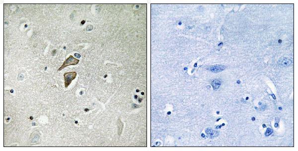
The selected antibody image shows paraffin-embedded human brain tissue, but its caption does not state a fixative (catalog antibody A30667 caption). Consequently, RNF149-specific sensitivity to fixation is unknown; neither tissue staining patterns nor protein topology establish how fixation changes this antibody’s epitope (HPA tissue IHC; UniProt Q8NC42). Record the fixative, fixation duration and processing history for each specimen, then compare sections from similarly handled material using the same retrieval and detection conditions (standard IHC practice). If signal varies across blocks, assess tissue preservation and internal staining patterns alongside control sections before attributing the difference to RNF149 abundance (standard IHC practice).
Which RNF149 staining pattern should I accept as plausible?
Look for membrane-associated staining with possible cytoplasmic signal: RNF149 has a transmembrane segment at residues 201–221, while tissue IHC reports cytoplasmic expression most prominent at membranes (UniProt Q8NC42 topology; HPA tissue IHC). Vesicles and the plasma membrane are approved locations in the subcellular atlas, although those observations come from ICC/IF images rather than this paraffin assay (HPA subcellular). In a kidney section, assess tubular cells; in liver, assess cholangiocytes, both reported as strongly stained tissue-IHC populations (HPA tissue IHC). Treat a solely nuclear or diffuse extracellular deposit as suspect, and compare it with section morphology and omission controls (standard IHC practice).
How do RNF149 processing and epitope position affect antibody interpretation?
RNF149 has a predicted signal sequence at residues 1–32, a mature chain spanning 33–400, and a transmembrane segment at 201–221 (UniProt Q8NC42). Its annotated glycosylation sites are residues 52 and 145, and phosphoserine 345 is listed among modified residues (UniProt Q8NC42). The record lists 0 isoforms, but no epitope position is supplied here, so these features cannot establish what the catalog antibody recognizes after tissue processing (UniProt Q8NC42; supplied antibody evidence). Use the peptide-blocked companion image as a useful specificity comparison, then seek an independently validated antibody or orthogonal evidence if compartmental staining remains uncertain (catalog antibody A30667 caption; standard IHC practice).
How can IF help resolve ambiguous RNF149 staining in tissue?
For a secondary IF experiment, multiplex RNF149 with a marker identifying the cell population being assessed, such as an established marker for tubular cells when examining kidney (HPA tissue IHC; standard IF practice). Choose a fluorophore channel with low tissue autofluorescence and inspect an unstained or no-primary section at identical acquisition settings before assigning puncta to RNF149 (standard IF practice). Because the transmembrane segment lies at residues 201–221 but the antibody epitope is unspecified, compare mild permeabilisation with no permeabilisation only where antibody access and specimen preparation make that comparison interpretable (UniProt Q8NC42 topology; supplied antibody evidence; standard IF practice). Judge colocalisation against cell boundaries and the reported vesicle and plasma-membrane locations, without transferring IF validation to chromogenic IHC (HPA subcellular; standard IF practice).
What should I check when RNF149 chromogenic staining looks diffuse?
Inspect a no-primary control for secondary-reagent or detection background, and include a peroxidase-blocking step before chromogen development when using an enzyme-based detection system (standard IHC practice). Shorten chromogen development or titrate the IHC-validated antibody if deposits obscure cellular boundaries; use identical processing across comparison sections (standard IHC practice). RNF149 tissue staining is reported as cytoplasmic with membrane prominence, so uniform extracellular colour is a reason to investigate background rather than score positive cells (HPA tissue IHC; standard IHC practice). The selected brain image includes a synthesized-peptide-blocked comparison, which can support assessment of antibody-dependent staining but does not by itself prove every deposit is specific (catalog antibody A30667 caption; standard IHC practice).
How should I quantify RNF149 across paraffin sections? ⚠ ANSWER MARKED FOR VERIFICATION
Define a tissue compartment and cell population before scoring, then report the percentage of positive cells and staining intensity as an H-score using one consistent threshold (standard IHC practice). For structures better represented by area, measure positive-cell density per mm² or positive area relative to viable tissue area, with the denominator stated explicitly (standard IHC practice). Score membrane-associated and cytoplasmic signal separately if both are visible, because the tissue profile emphasizes cytoplasmic expression with membrane prominence (HPA tissue IHC). Keep section thickness, retrieval, detection, imaging and exclusion rules consistent, and normalize comparisons to the same cell population or viable area rather than total section area (standard IHC practice).
How can I distinguish RNF149-positive cells from staining artefacts?
A convincing result has cellular boundaries and a plausible membrane-associated or cytoplasmic distribution, consistent with the transmembrane annotation and tissue-IHC profile (UniProt Q8NC42 topology; HPA tissue IHC). Check cell identity: kidney tubular cells and liver cholangiocytes are reported strongly positive, whereas bone-marrow hematopoietic cells are reported not detected in the tissue atlas (HPA tissue IHC). Discount signal confined to cut edges, folds or necrotic regions, and use no-primary and peroxidase-block controls to investigate endogenous-enzyme colour (standard IHC practice). Interpret a peptide-block-sensitive signal alongside morphology and independent evidence, since the selected brain caption documents peptide blocking while the tissue atlas reports low consistency between staining and RNA expression (catalog antibody A30667 caption; HPA tissue IHC).
Boster reagents

Best RNF149 / E3 ubiquitin-protein ligase RNF149 IHC Antibodies

A30667 is listed for IHC and IF in human and mouse (catalog: applications and reactivity). Its IHC figure shows paraffin-embedded human brain tissue (catalog: A30667 IHC image caption).

Real IHC data Immunohistochemistry analysis of paraffin-embedded human brain tissue, using RNF149 Antibody. The picture on the right is blocked with the synthesized peptide.
Anti-RNF149 Antibody
Cat # A30667

A30667 is listed for IHC, IF, ICC, and ELISA, with human and mouse reactivity (catalog: A30667 applications and reactivity). Its IHC figure shows paraffin-embedded human brain tissue with a peptide-blocked comparison; no IF figure is supplied (catalog: A30667 IHC and IF image captions).

Which to pick: For tissue IHC, choose A30667 for paraffin sections based on its human brain IHC figure; the fixative is unreported (catalog: A30667 IHC image caption). For IF/ICC, A30667 is listed for both applications, with an IF dilution of 1:50, but has no IF figure (catalog: A30667 applications, IF dilution, and IF image captions). For human or mouse samples, A30667 is the listed rabbit polyclonal option; its IHC figure documents human tissue only (catalog: A30667 reactivity, host, dilution_raw, and IHC image caption).

Each figure is that product's own IHC / IF validation image from its datasheet.

References

  1. UniProt Consortium. UniProt entry Q8NC42 (RN149_HUMAN, E3 ubiquitin-protein ligase RNF149).
  2. Human Protein Atlas. RNF149 tissue IHC expression (reliability: Approved).
  3. Human Protein Atlas. RNF149 subcellular location (ICC-IF): Localized to the plasma membrane and vesicles..
  4. Human Protein Atlas. RNF149 antibody validation summary (1 antibodies).
  5. Spatiotemporal evolution of AML immune microenvironment remodeling and RNF149-driven drug resistance through single-cell multidimensional analysis. Journal of translational medicine 2023 — PMC10612211.
  6. Role of the Ubiquitin Ligase RNF149 in the Development of Rat Neonatal Gonocytes. Frontiers in endocrinology 2022 — PMC9136010.
  7. RNF149 Promotes HCC Progression through Its E3 Ubiquitin Ligase Activity. Cancers 2023 — PMC10648572.
  8. PubMed PMID:14702039 — UniProt-cited evidence.
  9. PubMed PMID:17974005 — UniProt-cited evidence.
  10. PubMed PMID:15815621 — UniProt-cited evidence.