RNF2 / E3 ubiquitin-protein ligase RING2 · IHC design guide

Design Immunohistochemistry for RNF2

Plan RNF2 staining in paraffin sections with the IHC-validated antibody at a 2–5 μg/ml starting range (datasheet A01209-3). Expect nuclear and cytoplasmic staining across several tissues, while accounting for the uncertain tissue-IHC evidence (HPA tissue IHC).

Evidence assembled Oct 2026 · For research use; verify linked source records and product datasheet before use
Immunohistochemistry protocol sheet for RNF2 (IHC for RNF2): expected localisation Nuclear and cytoplasmic tissue staining (HPA tissue IHC), antibody A01209-3, validated IHC image, and IHC protocol steps
Printable RNF2 IHC protocol sheet — expected localisation Nuclear and cytoplasmic tissue staining (HPA tissue IHC), antibody A01209-3, controls and protocol steps. Open the full RNF2 IHC guide →

RNF2 Immunohistochemistry Experimental Design Guide

Expected localisation, validated protocols, controls and antibodies — the at-a-glance facts below, then the full design guide.

Must know before staining
Expected localisation Nuclear and cytoplasmic tissue staining (HPA tissue IHC)
Staining pattern Nuclear and cytoplasmic staining in several tissues (HPA tissue IHC)
Antigen retrieval EDTA pH 8.0 HIER, heat-mediated (datasheet A01209-3)
Positive control ⓘ Bronchus+4 more · see all
Negative control ⓘ Adipose tissue+4 more · see all
Important caveats
Reasons your staining may differ from the expected pattern.
Fixation Keep fixation conditions consistent across sections (standard IHC practice; not target-specific)
Caveat Staining has low concordance with RNA expression (HPA tissue IHC)
Regulation Expression regulation is unreported (UniProt)
Isoform / epitope 2 isoforms; epitope effects are unknown (UniProt)
Section 1

Recommended RNF2 IHC & IF Protocols

The catalog antibody’s IHC-P protocol is followed by published RNF2 chromogenic IHC methods for cutaneous melanoma, colorectal cancer, and skin squamous cell carcinoma (PMC5915146; PMC7803491; PMC7377046).

Recommended immunohistochemistry (IHC-P) protocol parameters
SampleParaffin-embedded human laryngeal squamous carcinoma tissue; fixative not specified (datasheet A01209-3)
FixationImage fixative and duration unreported (datasheet A01209-3); verify before use.
Sectioning4–5 µm sections on charged slides (standard)
DeparaffinisationXylene, graded ethanol series to water (standard)
Antigen retrievalHeat retrieval: EDTA pH 8.0 (datasheet A01209-3); 20 min, 95–100 °C (standard)
Peroxidase block3% H2O2, 10 min, room temperature (standard)
Blocking10% goat serum (datasheet A01209-3)
Primary antibodyRabbit anti-RNF2, 2-5 μg/ml (datasheet A01209-3)
Primary incubationOvernight at 4 °C (datasheet A01209-3)
DetectionHRP-conjugated secondary, DAB chromogen (datasheet A01209-3)
CounterstainHematoxylin, blue, dehydrate and mount (standard)
Expected resultRNF2-positive staining in respiratory epithelial cells of bronchus (HPA tissue IHC: Medium). HPA tissue profile: Cytoplasmic and nuclear expression in several tissues. No signal in the no-primary control.
💡Decision noteStart with heat-mediated EDTA pH 8.0 retrieval (datasheet A01209-3). The melanoma and skin squamous cell carcinoma methods used citrate pH 6.0 at 90°C for 60 min (PMC5915146; PMC7377046).
Section 2

What Is the Expected RNF2 Staining Pattern?

RNF2 is a nuclear, chromatin-associated protein with reported cytoplasmic localisation and no transmembrane segment (UniProt Q99496). In paraffin sections, expect staining in cell populations such as bronchial respiratory epithelium, liver hepatocytes and heart cardiomyocytes (HPA tissue IHC: Medium). Interpret intensity cautiously: HPA describes nuclear and cytoplasmic staining across several tissues, but rates tissue IHC reliability Uncertain because staining and RNA expression show low consistency (HPA tissue IHC).

What am I looking at on my slide?
Nuclear staining in hepatocytes, cardiomyocytes or bronchial respiratory epithelial cells (HPA tissue IHC: Medium).This fits the expected cell populations and RNF2’s nuclear, chromatin-associated role (HPA tissue IHC; UniProt Q99496). Cytoplasmic staining can coexist: HPA also reports it in tissue IHC (HPA tissue IHC).
Predominant staining at cell membranes or outside cells, with no convincing nuclear signal.This does not match the reported nuclear and cytoplasmic locations or the absence of a transmembrane segment (UniProt Q99496; HPA tissue IHC). Check section morphology and detection background before assigning the signal to RNF2 (standard IHC practice).
Strong signal in a cell population HPA lists as not detected, such as adipocytes or esophageal squamous epithelial cells (HPA tissue IHC).Treat the result as unexpected for that cell population, not proof of absence or presence: HPA tissue IHC is Uncertain (HPA tissue IHC). Compare a no-primary control and an HPA Medium population; cross-reactivity or endogenous detection activity may explain the signal (standard IHC practice).
Diffuse colour across nuclei, cytoplasm and tissue spaces, without clear cell boundaries.This distribution cannot establish RNF2 localisation (UniProt Q99496; standard IHC practice). Inspect the no-primary control, blocking and wash performance, and counterstained morphology for nonspecific background (standard IHC practice).
No staining in a section containing an HPA Medium population, such as liver hepatocytes (HPA tissue IHC).A negative result is inconclusive until the staining run and tissue quality are checked (standard IHC practice). The HPA tissue profile is Uncertain, and the listed level describes an observation rather than a guaranteed positive control (HPA tissue IHC).
💡Expected RNF2 appearanceA plausible positive shows discernible nuclear staining in HPA Medium populations, with possible cytoplasmic signal (HPA tissue IHC; UniProt Q99496); diffuse extracellular or membrane-only colour is suspect (UniProt Q99496; standard IHC practice).
How each factor affects the staining
Compartment and cell populationRNF2 is reported in the nucleus, cytoplasm and on chromosomes (UniProt Q99496). HPA reports cytoplasmic and nuclear tissue staining, with Medium staining in specified populations rather than every cell in a tissue (HPA tissue IHC).
Strength of tissue evidenceHPA assigns the tissue IHC profile Uncertain because antibody staining and RNA expression have low consistency (HPA tissue IHC). Use its Medium and Not detected entries as comparison points, not absolute acceptance thresholds (HPA tissue IHC; standard IHC practice).
Antibody validationHPA026803 is rated Uncertain for IHC and Enhanced for ICC (HPA antibodies). The ICC rating should not be transferred to paraffin IHC; judge the chromogenic slide against its controls and the tissue IHC profile (HPA antibodies; standard IHC practice).
IF/ICC Q: Where should RNF2 appear?A: Mainly in the nucleoplasm, with additional nuclear bodies (HPA subcellular ICC-IF). UniProt also reports colocalisation with SAMD7 in nuclear polycomb bodies; tissue IHC need not resolve those structures (UniProt Q99496; standard IHC practice).
Molecular forms and epitope uncertaintyUniProt lists two isoforms and four modified residues, including phosphoserines at positions 41, 143 and 168 (UniProt Q99496). The payload gives no antibody epitope or evidence that these features alter IHC staining; avoid assigning a staining difference to them.
Processing and detectionUniProt lists no signal peptide, propeptide, transmembrane segment or glycosylation site (UniProt Q99496). Endogenous enzyme activity or other detection background can still produce chromogenic colour; a no-primary control helps assess it (standard IHC practice).
Why is my staining missing, weak or wrong?
SituationLikely causeNext action
Nuclear signal is weak in hepatocytes or bronchial respiratory epithelial cells (HPA tissue IHC: Medium).The run may lack sensitivity, or that specimen may differ from the HPA observation; the tissue profile is Uncertain (standard IHC practice; HPA tissue IHC).Check a known working staining run, tissue morphology and the antibody’s documented IHC conditions before adjusting retrieval or dilution (standard IHC practice). No RNF2-specific retrieval effect is supplied.
Only cytoplasmic colour is visible.Cytoplasmic staining is reported by HPA, but RNF2 also has a nuclear location (HPA tissue IHC; UniProt Q99496). Background may obscure a nuclear component (standard IHC practice).Review counterstained nuclei alongside a no-primary control and an HPA Medium cell population before calling the result positive (standard IHC practice; HPA tissue IHC).
Membrane outlines or extracellular material stain prominently.That distribution conflicts with the reported RNF2 locations and lack of a transmembrane segment (UniProt Q99496). Nonspecific detection is a possibility (standard IHC practice).Compare the no-primary control, inspect morphology and repeat with adjusted blocking or washing if background persists (standard IHC practice).
Adipocytes stain strongly despite an HPA Not detected entry (HPA tissue IHC).The result may reflect background or antibody cross-reactivity; the HPA tissue IHC assessment itself is Uncertain (HPA tissue IHC; standard IHC practice).Compare adipocytes with an HPA Medium population in the same run and inspect the no-primary control; do not treat either HPA level as an absolute cutoff (HPA tissue IHC; standard IHC practice).
Colour appears in the no-primary control.The primary antibody cannot explain that control’s signal; endogenous detection activity or reagent background is plausible (standard IHC practice).Resolve the control background using the detection system’s blocking and washing checks before interpreting RNF2 staining (standard IHC practice).
IHC looks diffuse although ICC-IF shows nuclear bodies (HPA subcellular ICC-IF).HPA’s nuclear-body localisation comes from ICC-IF, while tissue IHC reports broader nuclear and cytoplasmic staining and has Uncertain reliability (HPA subcellular ICC-IF; HPA tissue IHC).Score identifiable cells and nuclear staining on the IHC slide; use a separate IF/ICC assessment if nuclear-body resolution is needed (standard IHC/IF practice).

Sample controls for RNF2 IHC & IF

🧪Start with bronchus: respiratory epithelial cells should show RNF2 staining (HPA: Medium in respiratory epithelial cells). Run adipose tissue as the negative comparator, assessing adipocytes (HPA: Not detected in adipocytes); on the bronchus slide, cells without nuclear chromogen can serve as internal background comparators, but the supplied HPA row does not identify a bronchial cell population as RNF2-negative (HPA: bronchus respiratory epithelial cells, Medium).
Positive control tissue: Bronchus (Respiratory epithelial cells, HPA Medium)
Negative control tissue: Adipose tissue (HPA Not detected)
ICC-IF cell lines (HPA subcellular resource): HPA ICC-IF images show RNF2 in A-431, U-251MG, U2OS, HeLa BAC 2473, HeLa , with annotated localisation: Nucleoplasm (enhanced) (HPA subcellular).
Technical controls: Include a no-primary, secondary-only control; a concentration-matched rabbit IgG isotype control with clonality matched if known (caption: rabbit primary antibody); and a matched RNF2 knockout specimen as a biological negative (standard IHC practice). For chromogenic bronchus sections, quench endogenous peroxidase and check inflammatory cells for background signal (standard IHC practice).
⚠️Feasibility: The selected A01209-3 paraffin-section caption does not report a fixative, and no target-specific fixation window or fixation effect is reported in the supplied evidence (caption: fixative not stated). Heat-mediated retrieval in EDTA at pH 8.0 was used for the illustrated IHC section, but the evidence does not establish that retrieval is required for every specimen (caption: EDTA retrieval, pH 8.0). ICC-IF images support checking nucleoplasmic signal, with possible nuclear-body signal (HPA: nucleoplasm enhanced; nuclear bodies enhanced); the supplied evidence does not establish whether frozen sections or IF are easier than paraffin IHC.

HPA tissue IHC evidence for RNF2

Comprehensive Human Protein Atlas IHC scoring per tissue (reliability: Uncertain — Low consistency between antibody staining and RNA expression data.). Rows are taken directly from the HPA tissue chart — click any row's HPA link to view the source.

Positive expression · recommended positive controls

TissueCell typeLevelEvidenceSource
Bronchus Respiratory epithelial cells Medium Protein (IHC) HPA →
Caudate Neuronal cells Medium Protein (IHC) HPA →
Duodenum Glandular cells Medium Protein (IHC) HPA →
Heart muscle Cardiomyocytes Medium Protein (IHC) HPA →
Liver Hepatocytes Medium Protein (IHC) HPA →

Undetected expression · recommended negative controls

TissueCell typeLevelEvidenceSource
Adipose tissue Adipocytes Not detected Protein (IHC) HPA →
Appendix Glandular cells Not detected Protein (IHC) HPA →
Cerebral cortex Endothelial cells Not detected Protein (IHC) HPA →
Cervix Glandular cells Not detected Protein (IHC) HPA →
Endometrium Cells in endometrial stroma Not detected Protein (IHC) HPA →
Section 3

Advanced RNF2 IHC Tips

Troubleshoot RNF2 staining in paraffin sections by checking retrieval, nuclear localisation, background and scoring against the available product and tissue evidence.

What retrieval should I try first when RNF2 staining is weak?
Start with heat-mediated antigen retrieval in EDTA at pH 8.0 for paraffin sections (datasheet A01209-3). The selected tissue image used this retrieval before 2 μg/ml primary antibody overnight at 4°C; its caption does not specify a heating time (datasheet A01209-3). If nuclear staining remains weak, compare modest changes in heating time on adjacent sections while keeping antibody concentration and detection conditions fixed (standard IHC practice). Include a section processed with the documented conditions in each comparison, and judge improvement by nuclear signal and background together (standard IHC practice; HPA subcellular).
Could fixation explain weak or uneven RNF2 staining?
RNF2-specific sensitivity to fixative type or fixation duration is unknown from the supplied evidence; the selected paraffin-section caption does not report its fixative (datasheet A01209-3). Record the actual fixative, time before fixation and fixation duration for each specimen before comparing staining across sections (standard IHC practice). Process adjacent sections together using EDTA retrieval at pH 8.0 and the documented 2 μg/ml primary concentration, so those variables remain controlled (datasheet A01209-3; standard IHC practice). If uneven staining tracks specimen edges or handling history, repeat the comparison on independently processed material before attributing it to RNF2 biology (standard IHC practice).
How should I assess RNF2 staining outside nuclei?
Prioritise nucleoplasmic staining when assessing RNF2, while allowing discrete nuclear-body signal: both locations are reported in subcellular images (HPA subcellular). UniProt also lists nuclear, cytoplasmic and chromosomal localisation, so cytoplasmic staining alone cannot establish an artefact or a true positive (UniProt Q99496 subcellular). Score nuclear and cytoplasmic compartments separately in matched sections, using the same EDTA pH 8.0 retrieval and 2 μg/ml primary conditions for comparison (datasheet A01209-3; standard IHC practice). Treat diffuse signal without a convincing nuclear component cautiously, especially because tissue staining has low agreement with RNA expression (HPA tissue IHC: Uncertain).
Could isoforms or epitope accessibility change my RNF2 IHC result?
RNF2 has 2 annotated isoforms, but the supplied product caption does not map the antibody epitope to either one (UniProt Q99496 isoforms; datasheet A01209-3). The record lists an acetylated residue at position 2 and phosphorylated residues at 41, 143 and 168; their effects on this antibody are unknown (UniProt Q99496 modified residues). Check the antibody's documented immunogen sequence, if available, against both isoforms before proposing isoform-specific staining (standard IHC practice). When signal varies, first reproduce the documented EDTA pH 8.0 retrieval and compare staining in the same cellular compartment (datasheet A01209-3; HPA subcellular).
How can IF help check the RNF2 pattern seen by chromogenic IHC?
Use IF as a separate localisation check: RNF2 is mainly nucleoplasmic, with additional nuclear-body localisation in subcellular images (HPA subcellular). Multiplex RNF2 with a validated marker for the cell type under examination, such as respiratory epithelial cells when examining bronchus, where medium tissue staining is reported (HPA tissue IHC: Bronchus; standard IF practice). Choose a fluorophore channel after checking tissue autofluorescence and include single-stain controls when separating the signals (standard IF practice). Because RNF2 has no transmembrane segment and its expected signal is nuclear, use controlled permeabilisation to permit nuclear antibody access without assuming an epitope-specific requirement (UniProt Q99496 topology; HPA subcellular; standard IF practice).
What should I check when RNF2 DAB staining is diffuse?
The selected paraffin-section example used 10% goat serum blocking, 2 μg/ml primary antibody overnight at 4°C, and DAB detection (datasheet A01209-3). Compare a no-primary section and a shorter chromogen development alongside the stained section to locate secondary or detection background (standard IHC practice). Check peroxidase blocking as a general chromogenic workflow step, particularly when pigment-like signal persists outside cells (standard IHC practice). Reassess whether diffuse staining follows tissue edges or obscures nucleoplasmic detail before increasing antibody concentration; nucleoplasm is the main reported subcellular location (standard IHC practice; HPA subcellular).
How should I quantify RNF2 staining across paraffin sections? ⚠ ANSWER MARKED FOR VERIFICATION
Define viable cells and the cellular compartment before scoring, because RNF2 tissue staining includes nuclear and cytoplasmic signal while subcellular images emphasise nucleoplasm (HPA tissue IHC; HPA subcellular). For nuclear staining, report the percentage of positive viable nuclei and an H-score from 0–300 based on intensity-weighted percentages (standard IHC practice). If cell density differs, express positive cells per mm² of viable tissue and also report the percentage relative to all eligible nuclei (standard IHC practice). Keep retrieval, imaging or visual scoring thresholds, and DAB development consistent across cases; interpret tissue comparisons cautiously given the reported uncertain staining reliability (standard IHC practice; HPA tissue IHC: Uncertain).
How do I distinguish convincing RNF2 staining from artefact?
Look for reproducible nuclear staining with discernible nucleoplasmic detail, consistent with RNF2's main reported subcellular location (HPA subcellular). Check cell identity against the tissue context: bronchial respiratory epithelial cells have medium reported staining, while adipocytes are listed as not detected, although tissue-level reliability is uncertain (HPA tissue IHC: Bronchus, Adipose tissue, Uncertain). Exclude edge effects, necrotic areas and residual endogenous peroxidase signal using morphology and a no-primary control before calling DAB-positive cells (standard IHC practice). A predominantly diffuse cytoplasmic pattern deserves separate reporting and confirmation, since UniProt permits cytoplasmic localisation but the subcellular images chiefly support nucleoplasmic signal (UniProt Q99496 subcellular; HPA subcellular).
Boster reagents

Best RNF2 / E3 ubiquitin-protein ligase RING2 IHC Antibodies

Anti-RNF2 IHC images show human paraffin sections; IF/ICC and mouse/rat reactivity are listed for a second antibody, without supplied IF images (A01209-3 IHC captions; M01209 catalog).

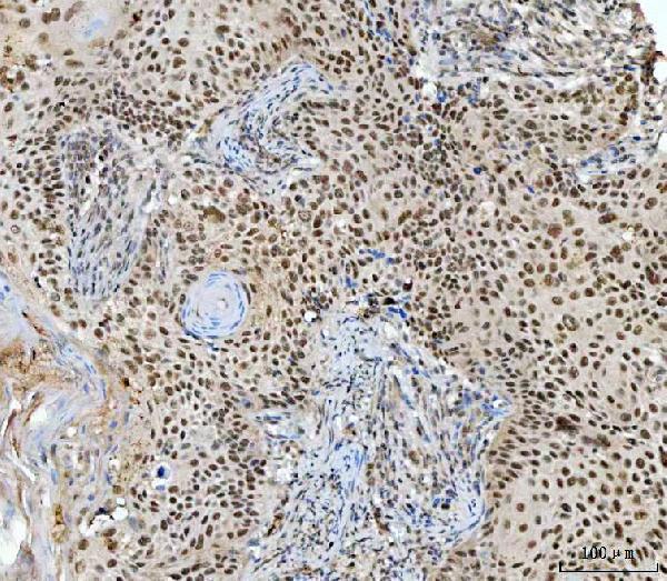
Real IHC data IHC analysis of RING2/RING1B/RNF2 using anti-RING2/RING1B/RNF2 antibody (A01209-3). RING2/RING1B/RNF2 was detected in a paraffin-embedded section of human laryngeal squamous carcinoma tissue. Heat mediated antigen retrieval was performed in EDTA buffer (pH 8.0, epitope retrieval solution). The tissue section was blocked with 10% goat serum. The tissue section was then incubated with 2 μg/ml rabbit anti-RING2/RING1B/RNF2 Antibody (A01209-3) overnight at 4°C. Peroxidase Conjugated Goat Anti-rabbit IgG was used as secondary antibody and incubated for 30 minutes at 37°C. The tissue section was developed using HRP Conjugated Rabbit IgG Super Vision Assay Kit (Catalog # SV0002) with DAB as the chromogen.
Anti-RING2/RING1B/RNF2 Antibody ®
Cat # A01209-3

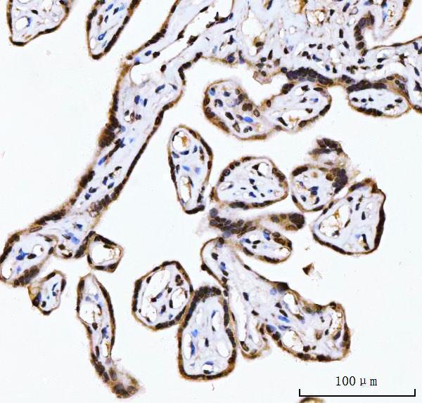
A01209-3 will render with an IHC figure from a human paraffin section of laryngeal squamous carcinoma; its captions also describe human placenta and thyroid cancer sections (A01209-3 IHC captions). M01209 will render with listed human, mouse and rat reactivity and IHC/IF/ICC applications, but no supplied IHC or IF image (M01209 catalog).

Which to pick: For human paraffin-section IHC, choose polyclonal A01209-3: its own captions document staining at 2 μg/ml after EDTA pH 8.0 retrieval; the fixative is unreported (A01209-3 dilution data and IHC captions). For IF/ICC, choose monoclonal M01209 on the basis of its application list, while treating its performance as unillustrated here (M01209 catalog). For mouse or rat IHC, M01209 is the listed option, though the payload supplies no species-specific IHC figure (M01209 catalog).

Each figure is that product's own IHC / IF validation image from its datasheet.

References

  1. UniProt Consortium. UniProt entry Q99496 (RING2_HUMAN, E3 ubiquitin-protein ligase RING2).
  2. Human Protein Atlas. RNF2 tissue IHC expression (reliability: Uncertain).
  3. Human Protein Atlas. RNF2 subcellular location (ICC-IF): Mainly localized to the nucleoplasm. In addition localized to the nuclear bodies..
  4. Human Protein Atlas. RNF2 antibody validation summary (1 antibodies).
  5. Enhanced intratumoral expression of RNF2 is a favorable prognostic factor for patients with cutaneous melanoma?. Oncotarget 2018 — PMC5915146.
  6. Ring finger protein 2 promotes colorectal cancer progression by suppressing early growth response 1. Aging 2020 — PMC7803491.
  7. Clinical significance of ring finger protein 2 high expression in skin squamous cell carcinoma. Oncology letters 2020 — PMC7377046.
  8. Ring Finger Protein 2 Promotes Oxidative Stress and Mitochondrial Dysfunction in Doxorubicin-Induced Cardiotoxicity Via the Mercaptopyruvate Sulfurtransferase/Hydrogen Sulfide Pathway. Journal of the American Heart Association 2025 — PMC12748078.
  9. PubMed PMID:14702039 — UniProt-cited evidence.
  10. PubMed PMID:16710414 — UniProt-cited evidence.
  11. PubMed PMID:15489334 — UniProt-cited evidence.