This website uses cookies to ensure you get the best experience on our website.
- Table of Contents
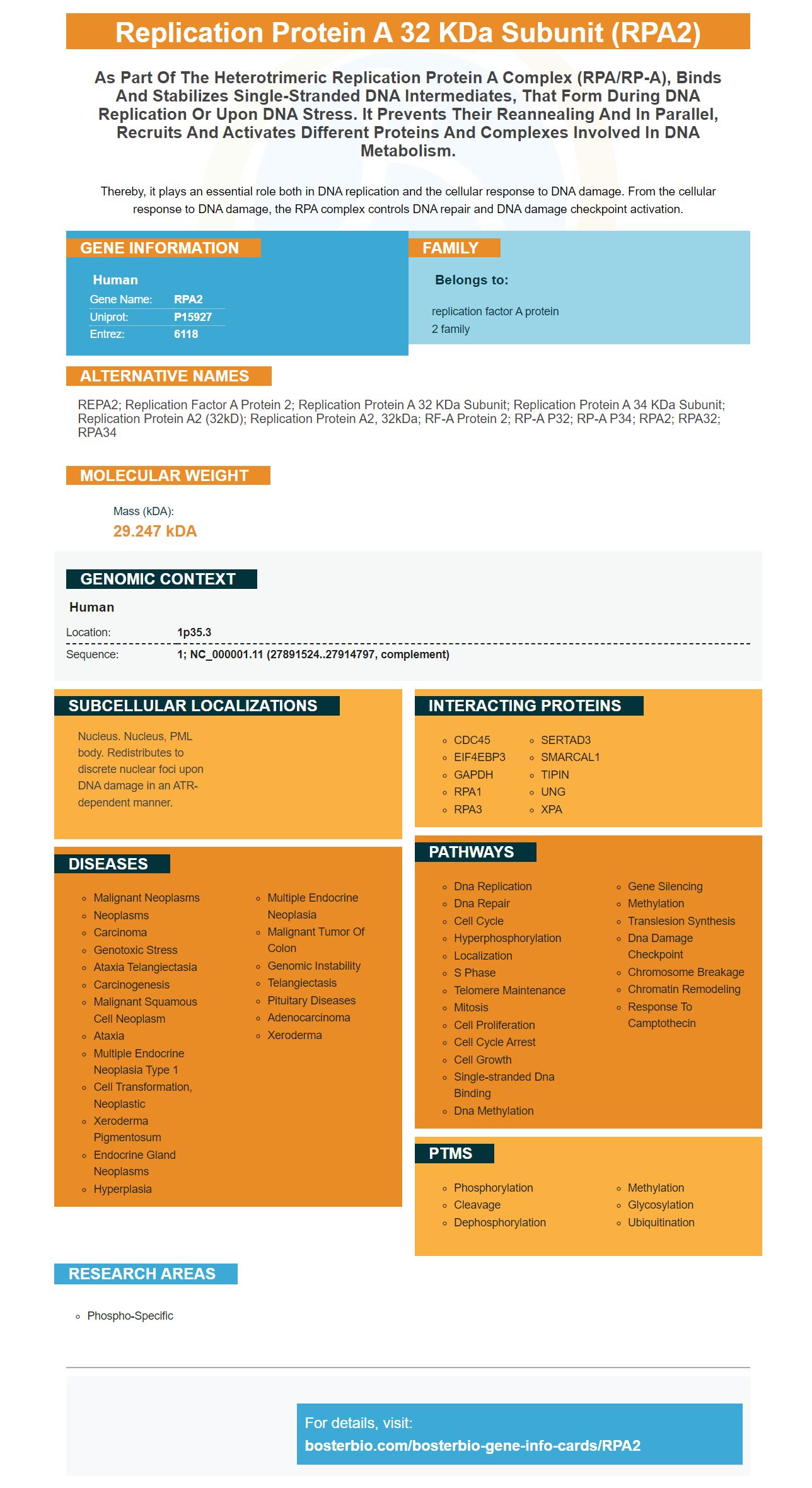
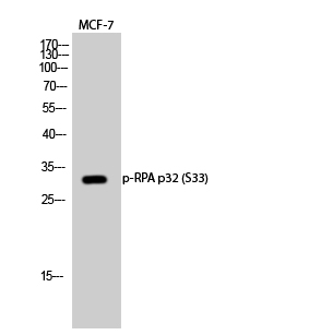
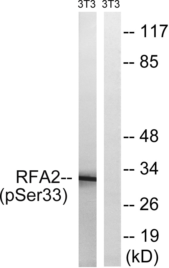
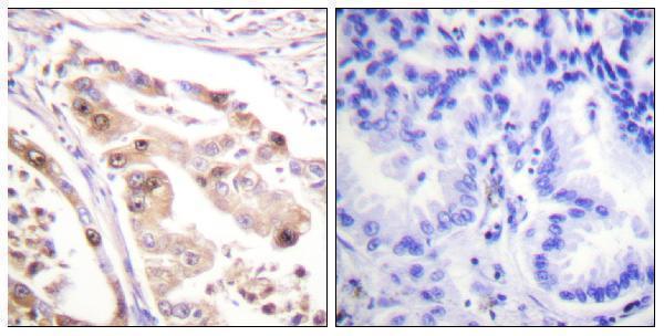
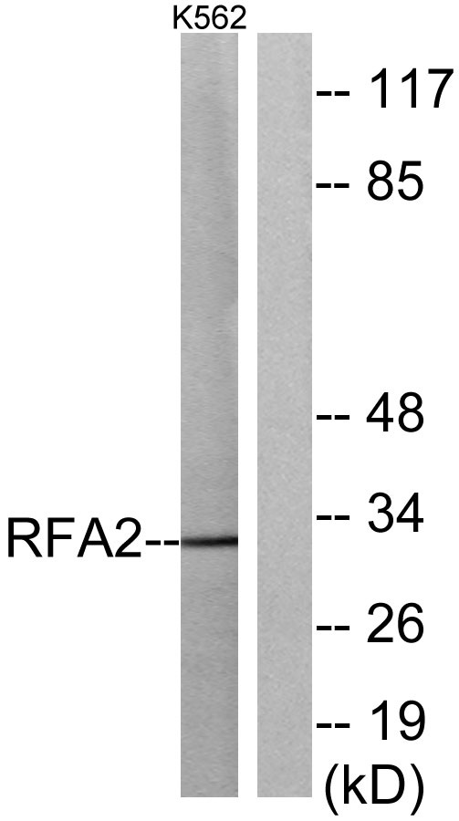
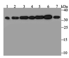
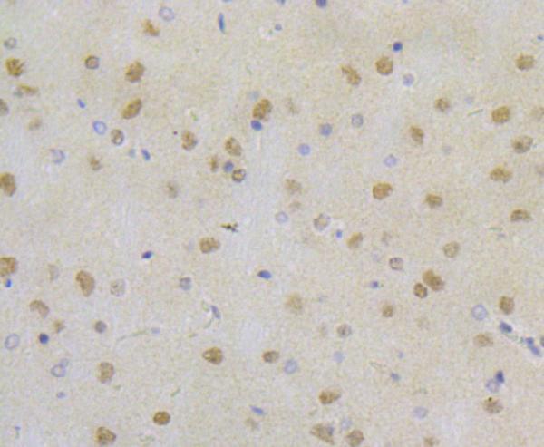
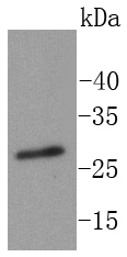
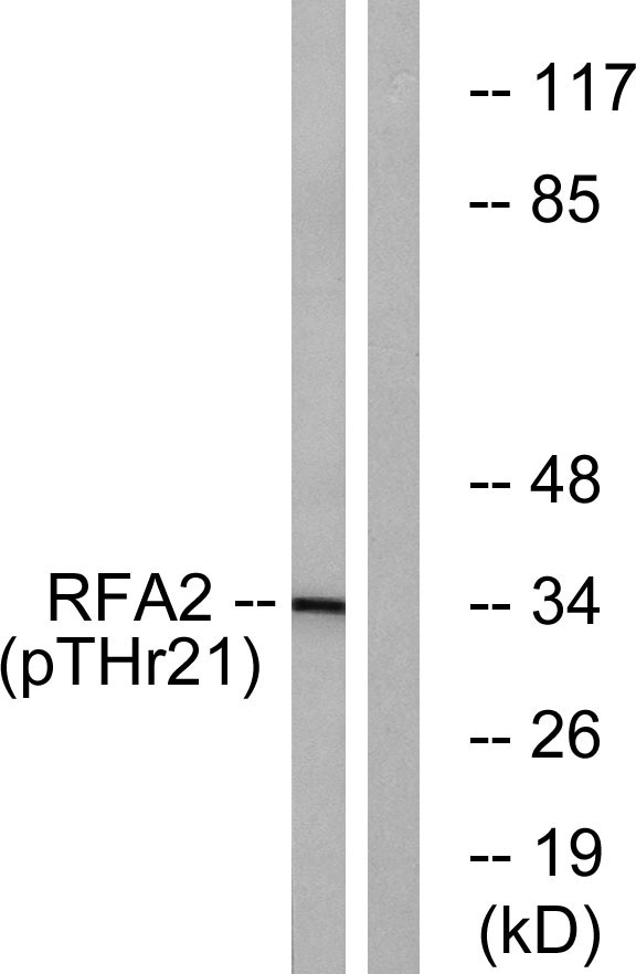
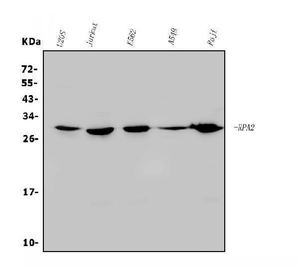
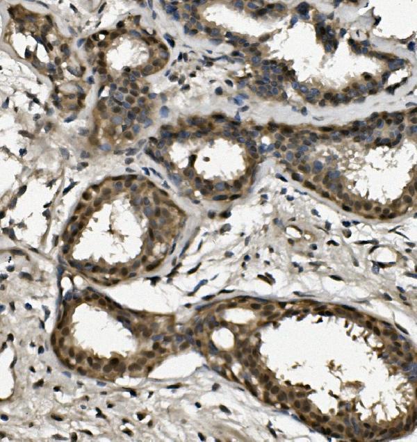
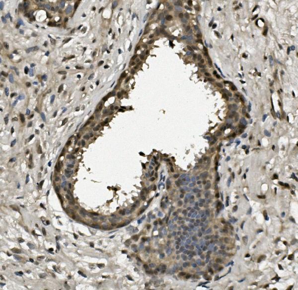
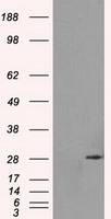
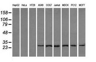
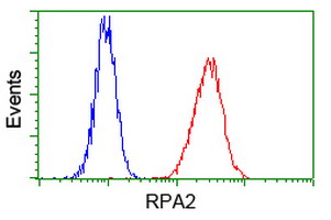
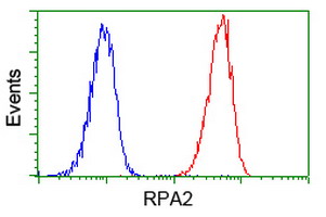
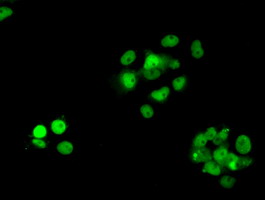
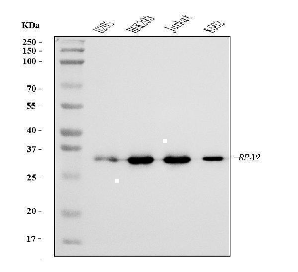
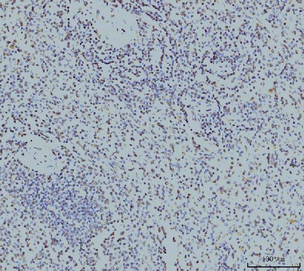
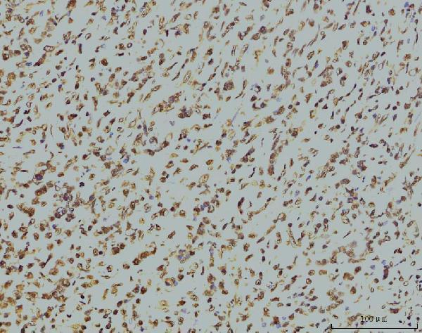
Facts about Replication protein A 32 kDa subunit.
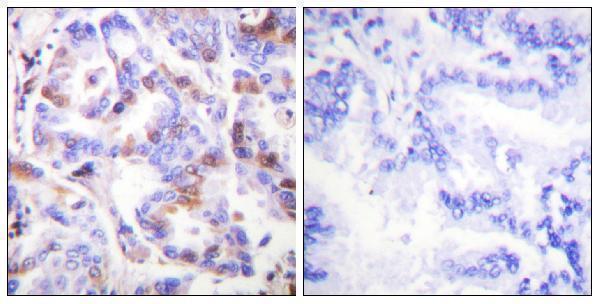
Thereby, it plays an essential role both in DNA replication and the cellular response to DNA damage. From the cellular response to DNA damage, the RPA complex controls DNA repair and DNA damage checkpoint activation.
Human | |
---|---|
Gene Name: | RPA2 |
Uniprot: | P15927 |
Entrez: | 6118 |
Belongs to: |
---|
replication factor A protein 2 family |
REPA2; Replication factor A protein 2; replication protein A 32 kDa subunit; Replication protein A 34 kDa subunit; replication protein A2 (32kD); replication protein A2, 32kDa; RF-A protein 2; RP-A p32; RP-A p34; RPA2; RPA32; RPA34
Mass (kDA):
29.247 kDA
Human | |
---|---|
Location: | 1p35.3 |
Sequence: | 1; NC_000001.11 (27891524..27914797, complement) |
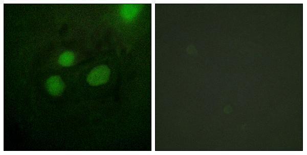
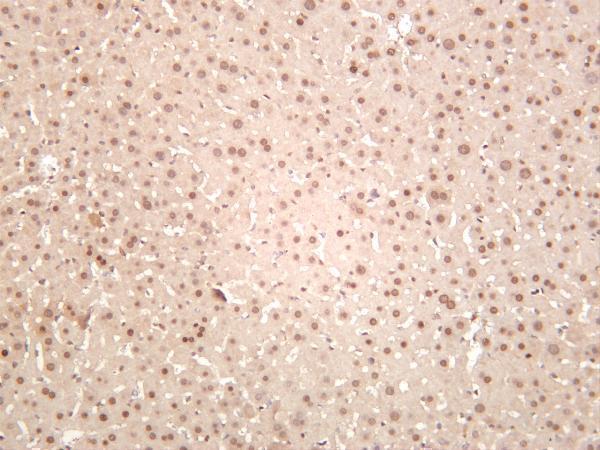
Nucleus. Nucleus, PML body. Redistributes to discrete nuclear foci upon DNA damage in an ATR-dependent manner.
Researchers have discovered that detecting the presence of RPA2 within cells is an essential research tool. The RPA2 marker is also able to be detected in damaged DNA cells by using a flow cytometry test. Researchers can make use of quantitative analysis of RPA2 signals within cells to assess the effects of DNA-end resection. Check out the following article for a few of the most exciting applications of the RPA2 marker.
The RPA2 marker is used to test cells to determine the replication and repair of DNA. RPA's role is to faithfully replicate billions of base pairs per cell cycle, as well as repair DNA lesions. If any of these processes is impaired cells show an abnormal growth pattern and may not survive. Replication protein A is a significant eukaryotic ssDNA-binding protein. It is involved in all the major DNA metabolic pathways, including repair of DNA and is regulated in certain cancers and other diseases.
Genomic instability is caused by infection with the PrimPolS538A mutant. The loss of both RPA binding sites (RPA-A and RPA-B) eliminates these manifestations. In the absence of PrimPol, RPA-mediated chromatin recruitment results in DNA damage. In response to DNA damage, the phosphorylation dependent on RPA of PrimPol is inhibited.
RPA2 can be used to determine the formation of gH2AX foci by using an enhanced green fluorescent protein tagged RPA2 expression plasmid. Flow cytometry with the RPA2 marker is much easier. The expression plasmid for RPA2 facilitates easy cell sorting and identification. The elution volumes of this RPA2 marker have been optimized for flow cytometry analysis.
The RPA2 MFI, which is highly sensitive, has been shown to be a reliable tool in identifying DNA damage in the SWI/SNF NHEJ and MMR pathways. The RPA2 MFI can be used to identify FA cell lines and patients with ICF syndromes. However, it is apparent that flow cytometry with the marker RPA2 is more efficient and more precise than staining using immunofluorescent.
The use of this marker can also help distinguish patients with Fanconi's anemia. This marker has been successfully utilized to identify patients suffering from this disease using flow cytometry. It has been used in clinical studies to identify DNA repair defects, such as Fanconi's. It could also be utilized as a biomarker to evaluate the role of RPA2 in cancer.
The discovery of RPA2 foci within DNA-damaged cells can have significant implications for understanding the biology of cell function. RPA foci are created within compact chromatin compartments that are formed by subtelomeric VSG genes that are silent. After a ribosomal DNA break, RPA foci accumulate adjacent to silent and transcribed VSG genes. As such, the foci are formed during S phase and remain in the cell cycle after mitosis. This suggests that the damage foci could enhance virulence and genetic diversity by helping to repair DNA.
RPA2 signals are amplified in response to DNA damage, as shown by the results. RPA2 foci can appear throughout the cell cycle and are associated with cells that are subject to induced DSBs. Three independent experiments were conducted to determine the presence of RPA2 foci in damaged DNA cells. The average of the three experiments was utilized. The percent of cells with positive RPA2 signals is represented by a dashed square.
The authors found that the RPA2 mechanism's cleavage site for meganuclease-induced RPA was associated both with active VSGES in bloodstream cells as well as TRF in insect stage cells. These RPA foci were also found to be colocalized with TRF Telomeric clusters. The foci of RPA2 in both cell types were situated on the nuclear periphery and were located in colocalization with TRF-associated telomeric clusters in the insect stage.
LAP2b, RPA2a and LAP2b are also implicated in the destruction of DNA. The RPA complex is a stable protein complex that is composed of heterotrimeric subunits. These heterotrimeric subunits collaborate and remove secondary structures from ssDNA, making it extremely stable. Six OB folds are observed in each of these molecules that allow the proteins to join with ssDNA. Flexible linkers are utilized to connect the DBDs.
Replication protein A (RPA) that connects to the cell end, triggers a series of events that recruit proteins for repair of HR. RPA2 is an heterotrimeric protein that requires phosphorylation at the N end to activate the human HR. Phosphorylation of RPA2 promotes its association with Rad51, an antigen-specific DNA repair protein.
Using a fluorescent protein, we measured the amount of pS33-RPA2 in a cell line treated with 680C91, KYN or BCNU. The quantitative analysis was conducted using 130 cells collected from three biological replicates. We conducted one-way ANOVA and calculated P values ranging from 0.05 to 100%. We also probed for PChk1, pChk2, yH2AX, and the yH2AX. GAPDH was also tested for cellular survival.
TDO inhibition led to a decrease in Chk1 activity and increased serine 33 phosphorylation at replication protein A. This suggests that TDO inhibition triggers a lower replication stress response and enhances the cytotoxicity of BCNU. Other results include decreased activation and MRN complex activation, as well as reduced sirtuin signaling. Additionally, the loss TDO activity reduces the capacity of GBM cells to recruit 53BP1, a DNA repair factor.
Our results reveal that polg influences the process of replication forks in vivo. Human cells without the polg gene are more susceptible to chemotherapy based on platinum. These findings support the hypothesis that polg is involved in preventing the formation of platinum-induced DNA lesions. Furthermore, we observe that severe inhibition of the progression of replication forks is associated with increased phosphorylation of RPA2 subunit.
We have previously shown that inhibition of the KP gene increases the activity of gH2AX response to oxidative stress. This indicates that UPR activity is enhanced when KP is blocked. The cells that are deficient in TyrRS have a higher levels of gH2AX-activation. This suggests that RPA2 could hinder cell proliferation, thereby reducing the risk of cancer.
The detection of RPA2 foci as a way to determine the status of DNA-end resection has been a long-standing readout for HR repair in cells. However, the function and regulation of Mcl-1 transcription factor functions could affect DNA resection in cells. Therefore we treated MEFs lacking in Mcl1 with Hu and CPT for one hour prior to being exposed to IR (5 Gy) and observed decreased formation of RPA2 foci.
A DSB caused by the removal of a DNA fragment in a cell could lead to a large number of RPA foci. These foci can be identified throughout the course of cell division and are associated with breaks in the rDNA. DNA-end resection can also induce RPA foci in nucleolus-associated cells.
Recognizing RPA2 foci as a readout for DNA-end resection is a powerful method to recognize DNA-end resections in the context of genetic diversity and virulence enhancement. However, it is important to note that RPA foci form within compact chromatin compartments formed by the silent subtelomeric VSG genes. Thus, in cells with RPA2 foci, DNA resection is unlikely to trigger the cell's checkpoint.
SRCAP is a member of the INO80 ATPase family , and is a part of the to the SRCAP complex for chromatin remodeling in humans. Mutations in SRCAP can cause the rare genetic disorder called Floating-Harbor syndrome. DNA resection can be controlled by distinct single-strand (ssDNA-binding) proteins. The major ssDNA-binding proteins RAD51 coat DNA and control homologous recombination.
The RPA complex is heterotrimeric and consists of RPA1 subunits as well as RPA2 and RPA3 subunits. In mammals, RPA focus formation typically occurs in the S and G2 phases of the cell cycle. Detection of RPA2 foci as a signal for DNA-end resection has been previously observed in the cell lineage of T. brucei, but T. brucei HAT3 is present in mammalian trypanomatids.
NHEJ and HR are the most important mechanisms used to perform DSB repairs. HR uses identical sequences as templates and is thought of as an error-free procedure. To allow DNA resection it is necessary for the three-stranded DNA to be in S/G2 and the sister chromatid needs to be in good condition for HR to take place. When HR and NHEJ fail to function the DNA resection process is activated.
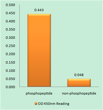
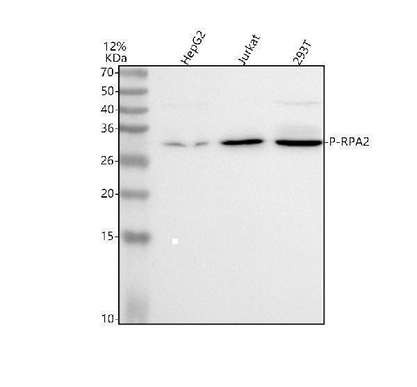
PMID: 2406247 by Erdile L.F., et al. The primary structure of the 32-kDa subunit of human replication protein A.
PMID: 2200738 by Din S., et al. Cell-cycle-regulated phosphorylation of DNA replication factor A from human and yeast cells.