This website uses cookies to ensure you get the best experience on our website.


- Table of Contents
Plan RPH3A paraffin-section IHC around strong cerebellar neuronal staining and moderate cortical neuropil staining (HPA tissue IHC). Use the guide to choose controls, optimize staining, and interpret regional differences.

Expected localisation, validated protocols, controls and antibodies — the at-a-glance facts below, then the full design guide.
| Expected localisation | Neuronal cytoplasm/membrane and neuropil (HPA tissue IHC) | |
| Staining pattern | Cerebellar neurons: cytoplasm/membrane; cortical neuropil (HPA tissue IHC) | |
| Antigen retrieval | EDTA pH 8.0 HIER, heat-mediated (datasheet A10247-2) | |
| Positive control | Cerebellum+1 more · see all | |
| Negative control | Adipose tissue+4 more · see all |
| Fixation | Keep paraffin-section fixation consistent across samples. (standard IHC practice; not target-specific) | |
| Caveat | Staining varies by brain region; hippocampus is low (HPA tissue IHC) | |
| Regulation | Expression regulation not annotated (UniProt) | |
| Isoform / epitope | 2 isoforms; epitope coverage unspecified (UniProt) |
The catalog antibody uses EDTA retrieval (datasheet A10247-2); the published RPH3A protocols below cover free-floating hippocampal slices (PMC10917878) and paraffin-embedded brain sections (PMC7923505).
| Sample | Paraffin-embedded mouse brain tissue; fixative not specified (datasheet A10247-2) |
| Fixation | Image fixative and duration unreported (datasheet A10247-2); verify before use. |
| Sectioning | 4–5 µm sections on charged slides (standard) |
| Deparaffinisation | Xylene, graded ethanol series to water (standard) |
| Antigen retrieval | Heat retrieval: EDTA pH 8.0 (datasheet A10247-2); 20 min, 95–100 °C (standard) |
| Peroxidase block | 3% H2O2, 10 min, room temperature (standard) |
| Blocking | 10% goat serum (datasheet A10247-2) |
| Primary antibody | Rabbit anti-RPH3A, 2-5 μg/ml (datasheet A10247-2) |
| Primary incubation | Overnight at 4 °C (datasheet A10247-2) |
| Detection | HRP-conjugated secondary, DAB chromogen (datasheet A10247-2) |
| Counterstain | Hematoxylin, blue, dehydrate and mount (standard) |
| Expected result | RPH3A-positive staining in gLUC cells - cytoplasm/membrane of cerebellum (HPA tissue IHC: High). HPA tissue profile: Cytoplasmic expression in neuronal cells and neuropil. No signal in the no-primary control. |
RPH3A is associated with synaptic vesicle membranes, dendritic spines and postsynaptic membranes, with no transmembrane segment (UniProt Q9Y2J0: location, topology). In tissue IHC, expect cytoplasmic staining in neuronal cells and neuropil (HPA: tissue IHC profile). HPA rates the tissue staining evidence Enhanced, citing consistency between antibody staining and RNA expression (HPA: reliability). Interpret the pattern by cell type and compartment as well as intensity.
| Strong cytoplasmic/membrane staining in cerebellar GLUC cells. | This matches a reported positive tissue and cell population (HPA: cerebellum, GLUC cells, High). Assess the cellular pattern across the section; RPH3A's vesicle and membrane associations make cytoplasmic and membrane-associated signal plausible, without requiring a continuous cell-outline stain (UniProt Q9Y2J0: location, topology). |
| Moderate staining in cerebral cortex neuropil; weaker neuronal staining in hippocampus or caudate. | These relative levels follow the tissue observations: cortical neuropil is Medium, while hippocampal and caudate neuronal cells are Low (HPA: tissue IHC). A weaker field in either Low category is therefore less informative than a failed High cerebellar control (HPA: tissue IHC levels). |
| Predominantly nuclear staining, with little cytoplasmic or neuropil signal. | This conflicts with the reported tissue profile and listed cellular locations (HPA: cytoplasmic neuronal and neuropil expression; UniProt Q9Y2J0: location). Treat it as suspect staining and compare with a known-positive section and a negative detection control before assigning it to RPH3A (general IHC practice). |
| Prominent signal in adipocytes or in adrenal glandular cells. | Those specific populations are reported as Not detected (HPA: adipose adipocytes; adrenal glandular cells). First check morphology and controls; staining there may reflect nonspecific antibody binding or endogenous detection activity, so appearance alone does not establish RPH3A expression (general IHC practice). |
| Diffuse color across tissue or no signal in cerebellar GLUC cells. | Diffuse deposition lacks the reported neuronal and neuropil distribution, while absent staining in a High population undermines a negative call elsewhere (HPA: tissue IHC profile; cerebellum, High). Review negative controls and the positive control on the same staining run before interpreting test sections (general IHC practice). |
| Tissue and cell selection | Cerebellar GLUC cells provide a High reference, cortical neuropil a Medium reference, and hippocampal or caudate neuronal cells Low references (HPA: tissue IHC). Adipocytes and adrenal glandular cells are reported Not detected; use these observations to choose comparisons, while keeping cell identity visible during scoring (HPA: tissue IHC). |
| Antibody evidence | HPA assigns antibody HPA002475 Enhanced IHC status and reports consistency between antibody staining and RNA expression (HPA: antibody validation; tissue reliability). That supports the observed IHC distribution; it does not specify the epitope, establish performance for every catalog antibody, or validate an IF appearance (HPA: supplied antibody and subcellular records). |
| Isoforms and epitope coverage | UniProt lists two RPH3A isoforms and a full-length chain spanning residues 1–694 (UniProt Q9Y2J0: isoforms, processing). The supplied records do not map the IHC antibody epitope, so isoform-specific staining or loss of a particular isoform cannot be inferred from a positive or negative section (HPA: supplied antibody record). |
| Can IF/ICC confirm the same pattern? | HPA supplies no ICC/IF cell images or main subcellular location for RPH3A, and lists no ICC validation for HPA002475 (HPA: subcellular record; antibody record). UniProt's vesicle and membrane locations offer a localisation expectation, but there is no supplied IF image pattern to use as an acceptance criterion (UniProt Q9Y2J0: location). |
| Situation | Likely cause | Next action |
|---|---|---|
| No chromogenic signal in the cerebellar positive control. | A High reference population should be visible: cerebellar GLUC cells are reported High (HPA: tissue IHC). A failed control can reflect a staining-run or detection problem (general IHC practice). | Confirm cell identity and section integrity, then review antibody dilution, antigen retrieval, detection reagents and counterstain against the run record (general IHC practice). Repeat the run with the known-positive control before interpreting negative test sections (general IHC practice). |
| Only nuclei stain in an otherwise intact neuronal section. | Nuclear-only staining disagrees with the cytoplasmic neuronal and neuropil tissue profile and UniProt cellular locations (HPA: tissue IHC; UniProt Q9Y2J0: location). | Compare the same run's cerebellar positive and negative detection controls; check whether the nuclear color persists without primary antibody (general IHC practice). Score the nuclear-only pattern as unconfirmed until the expected compartment is recovered (HPA: tissue IHC profile). |
| Strong color appears in adipocytes or adrenal glandular cells. | These cells are listed as Not detected, so strong staining needs a specificity check (HPA: tissue IHC negatives). Nonspecific binding or endogenous detection activity are possible technical causes (general IHC practice). | Inspect a no-primary control and the distribution of color within the cells; verify blocking and detection steps, then compare with the cerebellar positive control (general IHC practice; HPA: cerebellum High). Avoid labeling the unexpected color RPH3A on morphology alone (HPA: tissue IHC negatives). |
| Diffuse color obscures neuronal cytoplasm and neuropil. | The recorded pattern is cell and region associated, rather than uniform color across tissue (HPA: tissue IHC profile). General IHC causes include excessive antibody or detection background and inadequate washing (general IHC practice). | Inspect the negative detection control, then adjust antibody dilution, washes or detection development using the catalog antibody's validated IHC instructions (general IHC practice). Preserve a cerebellar positive control to check that background reduction retains the expected High signal (HPA: cerebellum High). |
| Cortical neuropil is visible, but hippocampal or caudate neurons seem negative. | The reported levels differ: cortical neuropil is Medium and hippocampal and caudate neuronal cells are Low (HPA: tissue IHC). A weak Low population may be hard to judge when staining contrast is limited (general IHC practice). | Compare sections stained in the same run and verify the High cerebellar reference first (HPA: tissue IHC levels; general IHC practice). Record Low populations as below detection when appropriate, without treating that result as evidence of global RPH3A absence (HPA: hippocampus and caudate Low). |
| An IF/ICC image is requested as proof of the IHC result. | The supplied HPA subcellular record has no ICC/IF images or main location, and HPA002475 has no listed ICC validation (HPA: subcellular record; antibody record). | Use the tissue IHC pattern and its positive and negative controls for this IHC interpretation (HPA: tissue IHC; general IHC practice). Treat any IF/ICC localisation assessment as a separate experiment requiring its own validation; do not infer an IF acceptance pattern from these HPA records (HPA: subcellular record). |
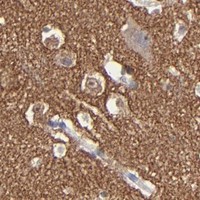


Comprehensive Human Protein Atlas IHC scoring per tissue (reliability: Enhanced — High consistency between antibody staining and RNA expression data.). Rows are taken directly from the HPA tissue chart — click any row's HPA link to view the source.
| Tissue | Cell type | Level | Evidence | Source |
|---|---|---|---|---|
| Cerebellum | GLUC cells - cytoplasm/membrane | High | Protein (IHC) | HPA → |
| Cerebral cortex | Neuropil | Medium | Protein (IHC) | HPA → |
| Tissue | Cell type | Level | Evidence | Source |
|---|---|---|---|---|
| Adipose tissue | Adipocytes | Not detected | Protein (IHC) | HPA → |
| Adrenal gland | Glandular cells | Not detected | Protein (IHC) | HPA → |
| Appendix | Glandular cells | Not detected | Protein (IHC) | HPA → |
| Bone marrow | Hematopoietic cells | Not detected | Protein (IHC) | HPA → |
| Breast | Glandular cells | Not detected | Protein (IHC) | HPA → |
Troubleshoot RPH3A staining in paraffin sections by checking retrieval, compartment, controls and scoring before interpreting chromogenic signal.
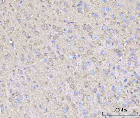
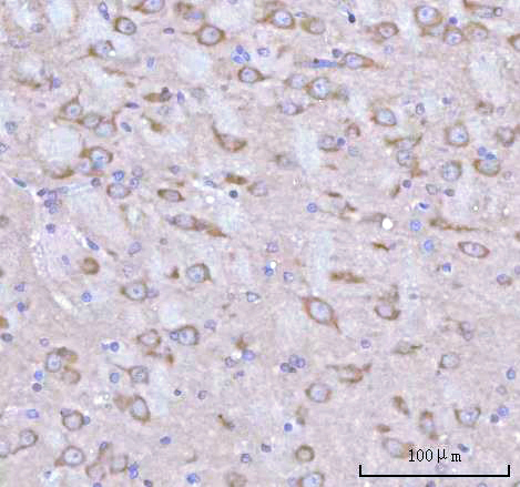
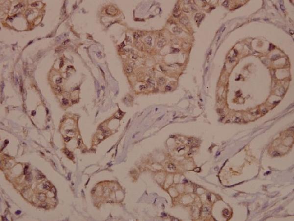
Two anti-RPH3A antibodies have real paraffin-section IHC images from mouse and rat brain and human breast carcinoma (catalog IHC captions). IF is listed for A10247-1, without an IF image (catalog A10247-1).
A10247-2 will render with mouse brain IHC; its catalog also documents rat brain IHC and lists human, mouse and rat reactivity (catalog A10247-2 IHC captions and reactivity). A10247-1 will render with human breast carcinoma IHC and lists IF plus human, mouse and rat reactivity (catalog A10247-1 IHC caption, applications and reactivity).
Which to pick: For paraffin-section tissue IHC, choose A10247-2 when the sample is mouse or rat brain: its own captions document EDTA pH 8 retrieval and DAB detection, with fixation unreported (catalog A10247-2 IHC captions). Choose A10247-1 for the pictured human breast carcinoma IHC example or as the IF candidate; its caption reports paraffin embedding but no fixative, and ICC is not listed (catalog A10247-1 IHC caption and applications). For cross-species planning, both list human, mouse and rat reactivity, while their pictured IHC evidence covers different species (catalog reactivity and IHC captions).