RPL17 / Large ribosomal subunit protein uL22 · IHC design guide

Design Immunohistochemistry for RPL17

Plan RPL17 paraffin IHC using colon glandular cells as a high-staining reference (HPA tissue IHC). This guide covers fixation, the catalog antibody’s 2–5 μg/ml IHC dilution, chromogenic detection, and the cross-gene staining caveat (datasheet A06980-2; HPA tissue IHC).

Evidence assembled Oct 2026 · For research use; verify linked source records and product datasheet before use
Immunohistochemistry protocol sheet for RPL17 (IHC for RPL17): expected localisation Cytoplasmic tissue staining (HPA tissue IHC), antibody A06980-2, validated IHC image, and IHC protocol steps
Printable RPL17 IHC protocol sheet — expected localisation Cytoplasmic tissue staining (HPA tissue IHC), antibody A06980-2, controls and protocol steps. Open the full RPL17 IHC guide →

RPL17 Immunohistochemistry Experimental Design Guide

Expected localisation, validated protocols, controls and antibodies — the at-a-glance facts below, then the full design guide.

Must know before staining
Expected localisation Cytoplasmic tissue staining (HPA tissue IHC)
Staining pattern Ubiquitous cytoplasmic staining across cell types (HPA tissue IHC)
Antigen retrieval EDTA pH 8.0 HIER, heat-mediated (datasheet A06980-2)
Positive control ⓘ Adrenal gland+4 more · see all
Negative control ⓘ None in HPA (detected in all 45 tissues); use no-primary + isotype controls
Important caveats
Reasons your staining may differ from the expected pattern.
Fixation Keep paraffin tissue fixation consistent (standard IHC practice; not target-specific)
Caveat Staining may reflect proteins from more than one gene (HPA tissue IHC)
Regulation Brain-enhanced RNA; IHC ubiquitous (HPA tissue RNA)
Isoform / epitope 3 isoforms; epitope coverage unreported (UniProt)
Section 1

Recommended RPL17 IHC & IF Protocols

The catalog antibody’s IHC-P protocol (datasheet A06980-2) is accompanied by published RPL17 protocols for zebra finch brain (PMC2991423), mouse carotid artery (PMC3502943), and rat hind-limb and clinical specimens (PMC9117278).

Recommended immunohistochemistry (IHC-P) protocol parameters
SampleParaffin-embedded human thyroid papillary carcinoma tissue; fixative not specified (datasheet A06980-2)
FixationImage fixative and duration unreported (datasheet A06980-2); verify before use.
Sectioning4–5 µm sections on charged slides (standard)
DeparaffinisationXylene, graded ethanol series to water (standard)
Antigen retrievalHeat retrieval: EDTA pH 8.0 (datasheet A06980-2); 20 min, 95–100 °C (standard)
Peroxidase block3% H2O2, 10 min, room temperature (standard)
Blocking10% goat serum (datasheet A06980-2)
Primary antibodyRabbit anti-RPL17, 2-5 μg/ml (datasheet A06980-2)
Primary incubationOvernight at 4 °C (datasheet A06980-2)
DetectionStreptavidin-biotin complex (SABC), DAB chromogen (datasheet A06980-2)
CounterstainHematoxylin, blue, dehydrate and mount (standard)
Expected resultRPL17-positive staining in glandular cells of adrenal gland (HPA tissue IHC: High). HPA tissue profile: Ubiquitous cytoplasmic expression. No signal in the no-primary control.
💡Decision noteStart with EDTA pH 8.0 heat retrieval for the catalog antibody (datasheet A06980-2). The mouse carotid study used 10 mM citrate pH 6.0 (PMC3502943).
Section 2

What Is the Expected RPL17 Staining Pattern?

RPL17 is a cytoplasmic large ribosomal subunit protein with no transmembrane segment (UniProt P18621: subunit, location, topology). In paraffin section IHC, expect cytoplasmic staining across many cell types, including high staining in colon glandular cells and bronchial respiratory epithelial cells (HPA: ubiquitous cytoplasmic expression; High in both). HPA rates the tissue pattern Approved, with medium agreement between staining and RNA data, pending external verification; its antibodies may recognize proteins from multiple genes (HPA: reliability description).

What am I looking at on my slide?
Clear cytoplasmic stain in colon glandular cells or bronchial respiratory epithelium, with cell outlines and nuclei readable.This fits the expected compartment and two reported high staining cell populations (HPA: ubiquitous cytoplasmic expression; High in colon glandular and bronchial respiratory epithelial cells). Compare intensity within the same run; HPA's categories are observations, not a numerical cutoff (HPA: High).
Strong nuclear only staining, or a sharply membrane restricted pattern with little cytoplasmic signal.Treat this as a possible localization artifact and review controls and detection conditions: UniProt places RPL17 in cytoplasm and reports no transmembrane segment (UniProt P18621: location, topology). HPA reports ubiquitous cytoplasmic tissue staining (HPA: profile).
The strongest signal falls in a low staining population, such as skeletal myocytes, while a high staining comparator is weak.Consider cross reactivity or endogenous detection activity; first compare matched controls (general IHC practice). Skeletal myocytes are reported Low, not absent (HPA: Low in skeletal muscle myocytes). HPA cautions that its antibodies target proteins from multiple genes (HPA: reliability description).
Uniform brown haze covers tissue and blank areas, obscuring cell boundaries.Interpret the run cautiously: diffuse background cannot establish RPL17 localization (general IHC practice). Check a no primary control and inspect washes, blocking, and chromogen development before scoring cellular staining (general IHC practice).
No cytoplasmic signal appears in a colon glandular or bronchial epithelial positive comparator.Treat the run as unresolved before calling the specimen negative: both comparators are reported High (HPA: colon glandular cells; bronchial respiratory epithelial cells). Review retrieval and primary and detection steps together (general IHC practice); HPA supplies no negative tissue in this payload (HPA: negative list empty).
💡Expected RPL17 appearanceA convincing positive is discernible cytoplasmic stain in high staining glandular or respiratory epithelial cells (HPA: High in colon glandular and bronchial respiratory epithelial cells; ubiquitous cytoplasmic profile), whereas nuclear only, membrane restricted, or cell independent haze should trigger control review (UniProt P18621: cytoplasmic location, no transmembrane segment; general IHC practice).
How each factor affects the staining
Tissue and cell choiceColon glandular and bronchial respiratory epithelial cells are reported High; skeletal myocytes and smooth muscle cells are Low (HPA: tissue IHC). Use a high staining population to check the run, and do not treat Low as a true negative (HPA: no negative populations listed).
IHC evidence strengthThe tissue profile is Approved, with medium staining to RNA consistency and external verification pending (HPA: reliability description). HPA043724 and HPA046385 are each IHC Approved, not listed as Enhanced (HPA: antibody validation). The multiple gene targeting caution limits attribution of an unexpected signal to RPL17 alone (HPA: reliability description).
Antigen retrieval in paraffin sectionsCheck the IHC validated antibody's supplied retrieval conditions, then optimize against a high staining comparator and a no primary control (general IHC practice; HPA: High in colon glandular cells). Neither UniProt nor the supplied HPA record establishes RPL17 specific fixation sensitivity or an optimal retrieval condition.
Isoforms and antigen regionUniProt lists three RPL17 isoforms and a mature chain spanning residues 2–184 (UniProt P18621: isoforms, processing). The supplied records do not map antibody epitopes or show isoform specific staining; avoid assigning a tissue pattern to one isoform.
IF/ICC Q&A: where should signal appear?HPA reports supported endoplasmic reticulum and cytosol localization in ICC IF, with images in A-431, U-251MG, U2OS, HEK293, and Hep-G2 (HPA: subcellular record). Apply its multiple gene targeting caution when interpreting either compartment (HPA: subcellular summary). IF/ICC protocol selection belongs in its own guide.
Why is my staining missing, weak or wrong?
SituationLikely causeNext action
High staining comparator has no visible stain.A failed retrieval, primary, or detection step is possible (general IHC practice); colon glandular cells are reported High (HPA: tissue IHC).Confirm the comparator contains glandular cells; review retrieval, primary incubation, and chromogen with controls before interpreting samples (general IHC practice).
Nuclei dominate and cytoplasm is faint.The pattern conflicts with cytoplasmic localization (UniProt P18621: location; HPA: tissue profile); nonspecific detection is possible (general IHC practice).Inspect the no primary control, adjust detection or blocking as indicated, and reassess cytoplasmic signal in a high staining comparator (general IHC practice; HPA: High in colon glandular cells).
Low staining muscle cells appear as intense as the positive comparator.Possible cross reactivity or endogenous detection activity (general IHC practice); muscle cells are Low (HPA: skeletal myocytes and smooth muscle cells).Compare cell matched controls and detection blanks; report the discrepancy without assigning it to RPL17 until specificity is supported (general IHC practice; HPA: multiple gene targeting caution).
Brown precipitate or haze extends outside cells.Excess chromogen development, inadequate washing, or background detection can obscure cellular localization (general IHC practice).Check blank areas and the no primary control; refine washing and development, then score only resolved cellular staining (general IHC practice).
A faint population is scored as completely negative.Low staining is being treated as absence, although HPA lists Low populations and no negative population here (HPA: tissue IHC).Record compartment and relative intensity, compare cells within the same run, and reserve a negative call for a validated decision threshold (general IHC practice).
A striking unexpected pattern persists after basic controls pass.HPA warns that the profiled antibodies target proteins from multiple genes; tissue staining has medium RNA agreement and awaits external verification (HPA: reliability description).Describe the observed cells and compartment, compare an independently validated antibody if available, and qualify attribution to RPL17 (general IHC practice; HPA: validation caution).

Sample controls for RPL17 IHC & IF

🧪Run bronchus first and look for staining in respiratory epithelial cells (HPA: High in bronchus respiratory epithelial cells). HPA detects RPL17 in all 45 scored tissues, so no negative tissue is established; use no-primary and isotype controls, and treat any unstained cells on the positive slide as unvalidated negative comparators rather than confirmed RPL17-negative cells (HPA: no negative rows; detected in all 45 tissues).
Positive control tissue: Adrenal gland (Glandular cells, HPA High)
Negative control tissue: None in HPA: RPL17 is detected in all 45 scored tissues. Use a no-primary (secondary-only) and an isotype control instead.
ICC-IF cell lines (HPA subcellular resource): HPA ICC-IF images show RPL17 in A-431, U-251MG, U2OS, HEK293, Hep-G2, with annotated localisation: Endoplasmic reticulum (supported), Cytosol (supported) (HPA subcellular).
Technical controls: Include a no-primary, secondary-only control and a concentration-matched rabbit IgG isotype control, plus RPL17 knockout tissue or a validated peptide-block control if available (caption: rabbit primary; standard IHC practice). Block endogenous peroxidase in bronchial sections and, if using the caption’s streptavidin–biotin detection, check endogenous biotin background (caption: SABC with DAB; standard IHC practice).
⚠️Feasibility: The selected paraffin-section caption uses heat retrieval in EDTA at pH 8.0, but it does not establish that retrieval is required; its fixative is unreported (A06980-2 tissue-IHC caption). A target-specific fixation window or fixation effect is unreported, and the supplied evidence does not establish whether frozen sections or IF are easier (supplied target/application evidence). Bronchial inflammatory cells can produce peroxidase background, so interpret DAB signal against the peroxidase-blocked controls (standard IHC practice).

HPA tissue IHC evidence for RPL17

Comprehensive Human Protein Atlas IHC scoring per tissue (reliability: Approved — Medium consistency between antibody staining and RNA expression data. Pending external verification. Caution, targets protein from more than one gene.). Rows are taken directly from the HPA tissue chart — click any row's HPA link to view the source.

Positive expression · recommended positive controls

TissueCell typeLevelEvidenceSource
Adrenal gland Glandular cells High Protein (IHC) HPA →
Appendix Glandular cells High Protein (IHC) HPA →
Bronchus Respiratory epithelial cells High Protein (IHC) HPA →
Caudate Neuronal cells High Protein (IHC) HPA →
Cerebellum Purkinje cells High Protein (IHC) HPA →

Undetected expression · recommended negative controls

TissueCell typeLevelEvidenceSource
None in HPA: RPL17 is detected in all 45 scored tissues. Use a no-primary (secondary-only) and an isotype control instead.
Section 3

Advanced RPL17 IHC Tips

Troubleshoot RPL17 chromogenic IHC by checking retrieval, cytoplasmic localization, detection controls, and cell-specific scoring (datasheet A06980-2; UniProt P18621; HPA: tissue IHC).

Which retrieval conditions should I try first for weak RPL17 staining?
Start with heat-mediated retrieval in EDTA at pH 8.0 for paraffin sections (datasheet A06980-2). The selected tissue image used this retrieval before 2 μg/ml primary antibody overnight at 4°C, so reproduce those linked conditions when investigating weak chromogenic signal (datasheet A06980-2). If staining remains weak, compare shorter and longer heating intervals on adjacent sections while holding detection and DAB development constant (standard IHC practice). Excessive heating can damage morphology and create apparent edge staining; judge any gain against preservation of cellular boundaries and a matched no-primary control (standard IHC practice).
Can I infer the best fixative for RPL17 from the available staining evidence?
The selected paraffin-section caption does not report its fixative, and target-specific RPL17 fixation sensitivity is unknown (datasheet A06980-2). Record fixative, fixation duration, and tissue processing for each block before comparing staining, because crosslinking and processing can change accessible epitopes in chromogenic IHC (standard IHC practice). Compare matched sections after EDTA retrieval at pH 8.0 and primary antibody at 2 μg/ml overnight at 4°C, varying only one processing factor at a time (datasheet A06980-2; standard IHC practice). Do not infer a preferred fixative from ubiquitous cytoplasmic tissue staining, absent glycosylation annotations, or the absence of a transmembrane segment (HPA: tissue profile; UniProt P18621).
Where should convincing RPL17 staining appear in a paraffin section?
Expect predominantly cytoplasmic signal in chromogenic sections because RPL17 is annotated in the cytoplasm and is a large ribosomal subunit component (UniProt P18621). The tissue atlas reports ubiquitous cytoplasmic staining, with high glandular-cell staining in colon and high neuronal-cell staining in cerebral cortex (HPA: tissue IHC). Its subcellular images describe supported endoplasmic-reticulum and cytosol locations, but those antibodies may recognize proteins from multiple genes (HPA: subcellular caution). Score nuclear-only or sharply membrane-restricted DAB as questionable until adjacent-section controls and cell morphology support it; the sequence has no annotated transmembrane segment (UniProt P18621; standard IHC practice).
Can staining intensity distinguish RPL17 isoforms or reveal epitope masking?
Do not assign an IHC signal to one RPL17 isoform from compartment or intensity alone: three isoforms are annotated (UniProt P18621). The record provides no mapped antibody epitope, so whether the catalog antibody recognizes all isoforms or distinguishes them is unresolved (record: RPL17 isoforms; datasheet A06980-2). Review the antibody immunogen or epitope documentation if available, then compare staining with an independent antibody directed at a documented different epitope using matched paraffin sections (standard IHC validation practice). Because the protein has no annotated glycosylation sites or modified residues, do not invoke either modification as a demonstrated explanation for differing staining (UniProt P18621).
How should I assess RPL17 localization in a complementary IF experiment?
For complementary IF, pair RPL17 with a neuronal marker when assessing cerebral cortex neurons (HPA: high neuronal-cell staining in cerebral cortex; standard IF practice). Select spectrally separated fluorophores and place the weaker signal in a channel with low tissue autofluorescence, checking single-stain and unstained controls before interpreting overlap (standard IF practice). Permeabilize enough to expose a cytoplasmic epitope; there is no annotated transmembrane segment and the precise antibody epitope is unreported (UniProt P18621; datasheet A06980-2). Compare IF distribution with chromogenic cytoplasmic staining, treating reported endoplasmic-reticulum and cytosol localization cautiously because the subcellular antibodies may target multiple genes (HPA: tissue IHC; HPA: subcellular caution).
How can I separate RPL17 staining from diffuse DAB background?
Begin by comparing a no-primary section with a complete stain, because tissue pigment and detection chemistry can generate DAB-like signal independently of RPL17 binding (standard IHC practice). The selected protocol used 10% goat serum, biotinylated goat anti-rabbit secondary antibody, a streptavidin–biotin complex, and DAB (datasheet A06980-2). For diffuse background, check serum blocking, primary incubation at 2 μg/ml overnight at 4°C, wash stringency, endogenous peroxidase blocking, and any endogenous biotin contribution with appropriate controls (datasheet A06980-2; standard IHC practice). Interpret stronger cytoplasmic staining only when it exceeds control background within intact cells, consistent with the reported tissue pattern (HPA: tissue IHC; standard IHC practice).
How should I quantify RPL17 IHC across sections with different cellularity? ⚠ ANSWER MARKED FOR VERIFICATION
Define cytoplasmic DAB positivity against a no-primary control and apply the same threshold and staining batch across compared paraffin sections (HPA: ubiquitous cytoplasmic expression; standard IHC practice). An H-score combines the percentage of cells at each intensity with its intensity grade; report it alongside % positive cells, or count positive cells per mm² when cell density varies (standard IHC practice). Normalize the numerator to viable cells of the same annotated compartment or histologic cell class, and exclude necrotic areas and cut edges from the denominator (standard IHC practice). Record whether glandular, neuronal, or other cells were scored, since atlas examples differ by cell class and tissue (HPA: tissue IHC).
What makes an RPL17 IHC signal credible rather than an artefact?
Call a section supportive of RPL17 only when intact cells show cytoplasmic DAB above matched controls; cytoplasmic localization is annotated and tissue staining is described as ubiquitous (UniProt P18621; HPA: tissue IHC). Nuclear-only, membrane-rim, cut-edge, or necrotic staining should prompt review of morphology, retrieval, and no-primary controls before biological interpretation (UniProt P18621; standard IHC practice). Check peroxidase and biotin-related background in the selected biotin-based DAB workflow (datasheet A06980-2; standard IHC practice). Even plausible staining is not definitive target identification: the atlas rates tissue evidence Approved but notes medium RNA agreement, pending external verification, and possible recognition of proteins from multiple genes (HPA: reliability description).
Boster reagents

Best RPL17 / Large ribosomal subunit protein uL22 IHC Antibodies

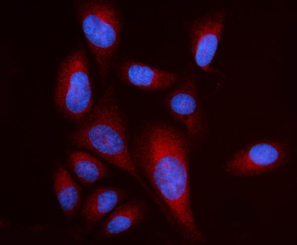
A06980-2 has IHC data from human paraffin sections and IF/ICC data from U2OS cells (A06980-2 image captions); listed reactivity covers human, mouse, and rat (catalog reactivity).

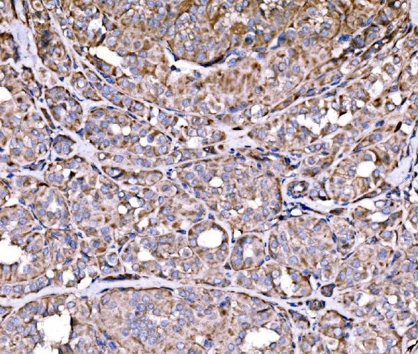
Real IHC data IHC analysis of RPL17 using anti-RPL17 antibody (A06980-2). RPL17 was detected in a paraffin-embedded section of human thyroid papillary carcinoma tissue. Heat mediated antigen retrieval was performed in EDTA buffer (pH 8.0, epitope retrieval solution). The tissue section was blocked with 10% goat serum. The tissue section was then incubated with 2 μg/ml rabbit anti-RPL17 Antibody (A06980-2) overnight at 4°C. Biotinylated goat anti-rabbit IgG was used as secondary antibody and incubated for 30 minutes at 37°C. The tissue section was developed using Strepavidin-Biotin-Complex (SABC) (Catalog # SA1022) with DAB as the chromogen.
Anti-RPL17 Antibody ®
Cat # A06980-2

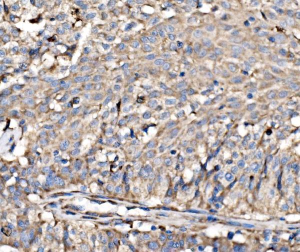
A06980-2 will render with its own IHC figure from a human thyroid papillary carcinoma paraffin section (A06980-2 IHC figure caption). Its additional IHC images show human laryngeal, liver, and stomach cancer paraffin sections, and its IF image shows U2OS cells (A06980-2 image captions).

Which to pick: For tissue IHC, choose A06980-2: its figure documents a human paraffin section, EDTA pH 8 retrieval, and 2 μg/ml primary antibody; the fixative is unreported (A06980-2 IHC figure caption). For IF/ICC, the same SKU lists both applications and shows U2OS cell staining at 5 μg/ml (catalog applications; A06980-2 IF caption). For mouse or rat samples, A06980-2 lists reactivity with both species; it is rabbit-hosted, and clonality is unreported (catalog reactivity, host, and clone).

Each figure is that product's own IHC / IF validation image from its datasheet.

References

  1. UniProt Consortium. UniProt entry P18621 (RL17_HUMAN, Large ribosomal subunit protein uL22).
  2. Human Protein Atlas. RPL17 tissue IHC expression (reliability: Approved).
  3. Human Protein Atlas. RPL17 subcellular location (ICC-IF): Mainly localized to the endoplasmic reticulum and cytosol. Caution: Based on antibodies targeting proteins from multiple genes..
  4. Human Protein Atlas. RPL17 antibody validation summary (2 antibodies).
  5. Sex- and age-related differences in ribosomal proteins L17 and L37, as well as androgen receptor protein, in the song control system of zebra finches. Neuroscience 2010 — PMC2991423.
  6. Effects of estradiol on incorporation of new cells in the developing zebra finch song system: potential relationship to expression of ribosomal proteins L17 and L37. Developmental neurobiology 2009 — PMC2692582.
  7. Ribosomal protein L17, RpL17, is an inhibitor of vascular smooth muscle growth and carotid intima formation. Circulation 2012 — PMC3502943.
  8. Comparative proteomic analysis identifies differentially expressed proteins and reveals potential mechanisms of traumatic heterotopic ossification progression. Journal of orthopaedic translation 2022 — PMC9117278.
  9. PubMed PMID:2402465 — UniProt-cited evidence.
  10. PubMed PMID:1793733 — UniProt-cited evidence.
  11. PubMed PMID:11875025 — UniProt-cited evidence.