This website uses cookies to ensure you get the best experience on our website.


- Table of Contents
Plan RPL18 staining in paraffin sections around the reported ubiquitous cytoplasmic tissue pattern (HPA tissue IHC). Compare cell types within each section, keeping in mind the reported low consistency between antibody staining and RNA expression (HPA tissue IHC).

Expected localisation, validated protocols, controls and antibodies — the at-a-glance facts below, then the full design guide.
| Expected localisation | Cytoplasmic tissue staining (HPA tissue IHC) | |
| Staining pattern | Cytoplasmic staining across many tissue cell types (HPA tissue IHC) | |
| Antigen retrieval | EDTA pH 8.0 HIER, heat-mediated (datasheet A07057-3) | |
| Positive control | Testis+4 more · see all | |
| Negative control | Adipose tissue+1 more · see all |
| Fixation | Keep paraffin-section fixation consistent. (standard IHC practice; not target-specific) | |
| Caveat | Antibody staining and RNA levels show low consistency (HPA tissue IHC) | |
| Regulation | Low tissue specificity; no regulator annotated (HPA tissue IHC; UniProt) | |
| Isoform / epitope | 2 isoforms; epitope differences are unspecified (UniProt) |
The catalog antibody’s IHC-P protocol uses EDTA retrieval (datasheet A07057-3). One published melanoma IHC protocol uses citrate retrieval (PMC12808902).
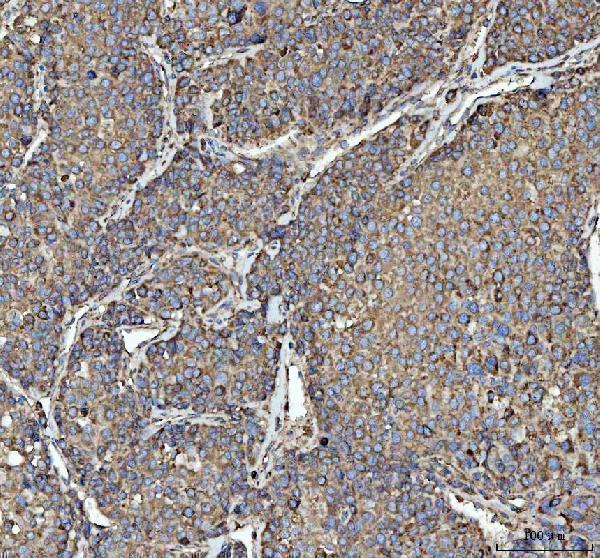
| Sample | Paraffin-embedded human liver cancer tissue; fixative not specified (datasheet A07057-3) |
| Fixation | Image fixative and duration unreported (datasheet A07057-3); verify before use. |
| Sectioning | 4–5 µm sections on charged slides (standard) |
| Deparaffinisation | Xylene, graded ethanol series to water (standard) |
| Antigen retrieval | Heat retrieval: EDTA pH 8.0 (datasheet A07057-3); 20 min, 95–100 °C (standard) |
| Peroxidase block | 3% H2O2, 10 min, room temperature (standard) |
| Blocking | 10% goat serum (datasheet A07057-3) |
| Primary antibody | Rabbit anti-RPL18, 2-5 μg/ml (datasheet A07057-3) |
| Primary incubation | Overnight at 4 °C (datasheet A07057-3) |
| Detection | HRP-conjugated secondary, DAB chromogen (datasheet A07057-3) |
| Counterstain | Hematoxylin, blue, dehydrate and mount (standard) |
| Expected result | RPL18-positive staining in elongated or late spermatids of testis (HPA tissue IHC: High). HPA tissue profile: Ubiquitous cytoplasmic expression. No signal in the no-primary control. |
RPL18 is a large ribosomal subunit protein found in the cytosol and on ribosomes associated with rough endoplasmic reticulum; it has no transmembrane segment (UniProt Q07020). In paraffin-section IHC, expect mainly cytoplasmic staining across many cell types, with stronger staining in some cells (HPA: ubiquitous cytoplasmic expression). Treat intensity comparisons cautiously: the tissue IHC assessment is Approved, with low consistency between antibody staining and RNA expression (HPA: tissue IHC reliability).
| Cytoplasmic staining appears in many cell types, with particularly strong signal in elongated or late spermatids. | This fits the reported distribution: RPL18 is cytosolic and associated with rough endoplasmic reticulum ribosomes (UniProt Q07020); HPA reports ubiquitous cytoplasmic expression and High staining in elongated or late spermatids (HPA: tissue IHC). Compare signal with the local negative control before scoring intensity (standard IHC practice). |
| Glandular or epithelial cytoplasm stains at a moderate intensity in appendix, breast, bronchus, cervix, or colon. | These are reported Medium patterns in the specified cell types (HPA: tissue IHC). Moderate signal can be a valid positive result; demanding uniformly strong staining would conflict with the observed tissue range (HPA: tissue IHC). Score the relevant cells and their cytoplasmic compartment separately from any diffuse slide background (standard IHC practice). |
| The dominant signal is nuclear, sharply membranous, or extracellular, with little cytoplasmic staining. | That distribution does not fit the expected tissue IHC pattern or UniProt localisation (HPA: ubiquitous cytoplasmic expression; UniProt Q07020). Investigate staining artefact and antibody specificity before calling it RPL18. Nucleolar localisation is an uncertain additional ICC-IF observation, so it does not by itself validate dominant nuclear IHC staining (HPA: subcellular). |
| Adipocytes or chondrocytes stain strongly while expected cytoplasmic staining in the same run is weak. | HPA reports RPL18 as Not detected in adipocytes and chondrocytes in its sampled tissues (HPA: tissue IHC). Strong staining there is a reason to check cross-reactivity, endogenous detection activity, and cell identification (standard IHC practice). These observations are comparison points, not proof that every such cell must always be negative. |
| Color covers tissue broadly, including spaces between cells, or a known-positive sample has no discernible signal. | Diffuse color without a cellular pattern suggests background; absence of signal in a reported positive sample leaves a negative result uninterpretable until controls pass (standard IHC practice). Use testis elongated or late spermatids as a reported High reference, or a listed Medium cell population, while allowing for HPA's low staining–RNA consistency (HPA: tissue IHC). |
| Cell and tissue context | HPA describes ubiquitous cytoplasmic expression but reports different levels among sampled cells, from High in elongated or late spermatids to Not detected in adipocytes and chondrocytes (HPA: tissue IHC). Interpret intensity within the identified cell population. |
| Compartment and topology | RPL18 occurs in cytosol and ribosomes associated with rough endoplasmic reticulum and has no transmembrane segment (UniProt Q07020). A predominantly cytoplasmic IHC pattern is therefore the relevant localisation check (HPA: tissue IHC). |
| Strength of tissue evidence | The listed antibody HPA046572 has Approved IHC validation, while HPA notes low consistency between antibody staining and RNA expression (HPA: antibody validation; HPA: tissue IHC reliability). Use the observed pattern as a guide, with controls for each staining run (standard IHC practice). |
| IF/ICC Q: What localisation should an image show? | A: Mainly endoplasmic reticulum and cytosol; nucleoli are an additional uncertain location (HPA: subcellular). HPA lists ICC-IF images from HEK293, MCF-7, and U2OS, and marks HPA046572 ICC as Supported (HPA: subcellular; HPA: antibody validation). |
| Situation | Likely cause | Next action |
|---|---|---|
| No cytoplasmic signal in a section containing a reported positive cell population. | The staining run may have failed, or the signal may be below the chosen detection threshold; absence alone does not establish lack of RPL18 (standard IHC practice). | Check a positive control and the negative detection control, then review antigen retrieval, antibody incubation, and detection steps using the validated IHC workflow (standard IHC practice). HPA reports High staining in elongated or late spermatids (HPA: tissue IHC). |
| Uniform brown haze obscures cell boundaries or covers areas outside cells. | Nonspecific antibody binding, excess detection signal, or residual endogenous detection activity can produce diffuse background (standard chromogenic IHC practice). | Inspect the negative detection control; review blocking, washes, antibody concentration, and detection time, then score only interpretable cellular staining (standard IHC practice). |
| Strong staining appears mainly in adipocytes or chondrocytes. | This conflicts with HPA's Not detected observations for those sampled cells and may reflect cross-reactivity or endogenous detection activity (HPA: tissue IHC; standard IHC practice). | Confirm cell identity and compare the signal with negative detection and reported positive tissue controls before assigning it to RPL18 (standard IHC practice; HPA: tissue IHC). |
| A crisp nuclear or membranous pattern dominates the section. | The dominant compartment differs from the reported cytoplasmic IHC pattern and the cytosolic, rough endoplasmic reticulum association (HPA: tissue IHC; UniProt Q07020). | Review the control slides and staining distribution; repeat with the IHC-validated antibody if the pattern persists (standard IHC practice; HPA: antibody validation). Do not treat the uncertain nucleolar ICC-IF observation as confirmation of nuclear IHC (HPA: subcellular). |
| A glandular or epithelial population stains only moderately. | Medium staining is the reported level for several sampled glandular and epithelial populations, including appendix glandular cells and bronchial respiratory epithelial cells (HPA: tissue IHC). | Assess cytoplasmic localisation and control performance before increasing signal development; moderate, well-localised staining can be the expected result (HPA: tissue IHC; standard IHC practice). |
| A negative tissue result disagrees with an RNA expression expectation. | HPA reports low consistency between antibody staining and RNA expression in its Approved tissue IHC assessment (HPA: tissue IHC reliability). | Report the observed cell type, compartment, intensity, and control results; avoid inferring protein absence from RNA alone (standard IHC practice; HPA: tissue IHC reliability). |


Comprehensive Human Protein Atlas IHC scoring per tissue (reliability: Approved — Low consistency between antibody staining and RNA expression data.). Rows are taken directly from the HPA tissue chart — click any row's HPA link to view the source.
| Tissue | Cell type | Level | Evidence | Source |
|---|---|---|---|---|
| Testis | Elongated or late spermatids | High | Protein (IHC) | HPA → |
| Appendix | Glandular cells | Medium | Protein (IHC) | HPA → |
| Breast | Glandular cells | Medium | Protein (IHC) | HPA → |
| Bronchus | Respiratory epithelial cells | Medium | Protein (IHC) | HPA → |
| Cerebellum | Purkinje cells | Medium | Protein (IHC) | HPA → |
Troubleshoot RPL18 staining in paraffin sections by checking retrieval, cytoplasmic distribution, controls, and how staining is scored.
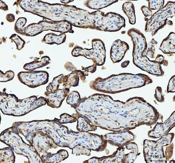
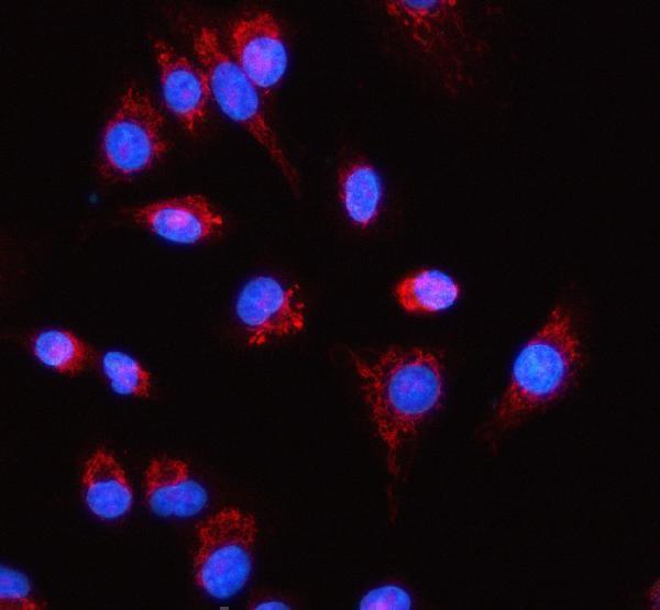
Anti-RPL18 A07057-3 has real IHC images from human paraffin sections and an IF image from A549 cells (catalog image captions); listed reactivity covers human, mouse, and rat (catalog reactivity).
A07057-3 is the sole card, with IHC images from paraffin sections of human liver cancer, placenta, rectum adenocarcinoma, and spleen (catalog IHC captions). Its IF image uses A549 cells (catalog IF caption), and its listed applications include IHC, ICC, and IF (catalog applications).
Which to pick: Choose A07057-3 for tissue IHC: its own paraffin-section captions document EDTA retrieval at pH 8.0 and 2 μg/ml primary antibody; the fixative is unreported (catalog IHC captions). For IF/ICC, the same SKU lists both applications and has an A549 IF image using 5 μg/ml primary antibody (catalog applications; catalog IF caption). For cross-species work, A07057-3 lists human, mouse, and rat reactivity, while the supplied IHC images show human tissue and the IF image shows A549 cells; clonality is unreported (catalog reactivity; catalog image captions; catalog clone field).