This website uses cookies to ensure you get the best experience on our website.


- Table of Contents
Plan RPL28 chromogenic IHC on paraffin sections using the reported cytoplasmic tissue pattern (HPA tissue IHC). This guide covers fixation, cell-type controls and the A10323Y77 IHC dilution range of 1:50–1:200 (datasheet).

Expected localisation, validated protocols, controls and antibodies — the at-a-glance facts below, then the full design guide.
| Expected localisation | Cytoplasmic tissue staining (HPA tissue IHC) | |
| Staining pattern | General cytoplasmic staining across multiple cell types (HPA tissue IHC) | |
| Antigen retrieval | Citrate pH 6.0 HIER, 95–98 °C, 20 min (rule: cytoplasmic / membrane antigen) | |
| Positive control | Appendix+4 more · see all | |
| Negative control | Adipose tissue+3 more · see all |
| Fixation | Matched tissue-IHC evidence does not establish the fixation claim. Validate the specimen-specific method before use. (selected-SKU IHC image A10323Y77) | |
| Caveat | Adipocytes lack detectable staining (HPA tissue IHC) | |
| Regulation | No specific regulator annotated (UniProt) | |
| Isoform / epitope | 5 isoforms; cytoplasmic target, epitope coverage unknown (UniProt) |
The catalog antibody IHC-P protocol is accompanied by a published RPL28 protocol using paraffin-embedded tissue sections (PMC13553147 methods).
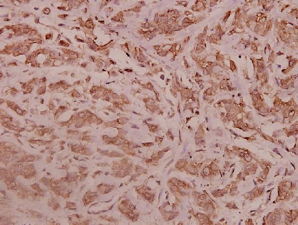
| Sample | Paraffin-embedded human breast carcinoma tissue; fixative not specified (datasheet A10323Y77) |
| Fixation | Image fixative and duration unreported (datasheet A10323Y77); verify before use. |
| Sectioning | 4–5 µm sections on charged slides (standard) |
| Deparaffinisation | Xylene, graded ethanol series to water (standard) |
| Antigen retrieval | Heat-induced epitope retrieval in citrate buffer, pH 6.0, 20 min at 95–98 °C (standard rule: cytoplasmic / membrane antigen) |
| Peroxidase block | 3% H2O2, 10 min, room temperature (standard) |
| Blocking | 10% normal serum of the secondary host, 30 min, room temperature (standard) |
| Primary antibody | Rabbit anti-RPL28, 1:50-1:200 (datasheet A10323Y77) |
| Primary incubation | Overnight at 4 °C (standard) |
| Detection | HRP-polymer secondary, DAB chromogen 5–10 min (standard) |
| Counterstain | Hematoxylin, blue, dehydrate and mount (standard) |
| Expected result | RPL28-positive staining in glandular cells of appendix (HPA tissue IHC: High). HPA tissue profile: General cytoplasmic expression. No signal in the no-primary control. |
RPL28 is a cytoplasmic component of the large ribosomal subunit and has no transmembrane segment (UniProt P46779). In paraffin section IHC, expect general cytoplasmic staining, including high staining in appendix and colon glandular cells, bone marrow hematopoietic cells, and cerebellar Purkinje cells (HPA: tissue IHC). HPA rates the tissue pattern Approved, with medium consistency between antibody staining and RNA expression (HPA: tissue IHC).
| Clear cytoplasmic staining in appendix or colon glandular cells, with tissue structures remaining easy to distinguish. | This matches the reported general cytoplasmic pattern and High staining in those cell populations (HPA: tissue IHC). Evaluate staining in identified cells; a positive area alone does not establish which cells carry the signal (general IHC practice). |
| Strong nuclear only staining, or a sharply membrane confined outline, with little cytoplasmic signal. | That distribution conflicts with the reported cytoplasmic IHC pattern (HPA: tissue IHC) and cytoplasmic, nontransmembrane protein annotation (UniProt P46779). Treat it as a possible artefact and check controls before assigning it to RPL28 (general IHC practice). |
| Prominent staining in adipocytes, ovarian follicle cells, smooth muscle cells, or chondrocytes. | HPA lists these specific cell populations as Not detected, so staining there warrants scrutiny; the designation is cell specific, not a claim that the whole tissue must be blank (HPA: tissue IHC). Cross reactivity or endogenous detection activity are possibilities to investigate with controls (general IHC practice). |
| Diffuse color covers cells and surrounding tissue, obscuring cytoplasmic boundaries. | A uniform deposit is difficult to reconcile with an interpretable cellular pattern (general IHC practice); HPA describes RPL28 staining as generally cytoplasmic (HPA: tissue IHC). Compare a no primary control and review blocking, washes, and chromogen development (general IHC practice). |
| No detectable signal in the expected cells of an appendix or colon section. | HPA reports High staining in glandular cells of both tissues, making a blank result a reason to examine assay performance (HPA: tissue IHC). Confirm that the expected cells are present, then review the antibody and detection workflow alongside a positive control (general IHC practice). |
| Cell population and tissue | HPA reports High staining in several defined populations but Not detected in adipocytes, follicle cells, smooth muscle cells, and chondrocytes; select and score controls by cell type (HPA: tissue IHC; general IHC practice). |
| Evidence strength | The tissue profile is Approved with medium staining to RNA consistency, and HPA050459 is Approved for IHC; these labels support use of the observed pattern while leaving ambiguous staining to be checked with controls (HPA: tissue IHC; HPA: HPA050459 validation). |
| Subcellular context | UniProt places RPL28 in the cytoplasm as a large ribosomal subunit component with no transmembrane segment; HPA describes IHC as generally cytoplasmic (UniProt P46779; HPA: tissue IHC). |
| Target specific fixation and retrieval response | No target specific fixation sensitivity or antigen retrieval condition is established by the supplied UniProt and HPA records; determine workable IHC conditions with assay controls (UniProt P46779; HPA: tissue IHC; general IHC practice). |
| Situation | Likely cause | Next action |
|---|---|---|
| Expected glandular cells are blank in appendix or colon. | These cells are reported High, so absence may reflect an assay or specimen problem; the pattern alone cannot identify which step failed (HPA: tissue IHC; general IHC practice). | Check tissue identity and cell preservation, then compare a working positive control; review retrieval, primary antibody application, and detection conditions as general IHC workflow checks (general IHC practice). |
| Color appears chiefly in nuclei or along cell membranes. | This differs from HPA's general cytoplasmic IHC pattern and UniProt's cytoplasmic, nontransmembrane annotation (HPA: tissue IHC; UniProt P46779). | Confirm the compartment with the counterstain and cell morphology, then inspect a no primary control and detection background before calling the signal specific (general IHC practice). |
| Cells reported Not detected show conspicuous staining. | HPA reports no detection in the specified adipocytes, follicle cells, smooth muscle cells, and chondrocytes; off target binding or endogenous activity could explain unexpected color (HPA: tissue IHC; general IHC practice). | Score the named cell type, compare an expected positive population, and use no primary and appropriate detection controls to investigate background (general IHC practice). |
| Diffuse background prevents cell level scoring. | Excess nonspecific binding or detection background can mask a cellular pattern (general IHC practice); the reported IHC distribution is generally cytoplasmic (HPA: tissue IHC). | Compare a no primary control, then review blocking, wash steps, detection reagent exposure, and chromogen development within the established IHC workflow (general IHC practice). |
| Weak staining in thyroid, parathyroid, lung alveolar, or placental trophoblastic cells is being called an assay failure. | HPA lists these cell populations as Low, so faint signal there is less informative about assay performance than the listed High populations (HPA: tissue IHC). | Judge the run against an expected High cell population and score each tissue's named cell type separately (HPA: tissue IHC; general IHC practice). |
| IF/ICC: what distribution should be compared with an image? | HPA reports supported endoplasmic reticulum and cytosol localization in ICC-IF, while its tissue IHC summary says general cytoplasmic expression (HPA: subcellular ICC-IF; HPA: tissue IHC). | Use the ER and cytosol annotation to interpret IF/ICC localization; evaluate paraffin section IHC against the tissue IHC pattern on this page (HPA: subcellular ICC-IF; HPA: tissue IHC). |


Comprehensive Human Protein Atlas IHC scoring per tissue (reliability: Approved — Medium consistency between antibody staining and RNA expression data.). Rows are taken directly from the HPA tissue chart — click any row's HPA link to view the source.
| Tissue | Cell type | Level | Evidence | Source |
|---|---|---|---|---|
| Appendix | Glandular cells | High | Protein (IHC) | HPA → |
| Bone marrow | Hematopoietic cells | High | Protein (IHC) | HPA → |
| Caudate | Neuronal cells | High | Protein (IHC) | HPA → |
| Cerebellum | Purkinje cells | High | Protein (IHC) | HPA → |
| Colon | Glandular cells | High | Protein (IHC) | HPA → |
| Tissue | Cell type | Level | Evidence | Source |
|---|---|---|---|---|
| Adipose tissue | Adipocytes | Not detected | Protein (IHC) | HPA → |
| Ovary | Follicle cells | Not detected | Protein (IHC) | HPA → |
| Smooth muscle | Smooth muscle cells | Not detected | Protein (IHC) | HPA → |
| Soft tissue | Chondrocytes | Not detected | Protein (IHC) | HPA → |
Use cytoplasmic staining, cell identity and matched controls to troubleshoot RPL28 chromogenic IHC in paraffin sections (HPA tissue IHC; selected image caption).
Catalog anti-RPL28 antibodies list human, mouse and rat reactivity; real IHC imagery shows paraffin-embedded human breast carcinoma tissue (catalog; A10323Y77 image caption).
A10323Y77 is the only card that will render, with an IHC image of paraffin-embedded human breast carcinoma tissue at 1:100 (A10323Y77 image caption). Its listed applications are IP, IHC and WB, and its listed reactivity is human, mouse and rat (catalog).
Which to pick: For tissue IHC, choose A10323Y77 because its own image documents paraffin-section staining; the fixative is unreported (A10323Y77 image caption). For IF/ICC, A10323-1 lists IF at 1:50, but has no IF figure and does not list ICC (catalog). Both list human, mouse and rat reactivity and a rabbit host; neither reports a clone, and the shown tissue IHC evidence is human only (catalog; A10323Y77 image caption).