RPL29 / Large ribosomal subunit protein eL29 · IHC design guide

Design Immunohistochemistry for RPL29

Plan RPL29 staining in paraffin sections around its cytoplasmic pattern in most tissues (HPA tissue IHC). Use the catalog antibody’s IHC conditions and interpret staining alongside the reported low consistency with RNA expression (HPA tissue IHC).

Evidence assembled Oct 2026 · For research use; verify linked source records and product datasheet before use
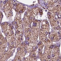
Immunohistochemistry protocol sheet for RPL29 (IHC for RPL29): expected localisation Cytoplasmic in most tissues (HPA tissue IHC), antibody A05949-1, validated IHC image, and IHC protocol steps
Printable RPL29 IHC protocol sheet — expected localisation Cytoplasmic in most tissues (HPA tissue IHC), antibody A05949-1, controls and protocol steps. Open the full RPL29 IHC guide →

RPL29 Immunohistochemistry Experimental Design Guide

Expected localisation, validated protocols, controls and antibodies — the at-a-glance facts below, then the full design guide.

Must know before staining
Expected localisation Cytoplasmic in most tissues (HPA tissue IHC)
Staining pattern Cytoplasmic staining across most tissues (HPA tissue IHC)
Antigen retrieval EDTA pH 8.0 HIER, heat-mediated (datasheet A05949-1)
Positive control ⓘ Adipose tissue+4 more · see all
Negative control ⓘ Heart muscle+2 more · see all
Important caveats
Reasons your staining may differ from the expected pattern.
Fixation Keep fixation consistent across sections. (standard IHC practice; not target-specific)
Caveat Antibody staining and RNA show low consistency (HPA tissue IHC)
Regulation Specific intensity regulators unreported (UniProt)
Isoform / epitope 0 annotated isoforms; chain spans residues 2–159 (UniProt)
Section 1

Recommended RPL29 IHC & IF Protocols

The catalog antibody’s IHC-P protocol (datasheet A05949-1) is followed by the published RPL29 TMA staining procedure (PMC8201403 methods).

Recommended immunohistochemistry (IHC-P) protocol parameters
SampleParaffin-embedded mouse brain tissue; fixative not specified (datasheet A05949-1)
FixationImage fixative and duration unreported (datasheet A05949-1); verify before use.
Sectioning4–5 µm sections on charged slides (standard)
DeparaffinisationXylene, graded ethanol series to water (standard)
Antigen retrievalHeat retrieval: EDTA pH 8.0 (datasheet A05949-1); 20 min, 95–100 °C (standard)
Peroxidase block3% H2O2, 10 min, room temperature (standard)
Blocking10% goat serum (datasheet A05949-1)
Primary antibodyRabbit anti-RPL29, 2-5 μg/ml (datasheet A05949-1)
Primary incubationOvernight at 4 °C (datasheet A05949-1)
DetectionStreptavidin-biotin complex (SABC), DAB chromogen (datasheet A05949-1)
CounterstainHematoxylin, blue, dehydrate and mount (standard)
Expected resultRPL29-positive staining in adipocytes of adipose tissue (HPA tissue IHC: High). HPA tissue profile: Cytoplasmic expression in most tissues. No signal in the no-primary control.
💡Decision noteStart with heat-mediated EDTA pH 8.0 retrieval for paraffin sections (datasheet A05949-1); the published TMA procedure does not specify retrieval conditions (PMC8201403 methods).
Section 2

What Is the Expected RPL29 Staining Pattern?

RPL29 is a cytoplasmic large ribosomal subunit protein with no transmembrane segment (UniProt P47914: localisation and topology). In paraffin sections, expect cytoplasmic staining in many cell types, including colon and duodenal glandular cells and bone marrow hematopoietic cells (HPA: tissue IHC, High). HPA rates the tissue staining Approved but reports low consistency with RNA expression, so interpret tissue comparisons cautiously (HPA: reliability).

What am I looking at on my slide?
Cytoplasmic staining is strong in colon or duodenal glandular cells.This matches the reported cell type, compartment and High staining level (HPA: colon and duodenum tissue IHC). Score the glandular cells themselves; signal elsewhere in the section does not establish the expected pattern.
Signal appears predominantly nuclear or along cell membranes, with little cytoplasmic staining.This conflicts with the cytoplasmic tissue IHC profile and UniProt localisation (HPA: tissue IHC; UniProt P47914: cytoplasm). Check whether counterstain, tissue pigment or detection artefact is being read as specific chromogen.
Strong stain appears in cardiomyocytes or smooth muscle cells.Those cells are reported Not detected in the listed tissues (HPA: heart muscle and smooth muscle tissue IHC). Consider cross-reactivity or endogenous detection activity, especially if the expected cytoplasmic pattern is absent; the HPA observations are comparators, not proof of universal negativity.
Color spreads across stroma, empty spaces and multiple compartments without clear cell boundaries.Treat this as diffuse background rather than a readable RPL29 pattern (HPA: cytoplasmic tissue IHC profile). General IHC practice: compare a no-primary control and inspect blocking, washing and chromogen development before scoring cells.
No staining is visible in colon glandular cells or bone marrow hematopoietic cells.Both are reported High by tissue IHC (HPA: colon and bone marrow). A blank result calls for a run-level check of the antibody, retrieval and detection steps; HPA does not establish an RPL29-specific retrieval condition or fixation sensitivity.
💡Expected RPL29 appearanceCall a positive result when cell-defined cytoplasmic chromogen is evident in a reported High population, such as colon glandular cells (HPA: colon tissue IHC, High); diffuse staining without cell boundaries or a predominantly nuclear pattern is suspect (HPA: cytoplasmic tissue IHC profile).
How each factor affects the staining
Tissue and cell selectionHPA reports High staining in adipocytes, hematopoietic cells, cerebral cortex glia and several glandular populations, but Not detected in listed cardiomyocytes and smooth muscle cells (HPA: tissue IHC). Choose comparators by cell type, not the tissue name alone.
Compartment evidence across applicationsParaffin tissue staining is described as cytoplasmic (HPA: tissue IHC; UniProt P47914: cytoplasm). ICC-IF also reports supported nucleolar, endoplasmic reticulum and cytosol localisation (HPA: subcellular ICC-IF); that cell-image result does not redefine the expected chromogenic tissue pattern.
Strength of tissue evidenceThe listed antibody HPA069064 is IHC Approved, while HPA flags low consistency between antibody staining and RNA expression (HPA: antibody validation and reliability). Treat High and Not detected labels as observed staining, with that validation caveat, rather than absolute expression calls.
Protein identity and processingUniProt describes RPL29 as a large ribosomal subunit component, with a 2–159 mature chain, no signal peptide and no transmembrane segment (UniProt P47914: subunit, processing and topology). These annotations support an intracellular interpretation; they do not specify an antigen retrieval method.
Why is my staining missing, weak or wrong?
SituationLikely causeNext action
The expected positive tissue is blank.A run-level failure is possible; colon glandular cells and bone marrow hematopoietic cells are reported High (HPA: tissue IHC).General IHC practice: check section integrity, antibody application, retrieval, secondary detection and chromogen with a working positive control. Do not infer a target-specific fixation effect from this result.
All cells have a brown haze.Diffuse color lacks the cell-defined cytoplasmic pattern reported for RPL29 (HPA: tissue IHC). Background from the detection workflow is possible.General IHC practice: inspect a no-primary control, then review blocking, washing, antibody concentration and development time. Score only distinguishable cellular staining.
Cardiomyocytes or smooth muscle cells stain strongly.These listed cell types are Not detected in HPA tissue images (HPA: heart muscle and smooth muscle tissue IHC); cross-reactivity or endogenous detection activity is possible.Compare the same run with a no-primary control and an expected positive cell population. Review detection blocking before assigning the signal to RPL29.
Most color is nuclear, while cytoplasm is faint.That differs from the tissue IHC profile and UniProt cytoplasmic annotation (HPA: tissue IHC; UniProt P47914: cytoplasm).Verify the chromogen against the counterstain and inspect a control section. Do not treat nuclear color alone as a positive tissue IHC result.
A reported Not detected cell type is blank.Absence there agrees with that specific HPA observation (HPA: cardiomyocytes, oral squamous epithelial cells or smooth muscle cells, Not detected).Use a reported High cell population in the same run to judge whether staining worked. HPA's low RNA–staining consistency limits conclusions from a negative cell type (HPA: reliability).
Does IF/ICC require the same compartment call?HPA's ICC-IF images support nucleoli, endoplasmic reticulum and cytosol, whereas tissue IHC is summarized as cytoplasmic (HPA: subcellular ICC-IF and tissue IHC).Interpret IF/ICC using its own localisation evidence and guide page (HPA: subcellular ICC-IF). For this paraffin IHC guide, judge the chromogenic tissue result against the cytoplasmic profile (HPA: tissue IHC).

Sample controls for RPL29 IHC & IF

🧪Run colon first: glandular cells should stain (HPA: High in colon glandular cells); use smooth muscle as the negative tissue (HPA: Not detected in smooth muscle cells). If the colon section includes smooth-muscle cells, expect counterstain with little or no specific chromogen there, while confirming their background against the control slides (HPA: smooth muscle cells not detected).
Positive control tissue: Adipose tissue (Adipocytes, HPA High)
Negative control tissue: Heart muscle (HPA Not detected)
ICC-IF cell lines (HPA subcellular resource): HPA ICC-IF images show RPL29 in Hep-G2, MCF-7, U2OS, with annotated localisation: Nucleoli (supported), Endoplasmic reticulum (supported), Cytosol (supported) (HPA subcellular).
Technical controls: Include a no-primary, secondary-only section and a concentration-matched rabbit IgG isotype control; use RPL29 knockout material or an immunizing-peptide block, if available, to test specificity (caption: rabbit primary antibody; standard IHC control practice). Quench endogenous peroxidase and check for endogenous biotin background in the positive tissue when using the caption’s biotin-based, DAB detection (caption: biotinylated secondary, SABC and DAB; standard IHC practice).
⚠️Feasibility: A target-specific fixation window or fixation effect is unreported, and the exact A05949-1 paraffin-section caption does not state the fixative (caption: fixative not stated). That caption uses heat retrieval in EDTA at pH 8.0; whether retrieval is required under other conditions, or whether frozen sections or IF are easier, is unreported (caption: heat-mediated EDTA retrieval; HPA: ICC-IF images in Hep-G2, MCF-7 and U2OS). In colon, distinguish glandular staining from luminal material and section-edge background when scoring (HPA: High in colon glandular cells; standard IHC interpretation practice).

HPA tissue IHC evidence for RPL29

Comprehensive Human Protein Atlas IHC scoring per tissue (reliability: Approved — Low consistency between antibody staining and RNA expression data.). Rows are taken directly from the HPA tissue chart — click any row's HPA link to view the source.

Positive expression · recommended positive controls

TissueCell typeLevelEvidenceSource
Adipose tissue Adipocytes High Protein (IHC) HPA →
Bone marrow Hematopoietic cells High Protein (IHC) HPA →
Cerebellum Cells in molecular layer High Protein (IHC) HPA →
Cerebral cortex Glial cells High Protein (IHC) HPA →
Colon Glandular cells High Protein (IHC) HPA →

Undetected expression · recommended negative controls

TissueCell typeLevelEvidenceSource
Heart muscle Cardiomyocytes Not detected Protein (IHC) HPA →
Oral mucosa Squamous epithelial cells Not detected Protein (IHC) HPA →
Smooth muscle Smooth muscle cells Not detected Protein (IHC) HPA →
Section 3

Advanced RPL29 IHC Tips

Troubleshoot RPL29 staining in paraffin sections by checking retrieval, compartment, cell identity, and detection controls before comparing staining intensity.

What retrieval should I try first when RPL29 staining is weak?
Start with heat-mediated retrieval in EDTA at pH 8.0 (datasheet A05949-1). The selected paraffin-section example used that retrieval before 10% goat serum block and 2 μg/ml primary antibody overnight at 4°C (datasheet A05949-1). If cytoplasmic staining remains weak, compare retrieval heating and cooling times on adjacent sections while keeping the antibody concentration and detection steps constant (standard IHC practice). Record tissue damage and background alongside signal, because stronger heating can make an apparent gain in staining difficult to interpret (standard IHC practice).
Could fixation explain weak or uneven RPL29 staining?
Target-specific sensitivity of RPL29 to fixation is unknown from the supplied evidence; the selected paraffin-section caption does not state a fixative (datasheet A05949-1). Record the fixative, fixation duration, section age, and retrieval conditions for each specimen before comparing staining intensity (standard IHC practice). Use adjacent sections to compare processing conditions, starting with EDTA retrieval at pH 8.0 and the reported 2 μg/ml primary antibody incubation overnight at 4°C (datasheet A05949-1). If signal differs, repeat with matched controls before attributing the difference to fixation, since section handling and detection can also affect chromogenic staining (standard IHC practice).
Where should I expect RPL29 staining in a tissue section?
Expect a mainly cytoplasmic pattern in tissue IHC (HPA: cytoplasmic expression in most tissues; UniProt P47914: cytoplasm). RPL29 is a large ribosomal subunit component and has no annotated transmembrane segment, so a crisp membrane-only DAB outline needs independent validation (UniProt P47914: subunit and topology). Nucleolar, endoplasmic-reticulum, and cytosolic locations are reported from cell imaging, but these compartments may be difficult to separate by chromogenic tissue IHC (HPA: subcellular locations; standard IHC practice). Check cellular morphology and a counterstain before scoring apparent nuclear or membrane signal, and compare with a no-primary control for detection artefacts (standard IHC practice).
How should I investigate an unexpected compartment-specific RPL29 stain?
The supplied record lists 0 isoforms and no annotated transmembrane segment, so an apparent membrane-restricted pattern cannot be assigned to a documented RPL29 isoform (UniProt P47914: isoforms and topology). The record lists modifications at residues 5, 31, 33, and 142, but the selected caption does not identify the antibody epitope (UniProt P47914: modified residues; datasheet A05949-1). Compare adjacent sections with and without primary antibody and review the expected cytoplasmic distribution (HPA: tissue profile; standard IHC practice). If staining differs across processing conditions, avoid attributing it to a particular modification until epitope information or an independent antibody supports that explanation (standard IHC practice).
How can IF help assess ambiguous RPL29 IHC staining?
Use IF as a separate localisation check and multiplex RPL29 with a validated glial-cell marker when examining cerebral cortex, where glial cells show high tissue staining (HPA: cerebral cortex glial cells, High). Choose a fluorophore channel with low tissue autofluorescence and include single-label and no-primary controls to distinguish bleed-through from RPL29 signal (standard IF practice). For intracellular RPL29, compare a mild detergent permeabilisation condition with an unpermeabilised control; the epitope is unmapped, so its accessibility at the endoplasmic reticulum cannot be assumed (UniProt P47914: topology; HPA: subcellular locations; datasheet A05949-1: epitope not stated). Interpret IF alongside morphology rather than transferring its intensity directly to DAB scores (standard IHC/IF practice).
What should I check if DAB background obscures RPL29?
First inspect a no-primary section for signal from the secondary, biotin-based detection, or endogenous peroxidase, then assess nonspecific primary staining on a matched section (standard IHC practice). The selected method used 10% goat serum block, biotinylated goat anti-rabbit secondary for 30 minutes at 37°C, and a streptavidin-biotin complex with DAB (datasheet A05949-1). Include a peroxidase block and, where needed, an endogenous-biotin control as general chromogenic workflow checks (standard IHC practice). Compare background in tissue edges and damaged areas with intact cells before adjusting antibody concentration or DAB development time (standard IHC practice).
How should I quantify RPL29 staining across tissue samples? ⚠ ANSWER MARKED FOR VERIFICATION
Define the cell population and tissue area before scoring, since HPA reports high staining in some cell types and no detected staining in others (HPA: tissue IHC profile). Within matched regions, record the percentage of positive cells and an intensity-based H-score, or count positive cells per mm² when cell density is the question (standard IHC practice). Normalise cell counts to viable tissue area and report intensity within the same cell type, using identical counterstain, illumination, and threshold settings across sections (standard IHC practice). Exclude folds, necrosis, and section edges by a prespecified rule, and keep no-primary controls with each staining run (standard IHC practice).
How do I distinguish true RPL29 staining from artefact?
Give most weight to staining in intact cytoplasm, consistent with the reported tissue pattern and RPL29's ribosomal role (HPA: cytoplasmic expression in most tissues; UniProt P47914: large ribosomal subunit). Check cell identity: HPA reports high staining in cerebral-cortex glial cells but no detected staining in cardiomyocytes, so those observations should not be treated as interchangeable controls (HPA: cerebral cortex and heart muscle). Discount isolated edge, necrotic, or vessel-associated DAB signal when it also appears in a no-primary or peroxidase control (standard IHC practice). HPA rates its tissue staining approved while reporting low consistency with RNA expression; confirm surprising compartment or cell-specific claims independently (HPA: reliability description).
Boster reagents

Best RPL29 / Large ribosomal subunit protein eL29 IHC Antibodies

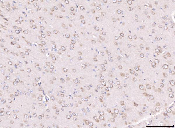
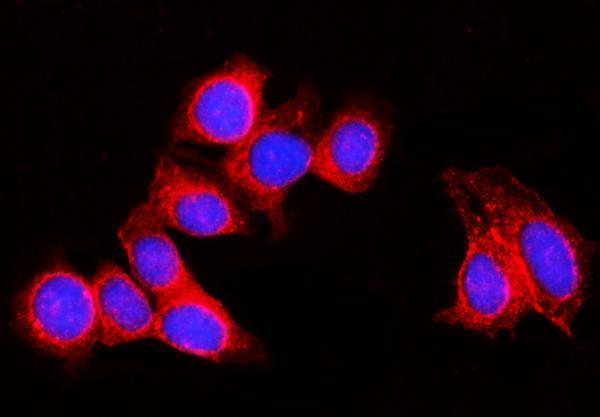
The IHC-validated antibody has paraffin-section images from mouse and rat brain and human tumors (catalog IHC captions); IF/ICC data cover MCF-7 cells (catalog IF caption).

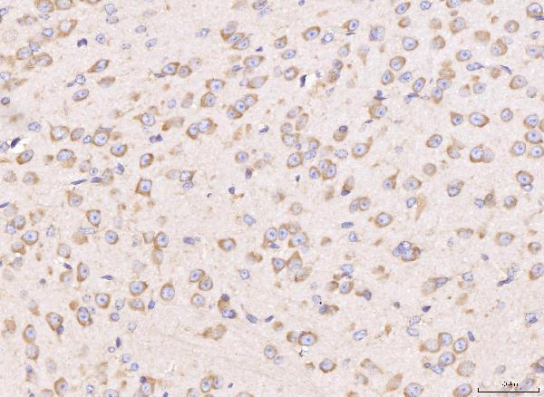
Real IHC data IHC analysis of RPL29 using anti-RPL29 antibody (A05949-1). RPL29 was detected in a paraffin-embedded section of mouse brain tissue. Heat mediated antigen retrieval was performed in EDTA buffer (pH 8.0, epitope retrieval solution). The tissue section was blocked with 10% goat serum. The tissue section was then incubated with 2 μg/ml rabbit anti-RPL29 Antibody (A05949-1) overnight at 4°C. Biotinylated goat anti-rabbit IgG was used as secondary antibody and incubated for 30 minutes at 37°C. The tissue section was developed using Strepavidin-Biotin-Complex (SABC) (Catalog # SA1022) with DAB as the chromogen.
Anti-RPL29 Antibody ®
Cat # A05949-1

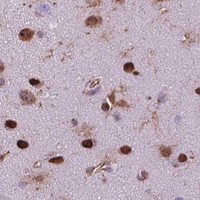
A05949-1 will render with an IHC image from paraffin-embedded mouse brain; additional IHC captions document rat brain, human appendiceal adenocarcinoma, and human breast cancer (catalog IHC captions). Its IF/ICC image documents MCF-7 cells (catalog IF caption), and its application list includes IHC, ICC, and IF (catalog applications).

Which to pick: Choose A05949-1 for paraffin-section IHC because its own caption documents mouse brain staining after EDTA pH 8.0 retrieval at 2 μg/ml (A05949-1 IHC caption); the fixative is unreported (A05949-1 IHC caption). Choose the same SKU for IF/ICC in MCF-7 cells at 5 μg/ml (A05949-1 IF caption). For work across species, its listed reactivity covers human, mouse, rat, and monkey (catalog reactivity), while the supplied IHC captions document human, mouse, and rat tissue only (catalog IHC captions).

Each figure is that product's own IHC / IF validation image from its datasheet.

References

  1. UniProt Consortium. UniProt entry P47914 (RL29_HUMAN, Large ribosomal subunit protein eL29).
  2. Human Protein Atlas. RPL29 tissue IHC expression (reliability: Approved).
  3. Human Protein Atlas. RPL29 subcellular location (ICC-IF): Localized to the nucleoli, endoplasmic reticulum and cytosol..
  4. Human Protein Atlas. RPL29 antibody validation summary (1 antibodies).
  5. Ribosomal Protein RPL29 Promotes Hepatocellular Carcinoma Progression Through Regulation the Expression of Exosome Component 4. Biological procedures online 2026 — PMC12866049.
  6. N-Myristoylation by NMT1 Is POTEE-Dependent to Stimulate Liver Tumorigenesis via Differentially Regulating Ubiquitination of Targets. Frontiers in oncology 2021 — PMC8201403.
  7. PubMed PMID:8597591 — UniProt-cited evidence.
  8. PubMed PMID:8662616 — UniProt-cited evidence.
  9. PubMed PMID:14702039 — UniProt-cited evidence.