This website uses cookies to ensure you get the best experience on our website.


- Table of Contents
Plan chromogenic RPL36 IHC around its general cytoplasmic tissue staining (HPA tissue IHC). Use strongly staining breast glandular cells and weakly staining adipocytes to help assess the staining range (HPA tissue IHC).

Expected localisation, validated protocols, controls and antibodies — the at-a-glance facts below, then the full design guide.
| Expected localisation | General cytoplasmic tissue staining (HPA tissue IHC) | |
| Staining pattern | Broad cytoplasmic staining; glandular cells can stain strongly (HPA tissue IHC) | |
| Antigen retrieval | EDTA pH 8.0 HIER, heat-mediated (datasheet A08922-3) | |
| Positive control | Appendix+4 more · see all | |
| Negative control | None in HPA (detected in all 45 tissues); use no-primary + isotype controls |
| Fixation | Keep fixation consistent across sections (standard IHC practice; not target-specific) | |
| Caveat | Adipocytes and ovarian stroma show low staining (HPA tissue IHC) | |
| Regulation | No expression regulator annotated (UniProt) | |
| Isoform / epitope | No isoforms annotated; mature chain spans residues 2–105 (UniProt) |
The catalog antibody’s paraffin-section IHC protocol (datasheet: A08922-3) is accompanied by one published frozen-section RPL36 IHC protocol (PMC5742712).
| Sample | Paraffin-embedded human breast cancer tissue; fixative not specified (datasheet A08922-3) |
| Fixation | Image fixative and duration unreported (datasheet A08922-3); verify before use. |
| Sectioning | 4–5 µm sections on charged slides (standard) |
| Deparaffinisation | Xylene, graded ethanol series to water (standard) |
| Antigen retrieval | Heat retrieval: EDTA pH 8.0 (datasheet A08922-3); 20 min, 95–100 °C (standard) |
| Peroxidase block | 3% H2O2, 10 min, room temperature (standard) |
| Blocking | 10% goat serum (datasheet A08922-3) |
| Primary antibody | Rabbit anti-RPL36, 2-5 μg/ml (datasheet A08922-3) |
| Primary incubation | Overnight at 4 °C (datasheet A08922-3) |
| Detection | HRP-conjugated secondary, DAB chromogen (datasheet A08922-3) |
| Counterstain | Hematoxylin, blue, dehydrate and mount (standard) |
| Expected result | RPL36-positive staining in endocrine cells of appendix (HPA tissue IHC: High). HPA tissue profile: General cytoplasmic expression. No signal in the no-primary control. |
RPL36 is a large ribosomal subunit protein detected on cytosolic polysomes, with no transmembrane segment (UniProt Q9Y3U8). In paraffin-section IHC, expect predominantly cytoplasmic staining across several cell types, including appendix endocrine cells, bone-marrow hematopoietic cells and breast glandular cells reported as High (HPA tissue IHC). HPA calls the tissue staining Supported, with medium consistency between antibody staining and RNA expression (HPA tissue IHC).
| Clear cytoplasmic staining in appendix endocrine cells, bone-marrow hematopoietic cells or breast glandular cells. | This agrees with the reported High staining in those cell populations and the general cytoplasmic profile (HPA tissue IHC). Interpret the compartment and the identified cell population together; a strong signal alone is less informative when surrounding tissue also stains. The cytosolic location is consistent with RPL36 on polysomes (UniProt Q9Y3U8). |
| Predominantly nuclear, membranous or extracellular staining, with little cytoplasmic signal. | This does not fit the reported cytoplasmic pattern (HPA tissue IHC) or cytosolic localization (UniProt Q9Y3U8). Check the corresponding no-primary control, tissue morphology and detection reagents before assigning the signal to RPL36 (general IHC practice). RPL36 has no annotated transmembrane segment (UniProt Q9Y3U8). |
| Signal is concentrated in adipocytes or ovarian stroma while expected High-staining cells appear weak. | HPA reports Low staining in adipocytes and ovarian stroma, versus High staining in the listed positive cell populations (HPA tissue IHC). This reversal warrants investigation for cross-reactivity, endogenous detection activity or cell-identification error (general IHC practice). Low does not mean absent, so neither population is a validated negative control (HPA tissue IHC). |
| Diffuse color covers cells and extracellular areas without a readable cytoplasmic pattern. | Treat the result as background until a no-primary control and the tissue morphology establish where signal originates (general IHC practice). Excess detection signal or inadequate blocking can obscure compartment boundaries (general IHC practice). Diffuse staining by itself cannot confirm the general cytoplasmic RPL36 profile reported by HPA (HPA tissue IHC). |
| No visible signal in a section containing an expected High-staining cell population. | First verify that the relevant cells are present and preserved; HPA reports High staining for the named populations, not for every cell in their tissues (HPA tissue IHC). Then review antibody application, retrieval, detection and control performance as general IHC variables (general IHC practice). A blank slide alone cannot establish RPL36 absence. |
| Cell population and tissue context | RPL36 has Low tissue RNA specificity, while HPA reports High staining in selected populations and Low staining in adipocytes and ovarian stroma (HPA tissue IHC). Compare like cell populations across sections; tissue-wide intensity averages can hide the expected pattern (general IHC practice). |
| Antibody evidence | HPA047153 is listed as Supported for IHC, and the tissue profile is Supported with medium staining–RNA consistency (HPA antibody record; HPA tissue IHC). These labels support pattern comparison but do not make every unexpected signal specific. Use controls when localization conflicts with the reported cytoplasmic profile (general IHC practice). |
| IF/ICC: where should RPL36 appear? | The expected location is the cytosol (HPA ICC-IF; UniProt Q9Y3U8). HPA lists ICC-IF images for HEK293, PC-3 and U2OS and marks HPA047153 Supported for ICC (HPA ICC-IF; HPA antibody record). This localization answers the IF/ICC question without implying that paraffin-section IHC settings transfer to IF. |
| Situation | Likely cause | Next action |
|---|---|---|
| A known High-staining population is blank. | The relevant cells may be absent from the section, or an IHC workflow step may have failed (HPA tissue IHC; general IHC practice). | Confirm cell identity and morphology, then check a positive control and the primary-antibody and detection steps (general IHC practice). Optimize retrieval empirically if needed; target-specific fixation sensitivity is unreported in the supplied evidence. |
| Signal is mostly nuclear or outlines cell membranes. | The distribution conflicts with cytosolic localization and the general cytoplasmic tissue profile (UniProt Q9Y3U8; HPA tissue IHC). | Inspect morphology and the no-primary control; reassess antibody and detection specificity before scoring those compartments as positive (general IHC practice). |
| Color appears where no primary antibody was applied. | The primary antibody cannot explain that control signal; endogenous detection activity or detection-reagent background is possible (general IHC practice). | Review blocking appropriate to the detection chemistry and the secondary or chromogen controls, then repeat with a clean no-primary control (general IHC practice). |
| Cytoplasmic boundaries disappear under widespread dark staining. | Overdeveloped chromogen or excessive assay signal can prevent compartment assessment (general IHC practice). | Reduce development or adjust assay conditions using matched controls, then score identifiable cells for a cytoplasmic pattern (general IHC practice; HPA tissue IHC). |
| Adipocytes or ovarian stroma look stronger than the selected positive cells. | That ranking differs from HPA's Low reports for those cells and High reports for selected positive populations (HPA tissue IHC). | Check cell identification, no-primary background and detection activity; compare matched sections before inferring cross-reactivity (general IHC practice). |
| Only weak staining is seen in a tissue outside HPA's listed High populations. | HPA reports general cytoplasmic expression and Low tissue RNA specificity, without a definitive negative tissue in this payload (HPA tissue IHC). | Score the identified cell population and compartment, and compare a listed High-staining population in the same run; avoid treating unlisted tissue as a negative control (HPA tissue IHC; general IHC practice). |




Comprehensive Human Protein Atlas IHC scoring per tissue (reliability: Supported — Medium consistency between antibody staining and RNA expression data.). Rows are taken directly from the HPA tissue chart — click any row's HPA link to view the source.
| Tissue | Cell type | Level | Evidence | Source |
|---|---|---|---|---|
| Appendix | Endocrine cells | High | Protein (IHC) | HPA → |
| Bone marrow | Hematopoietic cells | High | Protein (IHC) | HPA → |
| Breast | Glandular cells | High | Protein (IHC) | HPA → |
| Bronchus | Respiratory epithelial cells | High | Protein (IHC) | HPA → |
| Caudate | Glial cells | High | Protein (IHC) | HPA → |
| Tissue | Cell type | Level | Evidence | Source |
|---|---|---|---|---|
| None in HPA: RPL36 is detected in all 45 scored tissues. Use a no-primary (secondary-only) and an isotype control instead. | ||||
Use cytoplasmic localisation, matched controls and consistent staining conditions to troubleshoot RPL36 chromogenic IHC in paraffin sections.
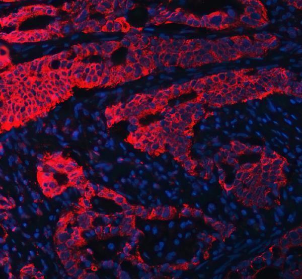
A08922-3 has IHC images from human paraffin-embedded breast, cervical, ovarian and endometrial cancer sections, and IF images from human paraffin-embedded colon and endometrial cancer sections (catalog image captions).
A08922-3 is the only rendered SKU; its IHC images show human paraffin-embedded breast, cervical, ovarian and endometrial cancer tissue (catalog IHC captions). Its IF images show human paraffin-embedded colon and endometrial cancer tissue (catalog IF captions).
Which to pick: For tissue IHC, choose A08922-3: its rabbit antibody was used on a human paraffin-embedded breast cancer section with EDTA retrieval at pH 8.0 and 2 μg/ml primary antibody (catalog IHC caption); the fixative is unreported (catalog IHC caption). For IF, the same SKU has paraffin-section images using 5 μg/ml primary antibody (catalog IF captions); ICC validation is unreported (catalog application list and IF captions). For cross-species planning, A08922-3 lists human, monkey, mouse and rat reactivity (catalog reactivity), while its IHC-P dilution entry specifies human and rat (catalog dilution line).