This website uses cookies to ensure you get the best experience on our website.


- Table of Contents
Plan RPLP2 paraffin IHC around general cytoplasmic staining and strong signal in kidney tubule cells (HPA tissue IHC). Start the catalog antibody at 1:100–1:300 (datasheet), and compare with adipocytes or myocytes, where staining was not detected (HPA tissue IHC).

Expected localisation, validated protocols, controls and antibodies — the at-a-glance facts below, then the full design guide.
| Expected localisation | General cytoplasmic staining (HPA tissue IHC) | |
| Staining pattern | Kidney tubule cells: high, generally cytoplasmic signal (HPA tissue IHC) | |
| Antigen retrieval | Citrate pH 6.0 HIER, 95–98 °C, 20 min (rule: cytoplasmic / membrane antigen) | |
| Positive control | Kidney+4 more · see all | |
| Negative control | Adipose tissue+3 more · see all |
| Fixation | Keep fixation consistent (standard IHC practice; not target-specific); Matched tissue-IHC evidence does not establish the fixation claim. Validate the specimen-specific method before use. (selected-SKU IHC image A05244-1) | |
| Caveat | Adipocytes and myocytes have no detected signal (HPA tissue IHC) | |
| Regulation | No expression regulator annotated (UniProt) | |
| Isoform / epitope | No isoforms; one 1–115 chain (UniProt) |
Compare the catalog antibody’s IHC-P protocol with three published RPLP2 IHC methods (PMC12189634; PMC10697958; PMC5911611).
| Sample | Paraffin-embedded human prostate carcinoma tissue; fixative not specified (datasheet A05244-1) |
| Fixation | Image fixative and duration unreported (datasheet A05244-1); verify before use. |
| Sectioning | 4–5 µm sections on charged slides (standard) |
| Deparaffinisation | Xylene, graded ethanol series to water (standard) |
| Antigen retrieval | Heat-induced epitope retrieval in citrate buffer, pH 6.0, 20 min at 95–98 °C (standard rule: cytoplasmic / membrane antigen) |
| Peroxidase block | 3% H2O2, 10 min, room temperature (standard) |
| Blocking | 10% normal serum of the secondary host, 30 min, room temperature (standard) |
| Primary antibody | Rabbit anti-RPLP2, 1:100 - 1:300 (datasheet A05244-1) |
| Primary incubation | Overnight at 4 °C (standard) |
| Detection | HRP-polymer secondary, DAB chromogen 5–10 min (standard) |
| Counterstain | Hematoxylin, blue, dehydrate and mount (standard) |
| Expected result | RPLP2-positive staining in cells in tubules of kidney (HPA tissue IHC: High). HPA tissue profile: General cytoplasmic expression. No signal in the no-primary control. |
In paraffin-section IHC, expect predominantly cytoplasmic RPLP2 staining, with kidney tubular cells a useful strong reference and several epithelial and neuronal populations showing moderate staining (HPA: general cytoplasmic expression; kidney tubules High; listed populations Medium). The protein has no transmembrane segment and is part of the ribosomal stalk (UniProt P05387 topology and subunit). HPA rates its tissue IHC profile Approved, pending external verification (HPA: reliability).
| Kidney tubular cells show strong cytoplasmic chromogen, while nearby adipocytes lack convincing signal (HPA: kidney tubules High; adipocytes Not detected). | This supports the expected cell and compartment pattern on the slide. Judge staining in identifiable cells rather than treating all tissue as equally positive (HPA: general cytoplasmic expression; kidney tubules High; adipocytes Not detected). |
| Signal is predominantly nuclear or confined to cell borders, with little cytoplasmic staining in otherwise positive kidney tubules (HPA: kidney tubules High; general cytoplasmic expression). | Review this IHC result for nonspecific staining or a processing artefact before scoring it as RPLP2. Nuclear speckles are reported by ICC-IF, so their presence in that assay does not make broad nuclear chromogen the expected tissue IHC pattern (HPA: subcellular ICC-IF; tissue IHC profile). |
| Adipocytes, skeletal myocytes, smooth muscle cells, or chondrocytes stain as strongly as kidney tubules (HPA: these cells Not detected; kidney tubules High). | That distribution conflicts with the reported tissue IHC profile. Consider antibody cross-reactivity or endogenous detection activity; confirm cell identity and assess controls before calling those cells positive (HPA: tissue IHC levels; standard IHC practice). |
| Chromogen coats stroma, vessel lumens, and cell-free areas without a discernible cellular cytoplasmic pattern (HPA: general cytoplasmic expression). | Treat this as background rather than RPLP2 expression. Review blocking, washes, chromogen development, and section quality using the same run's controls (standard IHC practice). |
| Kidney tubular cells show no convincing staining in a run expected to detect RPLP2 (HPA: kidney tubules High). | The absent reference signal makes a negative result elsewhere difficult to interpret. Check the positive-control section and detection run, then review antibody conditions and retrieval before drawing a biological conclusion (HPA: kidney tubules High; standard IHC practice). |
| Tissue and cell choice (HPA: tissue IHC) | Kidney tubules are reported High; glandular cells in adrenal gland, appendix, and breast are Medium, while adipocytes are Not detected. Score the identified cell population, since an overall tissue score can obscure this difference (HPA: listed tissue IHC levels). |
| Evidence strength (HPA: reliability and antibodies) | The tissue profile is Approved but pending external verification. HPA053635 has Approved IHC status; HPA075365 has Approved ICC status, which does not itself validate that antibody for paraffin-section IHC (HPA: tissue reliability; antibody statuses). |
| IF/ICC localisation? (HPA: subcellular ICC-IF) | In the separate IF/ICC guide, expect cytosol and nuclear speckles as approved main locations, with mitotic spindle as an additional location. These cell-image observations inform interpretation but are not an IHC-P protocol option (HPA: approved ICC-IF locations). |
| Protein context (UniProt P05387) | RPLP2 is a 115-residue ribosomal-stalk protein with no transmembrane segment, signal peptide, propeptide, or annotated isoforms. Those annotations support an intracellular interpretation but do not identify this antibody's epitope or predict antigen-retrieval behavior (UniProt P05387 topology, processing, isoforms, subunit). |
| Target-specific fixation sensitivity (HPA; UniProt P05387) | No target-specific fixation effect is supplied by the HPA tissue profile or UniProt record. Interpret retrieval changes as assay optimisation findings, without claiming that fixation alters RPLP2 epitope access or expression (HPA: tissue IHC scope; UniProt P05387 annotations). |
| Situation | Likely cause | Next action |
|---|---|---|
| Kidney tubular positive control is blank (HPA: kidney tubules High). | The run may have failed through antibody, retrieval, or detection conditions; the blank slide alone cannot identify which step failed (standard IHC practice). | Check control-section quality and reagent sequence, then compare a documented retrieval condition and antibody dilution on matched sections before interpreting study negatives (standard IHC practice). |
| All tissue, including cell-free areas, develops chromogen (standard IHC practice). | Excess detection signal, insufficient washing or blocking, or endogenous enzyme activity may contribute (standard IHC practice). | Review the no-primary control, detection chemistry, blocking, washes, and development time; require a cellular pattern before assigning RPLP2 positivity (standard IHC practice; HPA: general cytoplasmic expression). |
| Adipocytes or muscle cells appear strongly positive (HPA: adipocytes and skeletal and smooth muscle cells Not detected). | Cell misidentification, nonspecific antibody binding, or endogenous detection activity are possibilities; HPA's profile alone cannot distinguish them (HPA: tissue IHC levels; standard IHC practice). | Recheck morphology and a no-primary control, then compare with kidney tubules in the same run before accepting the unexpected signal (HPA: kidney tubules High; standard IHC practice). |
| Most signal is nuclear in paraffin-section IHC (HPA: general cytoplasmic tissue IHC expression). | Nonspecific nuclear chromogen or staining artefact may be present; approved ICC-IF nuclear speckles do not establish a predominantly nuclear tissue IHC pattern (HPA: tissue IHC; subcellular ICC-IF). | Inspect cellular detail and controls, then score cytoplasmic staining separately from nuclear signal rather than merging them into one positive call (HPA: tissue IHC profile; standard IHC practice). |
| Moderate reference tissues seem weaker than kidney tubules (HPA: listed Medium populations; kidney tubules High). | This can match the reported cell-level pattern, provided staining remains interpretable and controls pass (HPA: tissue IHC levels; standard IHC practice). | Score each identified population against its reported level and the run's kidney reference; avoid requiring equal intensity across tissues (HPA: tissue IHC levels; standard IHC practice). |
| Signal varies after changing retrieval conditions (standard IHC practice). | The assay conditions may change the observed staining; target-specific RPLP2 fixation sensitivity is unreported in the supplied HPA and UniProt evidence (HPA: tissue IHC scope; UniProt P05387 annotations). | Compare matched sections with positive and no-primary controls, document the condition that yields specific cytoplasmic staining, and avoid attributing the difference to a proven RPLP2 fixation effect (standard IHC practice; HPA: general cytoplasmic expression). |


Comprehensive Human Protein Atlas IHC scoring per tissue (reliability: Approved — Pending external verification.). Rows are taken directly from the HPA tissue chart — click any row's HPA link to view the source.
| Tissue | Cell type | Level | Evidence | Source |
|---|---|---|---|---|
| Kidney | Cells in tubules | High | Protein (IHC) | HPA → |
| Adrenal gland | Glandular cells | Medium | Protein (IHC) | HPA → |
| Appendix | Glandular cells | Medium | Protein (IHC) | HPA → |
| Breast | Glandular cells | Medium | Protein (IHC) | HPA → |
| Bronchus | Respiratory epithelial cells | Medium | Protein (IHC) | HPA → |
| Tissue | Cell type | Level | Evidence | Source |
|---|---|---|---|---|
| Adipose tissue | Adipocytes | Not detected | Protein (IHC) | HPA → |
| Skeletal muscle | Myocytes | Not detected | Protein (IHC) | HPA → |
| Smooth muscle | Smooth muscle cells | Not detected | Protein (IHC) | HPA → |
| Soft tissue | Chondrocytes | Not detected | Protein (IHC) | HPA → |
Troubleshoot RPLP2 staining in paraffin sections using the catalog antibody’s tissue image, HPA expression patterns, and standard IHC practice.
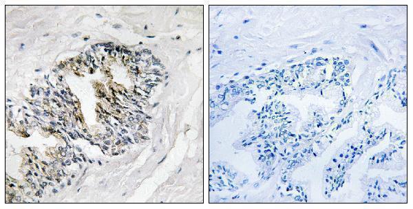
A05244-1 has a real IHC image from paraffin-embedded human prostate carcinoma tissue (catalog image caption); IHC, IF and ICC are listed for human, mouse and rat (catalog applications and reactivity).
A05244-1 will render with an IHC image of paraffin-embedded human prostate carcinoma tissue and a peptide-blocked comparison (catalog image caption). IF and ICC are listed applications, with human, mouse and rat reactivity, but no IF image is supplied (catalog applications, reactivity and image payload).
Which to pick: For tissue IHC, choose A05244-1: its image shows staining in a paraffin-embedded human prostate carcinoma section; the fixative is unreported (catalog image caption). For IF/ICC or work across human, mouse and rat, A05244-1 is the listed polyclonal option on the strength of its application and reactivity listings; no IF image is supplied (catalog dilution record, applications, reactivity and image payload).