This website uses cookies to ensure you get the best experience on our website.


- Table of Contents
Plan RPP30 paraffin IHC around nuclear staining that is most abundant in a subset of immune cells (HPA tissue IHC). Use bone marrow hematopoietic cells or spleen red-pulp cells as positive controls (HPA tissue IHC), and titrate the catalog antibody at 1:100–1:200 (datasheet).

Expected localisation, validated protocols, controls and antibodies — the at-a-glance facts below, then the full design guide.
| Expected localisation | Nuclear staining in tissue sections (HPA tissue IHC) | |
| Staining pattern | Nuclear staining, strongest in a subset of immune cells (HPA tissue IHC) | |
| Antigen retrieval | Tris-EDTA pH 9.0 HIER, 95–98 °C, 20 min (rule: nuclear antigen) | |
| Positive control | Bone marrow+4 more · see all | |
| Negative control | Adipose tissue+4 more · see all |
| Fixation | Keep fixation consistent across paraffin sections. (standard IHC practice; not target-specific) | |
| Caveat | Marrow and spleen peroxidase may mimic positive chromogen (HPA tissue IHC; standard IHC practice) | |
| Regulation | Specific expression regulator unreported (UniProt) | |
| Isoform / epitope | 2 isoforms; epitope impact unresolved (UniProt) |
The catalog antibody’s IHC-P protocol and published RPP30 staining workflows in gastric tissue (PMC9343801) and mouse growth plates (PMC5527100) provide starting points for paraffin sections.
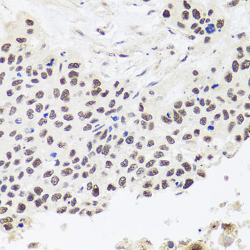
| Sample | Paraffin-embedded human lung cancer tissue; fixative not specified (datasheet A07553) |
| Fixation | Image fixative and duration unreported (datasheet A07553); verify before use. |
| Sectioning | 4–5 µm sections on charged slides (standard) |
| Deparaffinisation | Xylene, graded ethanol series to water (standard) |
| Antigen retrieval | Heat-induced epitope retrieval in Tris-EDTA buffer, pH 9.0, 20 min at 95–98 °C (standard rule: nuclear antigen) |
| Peroxidase block | 3% H2O2, 10 min, room temperature (standard) |
| Blocking | 10% normal serum of the secondary host, 30 min, room temperature (standard) |
| Primary antibody | Rabbit anti-RPP30, 1:100-1:200 (datasheet A07553) |
| Primary incubation | Overnight at 4 °C (standard) |
| Detection | HRP-polymer secondary, DAB chromogen 5–10 min (standard) |
| Counterstain | Hematoxylin, blue, dehydrate and mount (standard) |
| Expected result | RPP30-positive staining in hematopoietic cells of bone marrow (HPA tissue IHC: High). HPA tissue profile: Nuclear expression in several tissues, most abundant in a subset of immune cells. No signal in the no-primary control. |
RPP30 is a nuclear protein associated with RNase P and RNase MRP (UniProt P78346); it has no transmembrane segment (UniProt P78346 topology). In paraffin-section IHC, expect nuclear staining across several tissues, strongest in subsets of immune cells (HPA tissue IHC). HPA rates the tissue staining Approved, with medium agreement with RNA data and external verification pending (HPA tissue IHC).
| Distinct nuclear stain in bone marrow hematopoietic cells or spleen red-pulp cells (HPA tissue IHC). | This matches the reported high signal in those cell populations (HPA tissue IHC). Assess nuclei within the named population, since HPA's levels describe cells within a tissue rather than every cell on the section (HPA tissue IHC). |
| Stain is predominantly cytoplasmic, membranous or extracellular, with little nuclear signal. | That conflicts with the nuclear tissue profile (HPA tissue IHC) and nuclear location (UniProt P78346). Consider artefact or nonspecific detection; a minor cytosolic signal alone is less decisive because HPA also reports cytosol in ICC-IF (HPA subcellular). |
| Strong stain appears in adipocytes or cardiomyocytes, while the expected nuclei remain unstained. | HPA reports RPP30 as not detected in those specific cell types (HPA tissue IHC). Consider cross-reactivity or endogenous detection activity (general IHC practice); compare a known-positive section and an appropriate detection control before assigning target expression. |
| Broad, hazy chromogen covers cells and surrounding tissue without clear nuclear boundaries. | This does not resolve the expected nuclear pattern (HPA tissue IHC; UniProt P78346). Background from blocking, antibody concentration or detection chemistry can obscure localisation (general IHC practice); a darker field alone is insufficient evidence of RPP30. |
| No nuclear stain is visible in bone marrow hematopoietic cells on a candidate positive section. | HPA reports high staining in those cells (HPA tissue IHC), so review the run before interpreting a negative specimen. The absent signal does not identify which step failed; section quality, retrieval and detection require separate checks (general IHC practice). |
| Which cells provide an IHC reference? (HPA tissue IHC) | Bone marrow hematopoietic cells, spleen red-pulp cells and testis Leydig cells are reported High; adrenal and breast glandular cells are Medium (HPA tissue IHC). Compare the named cell population, since neighbouring cells need not share its score (HPA tissue IHC). |
| How firm is the tissue-pattern evidence? (HPA tissue IHC) | HPA marks the tissue result Approved, yet reports medium staining-to-RNA consistency and pending external verification (HPA tissue IHC). The listed HPA037578 antibody is IHC Approved; the supplied record does not mark it Enhanced (HPA antibodies). |
| Can IF/ICC help interpret compartment? (HPA subcellular) | Yes. HPA approves nucleoplasm and nucleoli as main ICC-IF locations, with cytosol and microtubule ends as additional locations (HPA subcellular). Use that as a localisation cross-check; the IHC tissue profile remains nuclear (HPA tissue IHC). |
| Do isoforms or processing specify a different IHC pattern? (UniProt P78346) | UniProt lists 2 isoforms and a chain spanning residues 2–268, with no signal peptide or propeptide (UniProt P78346). No antibody epitope or isoform-specific staining evidence is supplied, so these annotations cannot predict separate staining patterns. |
| Is RPP30 fixation sensitivity established? (supplied UniProt and HPA records) | No target-specific fixation effect is reported in the supplied records (UniProt P78346; HPA tissue IHC). Use the catalog antibody's validated IHC-P conditions as the starting point; assess retrieval performance with controls (general IHC practice). |
| Situation | Likely cause | Next action |
|---|---|---|
| Known-positive nuclei are blank. | A failed staining step or unsuitable section is possible (general IHC practice); high bone marrow hematopoietic staining is reported (HPA tissue IHC). | Check the catalog antibody's IHC-P conditions, retrieval, reagent activity and positive-section integrity one step at a time (general IHC practice). |
| Chromogen fills cytoplasm but leaves nuclei pale. | The pattern conflicts with nuclear tissue staining (HPA tissue IHC); cytosol is only an additional ICC-IF location (HPA subcellular). | Review morphology and counterstain, then compare an HPA-reported positive cell population and detection controls (HPA tissue IHC; general IHC practice). |
| Adipocytes, cardiomyocytes or liver cholangiocytes stain strongly. | These named cell types are reported Not detected (HPA tissue IHC); nonspecific binding or endogenous detection activity is possible (general IHC practice). | Confirm cell identity and examine a no-primary detection control before interpreting the stain as RPP30 (general IHC practice). |
| The whole section has a uniform brown haze. | Nuclear localisation is obscured (HPA tissue IHC); excessive antibody or detection background may contribute (general IHC practice). | Review blocking, washing and the catalog antibody's IHC-P dilution guidance; compare the no-primary control (general IHC practice). |
| A presumed negative tissue has scattered nuclear-positive cells. | HPA's Not detected calls apply to specified cell types, not necessarily every cell in that tissue (HPA tissue IHC). | Identify the stained cells before scoring. Compare their compartment and intensity with an HPA-reported positive population (HPA tissue IHC; general IHC practice). |
| Replicate sections yield inconsistent nuclear intensity. | Section or run variation is possible (general IHC practice); HPA reports only medium agreement between staining and RNA data (HPA tissue IHC). | Run comparable positive sections together, hold detection conditions constant and score the same cell population (general IHC practice). |


Comprehensive Human Protein Atlas IHC scoring per tissue (reliability: Approved — Medium consistency between antibody staining and RNA expression data. Pending external verification.). Rows are taken directly from the HPA tissue chart — click any row's HPA link to view the source.
| Tissue | Cell type | Level | Evidence | Source |
|---|---|---|---|---|
| Bone marrow | Hematopoietic cells | High | Protein (IHC) | HPA → |
| Spleen | Cells in red pulp | High | Protein (IHC) | HPA → |
| Testis | Leydig cells | High | Protein (IHC) | HPA → |
| Adrenal gland | Glandular cells | Medium | Protein (IHC) | HPA → |
| Breast | Glandular cells | Medium | Protein (IHC) | HPA → |
| Tissue | Cell type | Level | Evidence | Source |
|---|---|---|---|---|
| Adipose tissue | Adipocytes | Not detected | Protein (IHC) | HPA → |
| Duodenum | Glandular cells | Not detected | Protein (IHC) | HPA → |
| Fallopian tube | Glandular cells | Not detected | Protein (IHC) | HPA → |
| Heart muscle | Cardiomyocytes | Not detected | Protein (IHC) | HPA → |
| Liver | Cholangiocytes | Not detected | Protein (IHC) | HPA → |
Troubleshoot RPP30 staining in paraffin sections by checking retrieval, nuclear localisation, controls, and cell-specific scoring before interpreting chromogenic signal.
A07553 has a real paraffin-section IHC image from human lung cancer (catalog image caption) and lists IF/ICC use and Human, Mouse, and Rat reactivity (catalog applications and reactivity).
A07553 will render with an IHC image of paraffin-embedded human lung cancer stained at 1:100 after microwave retrieval in 10 mM PBS, pH 7.2 (catalog image caption). The catalog lists A07553 for IHC and IF/ICC in Human, Mouse, and Rat; it provides no IF image (catalog applications, reactivity, and image records).
Which to pick: For paraffin-section IHC, choose A07553 because its own image caption documents that preparation and staining conditions (catalog image caption); the fixative is unreported (catalog image caption). For IF/ICC or work across Human, Mouse, and Rat, A07553 is the listed option (catalog applications and reactivity), with IF/ICC at 1:50–1:200 (catalog dilution); its clone designation is unreported (catalog clone field).