RPRD1B / Regulation of nuclear pre-mRNA domain-containing protein 1B · IHC design guide

Design Immunohistochemistry for RPRD1B

Plan paraffin section IHC for RPRD1B using its nuclear tissue pattern (HPA tissue IHC) and a catalog antibody tested at 2–5 μg/mL (datasheet A07903-1). Compare staining across tissues with the reported medium consistency between antibody staining and RNA expression in mind (HPA tissue IHC).

Evidence assembled Oct 2026 · For research use; verify linked source records and product datasheet before use
Immunohistochemistry protocol sheet for RPRD1B (IHC for RPRD1B): expected localisation Nuclear staining in most tissues (HPA tissue IHC), antibody A07903-1, validated IHC image, and IHC protocol steps
Printable RPRD1B IHC protocol sheet — expected localisation Nuclear staining in most tissues (HPA tissue IHC), antibody A07903-1, controls and protocol steps. Open the full RPRD1B IHC guide →

RPRD1B Immunohistochemistry Experimental Design Guide

Expected localisation, validated protocols, controls and antibodies — the at-a-glance facts below, then the full design guide.

Must know before staining
Expected localisation Nuclear staining in most tissues (HPA tissue IHC)
Staining pattern Nuclear staining in most tissues, including glandular cells (HPA tissue IHC)
Antigen retrieval EDTA pH 8.0 HIER, heat-mediated (datasheet A07903-1)
Positive control ⓘ Adrenal gland+4 more · see all
Negative control ⓘ Adipose tissue
Important caveats
Reasons your staining may differ from the expected pattern.
Fixation Keep fixation consistent across sections. (standard IHC practice; not target-specific)
Caveat Staining and RNA expression have medium consistency (HPA tissue IHC)
Regulation Higher protein levels in tumors than adjacent tissue (UniProt)
Isoform / epitope No isoforms reported; mature chain spans residues 2–326 (UniProt)
Section 1

Recommended RPRD1B IHC & IF Protocols

Compare the catalog antibody’s IHC-P protocol (datasheet A07903-1) with the published kidney tissue protocol (PMC12767236).

Recommended immunohistochemistry (IHC-P) protocol parameters
SampleParaffin-embedded human esophageal squamous carcinoma tissue; fixative not specified (datasheet A07903-1)
FixationImage fixative and duration unreported (datasheet A07903-1); verify before use.
Sectioning4–5 µm sections on charged slides (standard)
DeparaffinisationXylene, graded ethanol series to water (standard)
Antigen retrievalHeat retrieval: EDTA pH 8.0 (datasheet A07903-1); 20 min, 95–100 °C (standard)
Peroxidase block3% H2O2, 10 min, room temperature (standard)
Blocking10% goat serum (datasheet A07903-1)
Primary antibodyRabbit anti-RPRD1B, 2-5 μg/ml (datasheet A07903-1)
Primary incubationOvernight at 4 °C (datasheet A07903-1)
DetectionHRP-conjugated secondary, DAB chromogen (datasheet A07903-1)
CounterstainHematoxylin, blue, dehydrate and mount (standard)
Expected resultRPRD1B-positive staining in glandular cells of adrenal gland (HPA tissue IHC: High). HPA tissue profile: Nuclear expression in most tissues. No signal in the no-primary control.
💡Decision noteStart with heat-mediated EDTA pH 8.0 retrieval (datasheet A07903-1); the kidney article reports retrieval but does not specify its conditions (PMC12767236).
Section 2

What Is the Expected RPRD1B Staining Pattern?

RPRD1B should appear predominantly in nuclei in paraffin-section IHC (UniProt Q9NQG5: nucleus; HPA tissue IHC: nuclear expression in most tissues). High staining is reported in several cell populations, including adrenal and breast glandular cells, bronchial respiratory epithelial cells, and bone-marrow hematopoietic cells (HPA tissue IHC: High). The protein has no transmembrane segment (UniProt Q9NQG5 topology). HPA rates the tissue staining profile Enhanced, with medium consistency between staining and RNA expression (HPA tissue IHC).

What am I looking at on my slide?
Distinct nuclear staining in breast glandular cells or bronchial respiratory epithelial cells.This fits reported high staining in those cell types (HPA tissue IHC: High). Assess the relevant cells, since an overall tissue impression can hide which population carries the signal (general IHC practice).
Predominantly membranous or cytoplasmic staining, with little nuclear signal.This conflicts with the expected nuclear pattern (UniProt Q9NQG5: nucleus; HPA tissue IHC: nuclear expression). Check localization against a known-positive section before interpreting that staining as RPRD1B (general IHC practice).
Strong staining in adipocytes while expected positive cells remain unstained.HPA reports RPRD1B as not detected in adipocytes (HPA tissue IHC: adipocytes, Not detected). Cross-reactivity or endogenous detection activity is possible; compare with appropriate reagent controls (general IHC practice).
Diffuse color across nuclei, cytoplasm, and tissue spaces.That distribution is less convincing than cell-associated nuclear staining (HPA tissue IHC: nuclear expression in most tissues). Background from detection or insufficient blocking is possible; inspect a control lacking primary antibody (general IHC practice).
No staining in a section expected to contain high-staining cells.First confirm that the sampled cell population matches an HPA high-staining entry, such as bone-marrow hematopoietic cells (HPA tissue IHC: High). If it does, review controls and the IHC detection workflow (general IHC practice).
💡Expected RPRD1B appearanceCall a result positive when the relevant cells show clear nuclear staining, potentially strong in HPA high-staining populations; isolated membranous, diffuse, or adipocyte staining warrants caution (HPA tissue IHC: nuclear profile, High populations, adipocytes Not detected; UniProt Q9NQG5 topology).
How each factor affects the staining
Cell compartment and topologyNuclear localization is supported by both sources, while HPA ICC-IF narrows the supported location to nucleoplasm (UniProt Q9NQG5: nucleus, no transmembrane segment; HPA subcellular: nucleoplasm). Score nuclear signal in IHC; a membrane-only pattern lacks support from these records.
Cell population within a tissueColon endothelial cells and duodenal glandular cells are high-staining examples (HPA tissue IHC: High). Identify the stained cell population before comparing tissues; HPA does not report the same staining level for every cell in an organ (HPA tissue IHC: cell-specific entries).
Lower and absent reference patternsHPA lists low staining in hepatocytes, hippocampal glial cells, and cerebellar molecular-layer cells, and no detected staining in adipocytes (HPA tissue IHC: Low; Not detected). Weak signal in a listed low population alone is a limited positive control.
RNA and protein interpretationLiver-enhanced RNA does not imply high hepatocyte IHC: HPA lists liver-enhanced RNA but low hepatocyte staining (HPA tissue IHC: RNA tissue enhanced in liver; hepatocytes Low). Use the protein staining entry when predicting the slide appearance.
Antibody evidence and IF/ICC Q&AQ: Can IF/ICC help check localization? A: HPA supports nucleoplasmic ICC-IF for HPA072645 and lists CACO-2 and Rh30 images (HPA antibodies: ICC Supported; HPA subcellular). Its IHC rating is Enhanced, as are HPA066290 and HPA070590 (HPA antibodies). These ratings do not establish that every assay condition will work.
Why is my staining missing, weak or wrong?
SituationLikely causeNext action
High-staining reference cells show no nuclear signal.The section may lack the intended cell population, or a workflow step may have failed (HPA tissue IHC: cell-specific High entries; general IHC practice).Verify cell identity, then inspect the positive control and the antibody, retrieval, and detection steps using the chosen assay instructions (general IHC practice). No RPRD1B-specific retrieval requirement is supplied.
Signal is mainly outside nuclei.The pattern disagrees with nuclear and nucleoplasmic localization (UniProt Q9NQG5: nucleus; HPA subcellular: nucleoplasm). Nonspecific signal or a detection artifact is possible (general IHC practice).Compare with a known-positive section and a control lacking primary antibody; judge whether the cell-associated nuclear pattern is reproducible (general IHC practice).
Adipocytes stain strongly.That finding conflicts with the HPA adipocyte entry (HPA tissue IHC: Not detected). Cross-reactivity or endogenous detection activity may explain unexpected color (general IHC practice).Check primary-omission and detection controls, including controls for endogenous activity appropriate to the chromogen system (general IHC practice). Do not use adipocyte staining alone as positive evidence.
Color is diffuse across the section.Diffuse signal obscures the reported nuclear pattern (HPA tissue IHC: nuclear expression in most tissues). Background from blocking, detection reagents, or washing is possible (general IHC practice).Compare with a control lacking primary antibody, then review blocking, washing, and detection conditions according to the assay instructions (general IHC practice).
Hepatocytes stain only weakly despite liver-enhanced RNA.These observations can coexist: the RNA category is tissue enhanced, while hepatocyte protein staining is low (HPA tissue IHC: liver RNA; hepatocytes Low).Use an HPA high-staining cell population for a stronger IHC reference and interpret hepatocytes against their reported low level (HPA tissue IHC: High entries; hepatocytes Low).
Different antibodies produce different staining patterns.An Enhanced IHC rating supports validation for each listed antibody, but does not prove identical results in every section or assay setup (HPA antibodies: HPA066290, HPA070590, HPA072645 IHC Enhanced).Compare nuclear localization and cell-specific staining with the HPA profile, then review each antibody's assay conditions and controls (HPA tissue IHC: nuclear, cell-specific profile; general IHC practice).

Sample controls for RPRD1B IHC & IF

🧪Run breast first: glandular cells are rated High (HPA: breast glandular cells, High). Use adipose tissue as the negative tissue (HPA: adipocytes, Not detected); adipocytes within the breast section, if present, should show no specific nuclear staining and can provide an internal negative comparison (HPA: adipocytes, Not detected).
Positive control tissue: Adrenal gland (Glandular cells, HPA High)
Negative control tissue: Adipose tissue (HPA Not detected)
ICC-IF cell lines (HPA subcellular resource): HPA ICC-IF images show RPRD1B in CACO-2, Rh30, with annotated localisation: Nucleoplasm (supported) (HPA subcellular).
Technical controls: Include no-primary (secondary-only) and matched rabbit IgG isotype controls alongside a RPRD1B knockout biological negative (caption: rabbit anti-RPRD1B antibody; standard IHC controls). For chromogenic detection, quench endogenous peroxidase and check that any signal in adipocyte-rich areas is not background staining (caption: HRP/DAB detection; HPA: adipocytes, Not detected).
⚠️Feasibility: A target-specific fixation window or fixation effect is unreported, and the selected A07903-1 paraffin-section caption does not state the fixative (caption: paraffin section; fixative unreported). Heat retrieval with EDTA at pH 8.0 was used in that caption, but a retrieval dependency is unreported; the supplied evidence does not establish whether frozen sections or IF are easier (caption: EDTA retrieval, pH 8.0). In breast sections, score staining in glandular cell nuclei rather than empty adipocyte spaces (HPA: breast glandular cells, High; HPA: adipocytes, Not detected; UniProt: nucleus).

HPA tissue IHC evidence for RPRD1B

Comprehensive Human Protein Atlas IHC scoring per tissue (reliability: Enhanced — Medium consistency between antibody staining and RNA expression data.). Rows are taken directly from the HPA tissue chart — click any row's HPA link to view the source.

Positive expression · recommended positive controls

TissueCell typeLevelEvidenceSource
Adrenal gland Glandular cells High Protein (IHC) HPA →
Appendix Glandular cells High Protein (IHC) HPA →
Bone marrow Hematopoietic cells High Protein (IHC) HPA →
Breast Glandular cells High Protein (IHC) HPA →
Bronchus Respiratory epithelial cells High Protein (IHC) HPA →

Undetected expression · recommended negative controls

TissueCell typeLevelEvidenceSource
Adipose tissue Adipocytes Not detected Protein (IHC) HPA →
Section 3

Advanced RPRD1B IHC Tips

Troubleshoot RPRD1B staining by checking nuclear localisation, matched controls and processing conditions before comparing signal across paraffin sections.

What retrieval should I start with for weak RPRD1B staining?
Start with heat-mediated antigen retrieval in EDTA at pH 8.0 (datasheet A07903-1). The catalog antibody produced staining in a paraffin section of human esophageal squamous carcinoma using that retrieval, followed by 2 μg/ml primary antibody overnight at 4°C (datasheet A07903-1). If nuclear staining is weak, vary heating time within a practical 10–20 minute starting range while keeping the buffer constant, and inspect section integrity. Compare each condition on adjacent sections with the same detection and development times. If signal remains weak, test another retrieval condition as a documented fallback rather than assuming RPRD1B is absent.
How should I troubleshoot fixation-related loss of nuclear staining?
The selected paraffin-section caption does not state a fixative, so target-specific RPRD1B fixation sensitivity is unknown (datasheet A07903-1). Record the actual fixative and fixation duration for each specimen, then compare similarly processed sections before attributing differences to biology. As general IHC practice, excessive fixation can reduce accessible epitopes and incomplete fixation can impair morphology; assess nuclear detail and tissue preservation alongside staining. Keep retrieval at EDTA pH 8.0 while investigating the fixation history, because that is the documented starting condition for this antibody (datasheet A07903-1). Include a concurrently processed positive control to distinguish a processing failure from a low-expressing sample.
Where should convincing RPRD1B staining appear in a tissue section?
Prioritise nuclear staining: RPRD1B is assigned to the nucleus (UniProt Q9NQG5), with supported nucleoplasmic localisation (HPA subcellular). HPA reports nuclear expression in most tissues, including high staining in adrenal gland glandular cells and bone marrow hematopoietic cells (HPA tissue IHC). Use a counterstain to judge whether chromogen lies within intact nuclei, and score the relevant cell population separately from neighbouring cells. Predominantly diffuse cytoplasmic or extracellular colour warrants checks of detection background, tissue preservation and antibody concentration before interpretation. The absence of a transmembrane segment gives no reason to expect a membrane outline (UniProt Q9NQG5 topology).
Could epitope accessibility explain discordant RPRD1B staining?
The supplied record lists 0 alternative isoforms, so an isoform-specific explanation for discordant staining is unsupported (UniProt Q9NQG5). It places a CID domain at residues 2–133 and lists phosphorylation at residues 132, 134, 161 and 166 (UniProt Q9NQG5). The antibody epitope is not specified in the supplied caption, so those features cannot establish whether retrieval or phosphorylation changes its binding (datasheet A07903-1). Compare adjacent sections across a controlled retrieval series, keeping primary concentration and chromogen development fixed. If results remain discordant, use an antibody with a disclosed, distinct epitope as an orthogonal check, without treating agreement alone as proof of specificity.
How can I assess RPRD1B by multiplex IF after IHC?
Treat IF as a separately optimised application; the supplied catalog example establishes paraffin-section chromogenic IHC conditions, not IF conditions (datasheet A07903-1). Pair RPRD1B with a marker identifying the cell population being evaluated, such as a glandular epithelial marker when examining glandular cells that HPA reports as positive (HPA tissue IHC). Choose a fluorophore channel with low tissue autofluorescence and include single-label and no-primary controls to assess bleed-through and background. For an intracellular nuclear target, permeabilise sufficiently to admit antibody through the plasma and nuclear membranes, then verify signal against a nuclear counterstain (UniProt Q9NQG5; HPA subcellular). Titrate permeabilisation and primary antibody independently, since the IHC concentration does not establish an IF dilution (datasheet A07903-1).
What causes widespread brown staining around RPRD1B-positive nuclei?
First separate nuclear signal from staining in stroma, vessels, damaged tissue and section edges; supported RPRD1B localisation is nucleoplasmic (HPA subcellular). The selected example used 10% goat serum block, 2 μg/ml primary antibody overnight at 4°C, and an HRP/DAB detection system (datasheet A07903-1). For HRP IHC, apply an appropriate peroxidase block and examine a no-primary control to identify endogenous enzyme or detection-system colour; these are general workflow checks. If background persists, titrate primary antibody below the documented concentration and shorten DAB development while retaining a positive control. Compare treated and untreated sections together so a cleaner slide is not mistaken for improved antigen detection.
How should I quantify RPRD1B across differently cellular specimens? ⚠ ANSWER MARKED FOR VERIFICATION
Define the cell population and nuclear positivity threshold before reading slides, consistent with the reported nucleoplasmic localisation (HPA subcellular). For intact tissue compartments, report the percentage of positive nuclei and, when intensity is reproducible, an H-score calculated from the percentages at intensity scores 0–3. Normalise cell-based scores to the number of evaluable nuclei in that compartment, rather than total tissue area; for spatial comparisons, report positive-cell density per mm² of evaluable tissue. Exclude folds, necrosis and poorly preserved regions by the same rules across specimens. Maintain matched retrieval, antibody concentration and DAB development, using the documented 2 μg/ml concentration as an IHC starting point (datasheet A07903-1).
How do I distinguish genuine RPRD1B positivity from artefact?
A credible result shows staining within intact nuclei in an appropriate cell population, consistent with nucleoplasmic localisation and nuclear expression across most tissues (HPA subcellular; HPA tissue IHC). High signal in adrenal gland glandular cells or bone marrow hematopoietic cells and undetected signal in adipocytes provide tissue-context checks, not absolute specificity tests (HPA tissue IHC). Question staining confined to section edges, necrotic regions, extracellular material or an unexpected membrane pattern; the record reports no transmembrane segment (UniProt Q9NQG5 topology). Use a no-primary control and peroxidase block to investigate endogenous colour in HRP/DAB sections. HPA calls its tissue-IHC reliability Enhanced while describing medium agreement with RNA, so interpret discordant specimens with matched controls (HPA tissue IHC).
Boster reagents

Best RPRD1B / Regulation of nuclear pre-mRNA domain-containing protein 1B IHC Antibodies

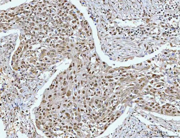
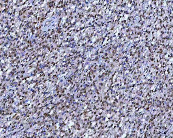
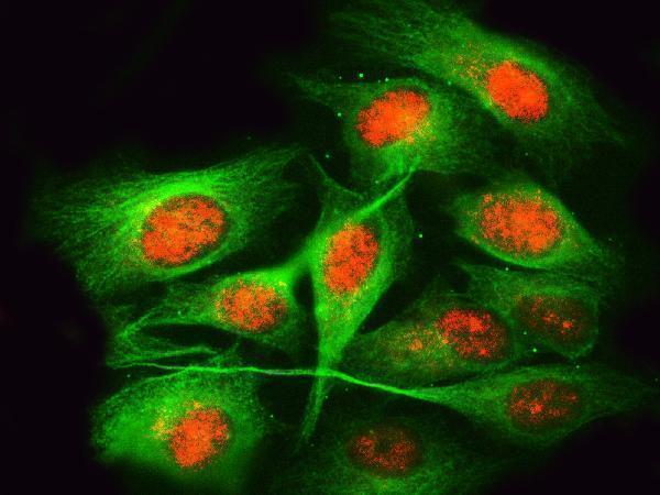
A07903-1 has real IHC data from human paraffin-embedded tissue and IF data from U2OS cells and human paraffin-embedded tissue (catalog IHC/IF captions).

Real IHC data IHC analysis of RPRD1B using anti-RPRD1B antibody (A07903-1). RPRD1B was detected in a paraffin-embedded section of human esophageal squamous carcinoma tissue. Heat mediated antigen retrieval was performed in EDTA buffer (pH 8.0, epitope retrieval solution). The tissue section was blocked with 10% goat serum. The tissue section was then incubated with 2 μg/ml rabbit anti-RPRD1B Antibody (A07903-1) overnight at 4°C. Peroxidase Conjugated Goat Anti-rabbit IgG was used as secondary antibody and incubated for 30 minutes at 37°C. The tissue section was developed using HRP Conjugated Rabbit IgG Super Vision Assay Kit (Catalog # SV0002) with DAB as the chromogen.
Anti-RPRD1B Antibody ®
Cat # A07903-1

A07903-1 is shown by chromogenic IHC in human paraffin-embedded esophageal squamous carcinoma, B cell lymphoma, and rectal cancer sections (catalog IHC captions). Its IF images show U2OS cells and human paraffin-embedded esophageal squamous cell carcinoma tissue (catalog IF captions).

Which to pick: Choose A07903-1 for human paraffin-section IHC: its image caption documents EDTA retrieval at pH 8.0, 2 μg/ml primary antibody, and peroxidase/DAB detection; the fixative is unreported (catalog IHC caption). The same rabbit antibody is listed for IF/ICC at 5 μg/ml, with IF images from U2OS cells and human paraffin-embedded tissue (catalog applications; catalog IF captions). Monkey reactivity is listed, but the supplied IHC and IF images show human tissue or U2OS cells (catalog reactivity; catalog IHC/IF captions).

Each figure is that product's own IHC / IF validation image from its datasheet.

References

  1. UniProt Consortium. UniProt entry Q9NQG5 (RPR1B_HUMAN, Regulation of nuclear pre-mRNA domain-containing protein 1B).
  2. Human Protein Atlas. RPRD1B tissue IHC expression (reliability: Enhanced).
  3. Human Protein Atlas. RPRD1B subcellular location (ICC-IF): Localized to the nucleoplasm..
  4. Human Protein Atlas. RPRD1B antibody validation summary (3 antibodies).
  5. Comprehensive pan-cancer analysis of RPRD1B as a promising diagnostic and prognostic biomarker. Discover oncology 2026 — PMC12873028.
  6. CREPT/RPRD1B associates with Aurora B to regulate Cyclin B1 expression for accelerating the G2/M transition in gastric cancer. Cell death & disease 2018 — PMC6281615.
  7. Long non-coding RNA NEAT1 mediated RPRD1B stability facilitates fatty acid metabolism and lymph node metastasis via c-Jun/c-Fos/SREBP1 axis in gastric cancer. Journal of experimental & clinical cancer research : CR 2022 — PMC9520879.
  8. Identification and Verification of Biomarkers Related to Polyamine Metabolism in Diabetic Nephropathy. Journal of diabetes research 2025 — PMC12767236.
  9. PubMed PMID:22264791 — UniProt-cited evidence.
  10. PubMed PMID:14702039 — UniProt-cited evidence.
  11. PubMed PMID:11780052 — UniProt-cited evidence.