This website uses cookies to ensure you get the best experience on our website.


- Table of Contents
Plan RPS11 paraffin IHC around cytoplasmic staining in several tissues (HPA tissue IHC). Start within the catalog antibody’s 1:100–1:300 IHC range (datasheet A07622S11), and interpret staining cautiously because HPA rates its tissue evidence uncertain (HPA tissue IHC).

Expected localisation, validated protocols, controls and antibodies — the at-a-glance facts below, then the full design guide.
| Expected localisation | Cytoplasmic in tissue (HPA tissue IHC); nucleolar location annotated (UniProt) | |
| Staining pattern | Cytoplasmic staining in several tissues, including glandular cells (HPA tissue IHC) | |
| Antigen retrieval | Tris-EDTA pH 9.0 HIER, heat-mediated (datasheet A07622S11) | |
| Positive control | Small intestine+4 more · see all | |
| Negative control | Adipose tissue+4 more · see all |
| Fixation | Keep fixation consistent across paraffin sections. (standard IHC practice; not target-specific) | |
| Caveat | Tissue staining has very low agreement with RNA expression (HPA tissue IHC) | |
| Regulation | No specific regulator annotated (UniProt) | |
| Isoform / epitope | No isoforms annotated; chain 2–158; no TM segment (UniProt) |
The catalog antibody’s IHC-P protocol (datasheet: A07622S11) is followed by published RPS11 staining protocols for glioblastoma, cardiac tissue and hepatocellular carcinoma (PMC4624638; PMC11922711; PMC7210141).
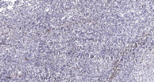
| Sample | Paraffin-embedded human tonsil tissue; fixative not specified (datasheet A07622S11) |
| Fixation | Image fixative and duration unreported (datasheet A07622S11); verify before use. |
| Sectioning | 4–5 µm sections on charged slides (standard) |
| Deparaffinisation | Xylene, graded ethanol series to water (standard) |
| Antigen retrieval | Heat retrieval: Tris-EDTA pH 9.0 (datasheet A07622S11); 20 min, 95–100 °C (standard) |
| Peroxidase block | 3% H2O2, 10 min, room temperature (standard) |
| Blocking | 10% normal serum of the secondary host, 30 min, room temperature (standard) |
| Primary antibody | Rabbit anti-RPS11, 1:100-1:300 (datasheet A07622S11) |
| Primary incubation | Overnight at 4 °C (standard) |
| Detection | HRP-polymer secondary, DAB chromogen 5–10 min (standard) |
| Counterstain | Hematoxylin, blue, dehydrate and mount (standard) |
| Expected result | RPS11-positive staining in glandular cells of small intestine (HPA tissue IHC: High). HPA tissue profile: Cytoplasmic expression in several tissues. No signal in the no-primary control. |
RPS11 is a small ribosomal subunit protein found in the cytoplasm and nucleolus, with no transmembrane segment (UniProt P62280). In paraffin-section IHC, expect chiefly cytoplasmic staining in positive epithelial cells, especially small-intestinal glandular cells (HPA tissue IHC). Interpret that expectation cautiously: HPA rates its tissue IHC pattern Uncertain because antibody staining has very low consistency with RNA expression data and awaits external verification (HPA tissue IHC).
| Distinct cytoplasmic staining in small-intestinal glandular cells. | This fits the strongest listed tissue IHC observation: High staining in those cells (HPA tissue IHC). Compare cell boundaries and counterstained nuclei before scoring. A matching pattern supports the stain, but does not resolve HPA's Uncertain tissue-IHC reliability rating (HPA tissue IHC). |
| Predominantly nuclear staining, or staining confined to tissue structures outside cells. | Predominantly nuclear chromogen departs from HPA's cytoplasmic tissue-IHC profile (HPA tissue IHC). RPS11 also has a nucleolar location in UniProt and approved ICC-IF data (UniProt P62280; HPA subcellular), so a discrete nucleolar signal alone is not proof of an artefact. Check morphology and controls. |
| Strong staining in a cell population listed as Not detected, such as adipocytes. | That conflicts with the reported adipocyte result (HPA tissue IHC). Consider antibody cross-reactivity or endogenous detection activity, using an appropriate no-primary control to investigate the latter (general IHC practice). Because the tissue profile is rated Uncertain, do not treat one discordant cell population as definitive proof of either cause (HPA tissue IHC). |
| Uniform haze across cells and surrounding tissue, without clear cytoplasmic borders. | A nonlocalized deposit is difficult to reconcile with the reported cytoplasmic cellular pattern (HPA tissue IHC). Review background in the no-primary control, blocking and detection steps, and wash conditions (general IHC practice). Avoid assigning an RPS11-positive score to diffuse haze alone. |
| No visible signal in small-intestinal glandular cells. | This misses the listed High IHC observation (HPA tissue IHC), but the Uncertain reliability rating prevents treating it as a guaranteed positive control (HPA tissue IHC). Check section integrity, antigen-retrieval and detection execution, and the IHC-validated antibody's documented conditions (general IHC practice). |
| Compartment and assay | UniProt lists cytoplasm and nucleolus (UniProt P62280). HPA tissue IHC reports cytoplasmic expression, while approved ICC-IF localization includes cytosol, nucleoli and endoplasmic reticulum (HPA tissue IHC; HPA subcellular). Use the ICC-IF observations to interpret localization cautiously; they do not establish a paraffin-IHC protocol. |
| Tissue and cell selection | HPA reports High staining in small-intestinal glandular cells, Medium staining in several listed epithelial or tubular populations, and Not detected staining in several other populations (HPA tissue IHC). Its low RNA tissue specificity and Uncertain IHC reliability limit how firmly any one listed tissue can serve as a control (HPA tissue IHC). |
| Antibody validation | HPA lists HPA049719 tissue IHC as Uncertain and its ICC as Approved; HPA032020 has Approved ICC with no tissue-IHC status listed (HPA antibodies). An ICC approval cannot establish IHC performance. Review the IHC-validated antibody's own paraffin-section instructions before comparing stains (general IHC practice). |
| Protein architecture | RPS11 has no annotated transmembrane segment, signal peptide, propeptide, domain or isoform in this record (UniProt P62280). These annotations support interpreting it as an intracellular target; they provide no basis for predicting a membrane staining pattern or a special processing-related IHC signal. |
| Antigen retrieval | Use the IHC-validated antibody's documented retrieval conditions and record them when comparing sections (general IHC practice). The supplied UniProt and HPA records do not report RPS11-specific fixation sensitivity or establish which retrieval condition exposes its epitope. |
| Situation | Likely cause | Next action |
|---|---|---|
| Small-intestinal glandular cells show no signal. | A failed staining step is possible; the listed High result is an uncertain tissue-IHC reference (HPA tissue IHC). | Verify tissue morphology, retrieval and detection execution, then compare with the IHC-validated antibody's documented paraffin-section conditions (general IHC practice). |
| Chromogen is predominantly nuclear. | This differs from the cytoplasmic tissue-IHC profile; nucleolar localization is separately supported by UniProt and ICC-IF (HPA tissue IHC; UniProt P62280; HPA subcellular). | Distinguish discrete nucleoli from broad nuclear deposit using the counterstain and matched controls; avoid calling broad nuclear staining expected (general IHC practice). |
| Adipocytes stain strongly. | Adipocytes are listed as Not detected, though the overall tissue-IHC profile is Uncertain (HPA tissue IHC). Cross-reactivity or endogenous detection activity is possible (general IHC practice). | Review a no-primary control and inspect whether the deposit follows cells or background; confirm the result with an independent validation approach if it matters to the conclusion (general IHC practice). |
| The whole section has diffuse brown haze. | Diffuse deposit obscures the cellular cytoplasmic pattern described by HPA (HPA tissue IHC); nonspecific detection or inadequate washing is possible (general IHC practice). | Compare the no-primary control, then review blocking, wash steps and detection exposure before scoring cellular signal (general IHC practice). |
| An ICC-IF image appears to show more compartments than the IHC section. | Approved ICC-IF localization includes nucleoli and endoplasmic reticulum, while the tissue-IHC summary emphasizes cytoplasm (HPA subcellular; HPA tissue IHC). | Interpret each assay against its own reported pattern; assess paraffin-IHC staining with tissue morphology and IHC controls (general IHC practice). |
| Staining differs between listed positive tissues. | HPA reports High small-intestinal glandular staining but Medium staining in several other listed cell populations, with Uncertain overall tissue-IHC reliability (HPA tissue IHC). | Score the named cell population and compartment in each tissue separately; document controls and avoid treating every tissue difference as a technical failure (general IHC practice). |


Comprehensive Human Protein Atlas IHC scoring per tissue (reliability: Uncertain — Very low consistency between antibody staining and RNA expression data. Pending external verification.). Rows are taken directly from the HPA tissue chart — click any row's HPA link to view the source.
| Tissue | Cell type | Level | Evidence | Source |
|---|---|---|---|---|
| Small intestine | Glandular cells | High | Protein (IHC) | HPA → |
| Appendix | Glandular cells | Medium | Protein (IHC) | HPA → |
| Cervix | Squamous epithelial cells | Medium | Protein (IHC) | HPA → |
| Duodenum | Glandular cells | Medium | Protein (IHC) | HPA → |
| Epididymis | Glandular cells | Medium | Protein (IHC) | HPA → |
| Tissue | Cell type | Level | Evidence | Source |
|---|---|---|---|---|
| Adipose tissue | Adipocytes | Not detected | Protein (IHC) | HPA → |
| Adrenal gland | Glandular cells | Not detected | Protein (IHC) | HPA → |
| Bone marrow | Hematopoietic cells | Not detected | Protein (IHC) | HPA → |
| Bronchus | Respiratory epithelial cells | Not detected | Protein (IHC) | HPA → |
| Caudate | Glial cells | Not detected | Protein (IHC) | HPA → |
Troubleshoot chromogenic RPS11 IHC in paraffin sections using the catalog antibody’s tissue image, protein annotations, and cautiously interpreted expression data.
A07622S11 has real IHC data from paraffin-embedded human tonsil (catalog image caption). IF and Human, Mouse, and Rat reactivity are listed, but no IF figure is supplied (catalog applications, reactivity, image alts).
A07622S11 will render with an IHC figure from paraffin-embedded human tonsil (catalog image caption). The catalog lists IHC and IF applications and Human, Mouse, and Rat reactivity; it supplies no IF image (catalog applications, reactivity, image alts).
Which to pick: Choose A07622S11 for paraffin-section IHC: its own caption reports human tonsil, antibody at 1:200 overnight at 4°C, Tris-EDTA retrieval at pH 9.0, and secondary antibody at 1:200 for 45 minutes at room temperature; the fixative is unreported (catalog image caption). For IF, A07622S11 lists 1:50, but has no IF figure or ICC listing (catalog dilution, applications, image alts). It is the only listed cross-species option, with Human, Mouse, and Rat reactivity; its host is rabbit and clonality is unreported (catalog reactivity, host, clone).