This website uses cookies to ensure you get the best experience on our website.


- Table of Contents
Plan chromogenic RPS12 IHC in paraffin sections using the catalog antibody's documented workflow (datasheet A01040-3). Expect a general cytoplasmic pattern, with high staining in selected epithelial and lymph node germinal center cells; interpret intensity in light of medium consistency with RNA expression (HPA tissue IHC).

Expected localisation, validated protocols, controls and antibodies — the at-a-glance facts below, then the full design guide.
| Expected localisation | General cytoplasmic tissue staining (HPA tissue IHC) | |
| Staining pattern | Cytoplasmic staining in epithelial and germinal center cells (HPA tissue IHC) | |
| Antigen retrieval | EDTA pH 8.0 HIER, heat-mediated (datasheet A01040-3) | |
| Positive control | Bronchus+4 more · see all | |
| Negative control | Adipose tissue+4 more · see all |
| Fixation | Keep fixation consistent across sections (standard IHC practice; not target-specific) | |
| Caveat | Staining has medium consistency with RNA expression (HPA tissue IHC) | |
| Regulation | Low tissue specificity; no regulator reported (HPA tissue IHC; UniProt) | |
| Isoform / epitope | No annotated isoforms; mature chain spans residues 2–132 (UniProt) |
The catalog antibody protocol uses EDTA pH 8.0 retrieval (datasheet: A01040-3); one published RPS12 IHC protocol uses citrate retrieval (PMC12291262).
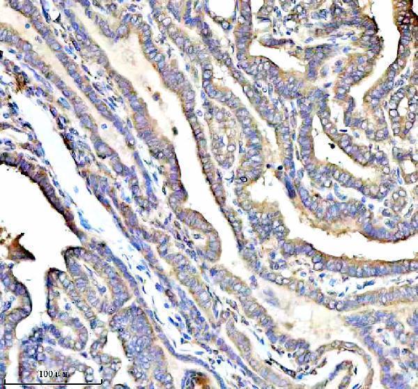
| Sample | Paraffin-embedded human thyroid cancer tissue; fixative not specified (datasheet A01040-3) |
| Fixation | Image fixative and duration unreported (datasheet A01040-3); verify before use. |
| Sectioning | 4–5 µm sections on charged slides (standard) |
| Deparaffinisation | Xylene, graded ethanol series to water (standard) |
| Antigen retrieval | Heat retrieval: EDTA pH 8.0 (datasheet A01040-3); 20 min, 95–100 °C (standard) |
| Peroxidase block | 3% H2O2, 10 min, room temperature (standard) |
| Blocking | 10% goat serum (datasheet A01040-3) |
| Primary antibody | Rabbit anti-RPS12, 1:50 recommended; image 1:100 (datasheet A01040-3) |
| Primary incubation | Overnight at 4 °C (datasheet A01040-3) |
| Detection | HRP-conjugated secondary, DAB chromogen (datasheet A01040-3) |
| Counterstain | Hematoxylin, blue, dehydrate and mount (standard) |
| Expected result | RPS12-positive staining in basal cells of bronchus (HPA tissue IHC: High). HPA tissue profile: General cytoplasmic expression. No signal in the no-primary control. |
RPS12 is a cytoplasmic protein that also localizes to the nucleus and nucleolus (UniProt P25398). It has no transmembrane segment (UniProt P25398 topology). In paraffin tissue sections, expect mainly cytoplasmic staining, including strong signal in bronchial basal cells and several epithelial and glandular cell populations (HPA tissue IHC: High). The tissue IHC profile is Approved, with medium consistency between antibody staining and RNA expression (HPA tissue IHC: reliability).
| Strong cytoplasmic staining in bronchial basal cells or duodenal glandular cells, with recognizable cell boundaries. | This fits the reported high staining in those cell populations (HPA tissue IHC: High in bronchial basal cells and duodenal glandular cells). Judge intensity within the named cells; neighboring cells need not stain equally. Cytoplasmic predominance also fits the reported tissue profile (HPA tissue IHC: general cytoplasmic expression). |
| Nuclear or nucleolar signal accompanies cytoplasmic staining; alternatively, staining is predominantly membranous or extracellular. | Nuclear and nucleolar localization is plausible (UniProt P25398: nucleus, nucleolus). A predominantly membranous or extracellular pattern is inconsistent with the reported cytoplasmic localization and absence of a transmembrane segment (HPA tissue IHC: cytoplasmic; UniProt P25398 topology). Check controls before calling it an artefact. |
| Strong staining appears in adipocytes or cardiomyocytes while a reported high staining population is weak. | Those specific cell populations are reported as not detected, whereas several epithelial and glandular populations stain strongly (HPA tissue IHC: adipocytes and cardiomyocytes Not detected; bronchial basal cells High). Consider cross-reactivity or endogenous detection activity; the mismatch alone does not identify its cause. |
| Brown deposit spreads across stroma, empty spaces, or many unrelated cell types, obscuring cytoplasm. | A diffuse deposit is hard to reconcile with cell-resolved cytoplasmic staining (HPA tissue IHC: general cytoplasmic expression). Review a no-primary control for detection background, then assess blocking and washes (general chromogenic IHC practice). Do not score deposit outside identifiable cells as RPS12-positive. |
| No signal is visible in bronchial basal cells or cervical squamous epithelial cells. | Both are reported high staining populations, so absence of signal calls for a technical check (HPA tissue IHC: High in bronchial basal cells and cervical squamous epithelial cells). The HPA profile has medium consistency with RNA data; a single blank section does not establish true absence (HPA tissue IHC: reliability). |
| Choice of comparison cells | Use named cells within a tissue: duodenal glandular cells are reported High, while adipocytes are Not detected (HPA tissue IHC). A negative call for cholangiocytes must not be extended to every cell in liver (HPA tissue IHC: cholangiocytes Not detected). |
| Strength of tissue evidence | The tissue IHC profile is Approved but has medium consistency with RNA expression (HPA tissue IHC: reliability). Interpret an unexpected result alongside cell identity, localization, and controls; do not treat an HPA staining category as an absolute threshold for every specimen. |
| Antibody validation | HPA006124 is listed as IHC Approved; HPA006365 is listed as ICC Supported, with no IHC status supplied for it (HPA antibodies). The supplied validation record does not label the tissue result Enhanced (HPA antibodies). Keep IHC and ICC evidence separate. |
| Protein localization and processing | Cytoplasmic and nucleolar signal can fit RPS12 biology (UniProt P25398: cytoplasm, nucleus, nucleolus). No transmembrane segment or signal peptide is annotated, and the listed chain spans residues 2–132 (UniProt P25398 topology and processing); a surface-only pattern warrants scrutiny. |
| IF/ICC Q&A: What pattern is expected? | Mainly cytosolic fluorescence, with additional Golgi apparatus and vesicle localization, is reported in ICC-IF (HPA subcellular: Cytosol Supported; Golgi apparatus and Vesicles Approved). This is localization context for IF/ICC; the IHC interpretation here relies on the tissue IHC record. |
| Situation | Likely cause | Next action |
|---|---|---|
| The reported high staining cells are blank. | The run may have lost signal during retrieval, antibody incubation, or detection (general chromogenic IHC practice); these data do not establish RPS12-specific fixation or retrieval sensitivity. | Check a reported high staining cell population on the same run, such as bronchial basal cells (HPA tissue IHC: High). Review the catalog antibody's IHC-P instructions and run controls before changing a condition (general IHC practice). |
| Nearly every area of the section turns brown. | Endogenous detection activity or nonspecific reagent binding can produce widespread color (general chromogenic IHC practice). This appearance cannot be assigned to RPS12 from color alone. | Compare a no-primary control and inspect the detection reagent and blocking steps (general chromogenic IHC practice). Reassess whether identifiable high staining cells retain stronger cytoplasmic signal (HPA tissue IHC: general cytoplasmic expression). |
| Strong color appears in adipocytes or cardiomyocytes. | Those named cells are reported Not detected (HPA tissue IHC). Cross-reactivity, background, or specimen variation could explain a discrepancy; the HPA tissue profile has medium consistency (HPA tissue IHC: reliability). | Confirm cell identity, compare a no-primary control, and inspect a reported high staining population in the same run (general IHC practice; HPA tissue IHC: High in bronchial basal cells). |
| Only cell edges or extracellular material stain. | That distribution conflicts with the reported cytoplasmic tissue profile and no-transmembrane topology (HPA tissue IHC: general cytoplasmic expression; UniProt P25398 topology). Nonspecific deposit is possible. | Review the counterstain and cell boundaries, compare the no-primary control, and reassess primary antibody concentration using its IHC-P instructions (general chromogenic IHC practice). |
| The section has widespread haze but little cell-resolved staining. | Excess primary reagent, incomplete blocking, or inadequate washing can obscure localization (general chromogenic IHC practice). Haze does not match the reported general cytoplasmic pattern (HPA tissue IHC). | Check blocking, washes, and detection exposure against the validated IHC-P workflow (general IHC practice). Score only interpretable cells after background is controlled. |
| Nuclear staining appears stronger than expected. | RPS12 can localize to the nucleus and nucleolus (UniProt P25398), while the reported tissue IHC profile is generally cytoplasmic (HPA tissue IHC). Nuclear signal alone is therefore insufficient to label a section false positive. | Examine whether cytoplasmic staining remains in reported high staining cells and whether the no-primary control is clean (HPA tissue IHC: High; general IHC practice). Record nuclear and cytoplasmic compartments separately. |
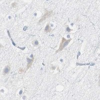


Comprehensive Human Protein Atlas IHC scoring per tissue (reliability: Approved — Medium consistency between antibody staining and RNA expression data.). Rows are taken directly from the HPA tissue chart — click any row's HPA link to view the source.
| Tissue | Cell type | Level | Evidence | Source |
|---|---|---|---|---|
| Bronchus | Basal cells | High | Protein (IHC) | HPA → |
| Cervix | Squamous epithelial cells | High | Protein (IHC) | HPA → |
| Duodenum | Glandular cells | High | Protein (IHC) | HPA → |
| Endometrium | Glandular cells | High | Protein (IHC) | HPA → |
| Epididymis | Glandular cells | High | Protein (IHC) | HPA → |
| Tissue | Cell type | Level | Evidence | Source |
|---|---|---|---|---|
| Adipose tissue | Adipocytes | Not detected | Protein (IHC) | HPA → |
| Heart muscle | Cardiomyocytes | Not detected | Protein (IHC) | HPA → |
| Liver | Cholangiocytes | Not detected | Protein (IHC) | HPA → |
| Skeletal muscle | Myocytes | Not detected | Protein (IHC) | HPA → |
| Smooth muscle | Smooth muscle cells | Not detected | Protein (IHC) | HPA → |
These questions address RPS12 staining in paraffin sections with chromogenic IHC; the immunofluorescence entry covers the separate IF/ICC application.
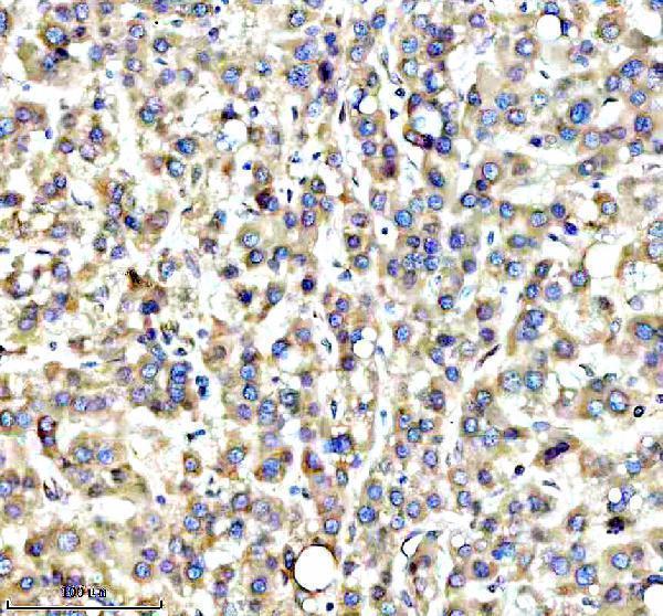
The catalog antibody has real IHC images from human paraffin sections of thyroid, liver, bladder, and clear cell renal cancers (catalog: A01040-3 IHC captions). No IF figure is supplied (catalog: if_image_alts).
A01040-3 is listed for IHC and Human, Mouse, and Rat reactivity (catalog: A01040-3 applications/reactivity). Its images document human paraffin-section IHC in thyroid, liver, bladder, and clear cell renal cancers (catalog: A01040-3 IHC captions).
Which to pick: Choose A01040-3 for tissue IHC: its own captions document paraffin sections, EDTA retrieval at pH 8.0, and 1:100 primary antibody incubation; the fixative is unreported (catalog: A01040-3 IHC captions). For IF/ICC, A01040-3 lists a 1:50–400 dilution range, but supplies no IF image (catalog: A01040-3 dilution_raw/if_image_alts). For cross-species studies, A01040-3 lists Human, Mouse, and Rat reactivity, while its IHC images show human tissue only (catalog: A01040-3 reactivity/IHC captions).