This website uses cookies to ensure you get the best experience on our website.


- Table of Contents
Plan RPS20 chromogenic IHC on paraffin sections using the reported general cytoplasmic tissue pattern (HPA tissue IHC). Compare high-staining glandular or neuronal cells with skeletal myocytes, where staining was not detected (HPA tissue IHC).

Expected localisation, validated protocols, controls and antibodies — the at-a-glance facts below, then the full design guide.
| Expected localisation | General cytoplasmic staining (HPA tissue IHC) | |
| Staining pattern | Cytoplasmic staining in glandular and neuronal cells (HPA tissue IHC) | |
| Antigen retrieval | Citrate pH 6.0 HIER, 95–98 °C, 20 min (rule: cytoplasmic / membrane antigen) | |
| Positive control | Appendix+4 more · see all | |
| Negative control | Skeletal muscle |
| Fixation | Keep fixation consistent across paraffin sections. (standard IHC practice; not target-specific) | |
| Caveat | Skeletal myocytes are not detected (HPA tissue IHC) | |
| Regulation | Specific expression regulators unannotated (UniProt) | |
| Isoform / epitope | 2 isoforms; epitope effect undetermined (UniProt) |
The catalog antibody’s IHC-P protocol (datasheet) is accompanied by published RPS20 IHC protocols for renal carcinoma and glioblastoma tissue (PMC9773015; PMC4624638).
| Sample | Paraffin-embedded human kidney tissue; fixative not specified (datasheet M06634) |
| Fixation | Image fixative and duration unreported (datasheet M06634); verify before use. |
| Sectioning | 4–5 µm sections on charged slides (standard) |
| Deparaffinisation | Xylene, graded ethanol series to water (standard) |
| Antigen retrieval | Heat-induced epitope retrieval in citrate buffer, pH 6.0, 20 min at 95–98 °C (standard rule: cytoplasmic / membrane antigen) |
| Peroxidase block | 3% H2O2, 10 min, room temperature (standard) |
| Blocking | 10% normal serum of the secondary host, 30 min, room temperature (standard) |
| Primary antibody | Rabbit monoclonal (clone FEH-18) anti-RPS20, 1:50 (datasheet M06634) |
| Primary incubation | Overnight at 4 °C (standard) |
| Detection | HRP-polymer secondary, DAB chromogen 5–10 min (standard) |
| Counterstain | Hematoxylin, blue, dehydrate and mount (standard) |
| Expected result | RPS20-positive staining in glandular cells of appendix (HPA tissue IHC: High). HPA tissue profile: General cytoplasmic expression. No signal in the no-primary control. |
RPS20 is a cytoplasmic component of the 40S ribosomal subunit and has no transmembrane segment (UniProt P60866). Expect cytoplasmic staining in many cell types, including appendix glandular cells, bone marrow hematopoietic cells and cortical neurons, where HPA reports High staining (HPA tissue IHC). HPA describes the overall pattern as general cytoplasmic expression and rates its IHC staining Approved, supported by external characterization data (HPA tissue IHC).
| Cytoplasmic staining appears in appendix glandular cells or bone marrow hematopoietic cells. | This matches the expected compartment (UniProt P60866; HPA tissue IHC). HPA reports High staining in both cell populations, making them useful positive references for slide review (HPA tissue IHC). |
| Signal is mainly nuclear or outlines the plasma membrane, with little cytoplasmic staining. | That distribution conflicts with UniProt's cytoplasmic assignment and HPA's general cytoplasmic IHC profile (UniProt P60866; HPA tissue IHC). Treat it as a possible staining artefact and assess controls before calling it RPS20. |
| Skeletal muscle myocytes stain strongly while expected positive cells stain weakly. | HPA reports RPS20 as Not detected in skeletal muscle myocytes (HPA tissue IHC). Unexpected signal may reflect cross-reactivity or endogenous chromogenic activity (general IHC practice); check controls before assigning target expression. |
| Brown signal spreads across tissue, empty spaces or slide background without clear cell boundaries. | A broad deposit cannot establish the reported cytoplasmic cell pattern (HPA tissue IHC). In chromogenic IHC, nonspecific binding, endogenous detection activity or excess detection reagent can cause background (general IHC practice). |
| No stain appears in appendix glandular cells or bone marrow hematopoietic cells. | Both are reported High by HPA, so an absent signal calls the staining run into question (HPA tissue IHC). Review tissue preservation, antibody application, retrieval and detection controls as general IHC checks; this record does not establish RPS20 fixation sensitivity. |
| Tissue and cell choice | HPA reports High staining in appendix and breast glandular cells, bone marrow hematopoietic cells and cortical neurons, but Not detected in skeletal muscle myocytes (HPA tissue IHC). Compare named cell populations rather than whole-section darkness. |
| Cytoplasmic target location | RPS20 is a cytoplasmic 40S subunit protein without a transmembrane segment (UniProt P60866). Interpret signal by its cellular compartment; this annotation does not predict a membrane pattern. |
| IHC evidence strength | HPA rates its tissue IHC profile Approved because external characterization supports the staining (HPA tissue IHC). That rating describes the reported antibody evidence; it does not validate every antibody or establish a fixation effect. |
| Isoforms and processing | UniProt lists 2 isoforms and annotates a chain spanning residues 2–119, with no signal peptide or propeptide (UniProt P60866). The payload gives no epitope mapping, so isoform recognition cannot be assigned. |
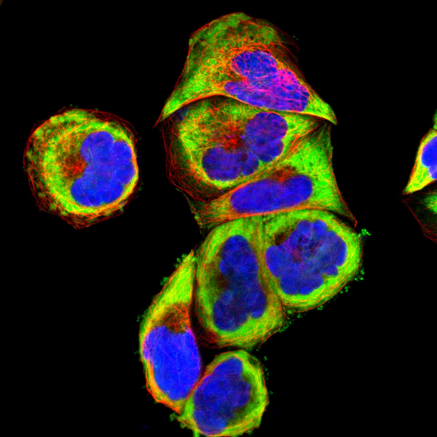
| IF/ICC cross-check — where should signal appear? | HPA reports supported cytosol and endoplasmic-reticulum localization in ICC-IF images from A-431, U-251MG and U2OS (HPA subcellular ICC-IF). Use this as compartment context; evaluate IHC with the tissue IHC profile. |
| Situation | Likely cause | Next action |
|---|---|---|
| An expected positive tissue is blank. | Appendix glandular cells and bone marrow hematopoietic cells are reported High, so a blank result may indicate an assay failure (HPA tissue IHC). | Confirm the intended cells are present, then review antibody application, retrieval and detection controls (general IHC practice). No RPS20-specific retrieval or fixation sensitivity is supplied. |
| Staining appears mainly in nuclei. | A dominant nuclear pattern differs from the reported cytoplasmic localization (UniProt P60866; HPA tissue IHC). | Check the positive reference and a control lacking primary antibody; reassess localization before scoring (general IHC practice). |
| Skeletal muscle myocytes appear positive. | HPA reports these myocytes as Not detected; cross-reactivity or endogenous detection activity is possible (HPA tissue IHC; general IHC practice). | Compare a control lacking primary antibody and confirm signal belongs to myocytes rather than neighboring cells (general IHC practice). |
| Diffuse brown background obscures cells. | Nonspecific antibody binding or endogenous detection activity can obscure the cytoplasmic pattern (general IHC practice; HPA tissue IHC). | Review blocking, washing and detection controls, then adjust routine IHC conditions using the antibody's instructions (general IHC practice). |
| Only some tissue compartments stain. | HPA reports different levels across named cells, including High in cortical neurons and Low in kidney tubule cells (HPA tissue IHC). | Identify and score the relevant cell population against its HPA entry; avoid treating all cells in an organ as one reference (HPA tissue IHC). |
| The slide shows a sharp membrane rim. | RPS20 has no annotated transmembrane segment, and HPA describes general cytoplasmic IHC expression (UniProt P60866; HPA tissue IHC). | Inspect controls and verify that cytoplasmic staining accompanies the rim before calling it target signal (general IHC practice). |



Comprehensive Human Protein Atlas IHC scoring per tissue (reliability: Approved — External characterization data supports antibody staining.). Rows are taken directly from the HPA tissue chart — click any row's HPA link to view the source.
| Tissue | Cell type | Level | Evidence | Source |
|---|---|---|---|---|
| Appendix | Glandular cells | High | Protein (IHC) | HPA → |
| Bone marrow | Hematopoietic cells | High | Protein (IHC) | HPA → |
| Breast | Glandular cells | High | Protein (IHC) | HPA → |
| Caudate | Neuronal cells | High | Protein (IHC) | HPA → |
| Cerebellum | Cells in molecular layer | High | Protein (IHC) | HPA → |
| Tissue | Cell type | Level | Evidence | Source |
|---|---|---|---|---|
| Skeletal muscle | Myocytes | Not detected | Protein (IHC) | HPA → |
Troubleshoot RPS20 staining in paraffin sections by checking retrieval, compartment, cell identity and controls before comparing signal intensity.
The catalog lists M06634 for IHC and IF/ICC in human, mouse, and rat (catalog applications/reactivity). Its image shows IHC on paraffin-embedded human kidney; no IF image is supplied (catalog image captions).
M06634 renders with an IHC image of paraffin-embedded human kidney (catalog IHC image caption). It lists IHC and IF/ICC applications and human, mouse, and rat reactivity; no IF image is supplied (catalog applications/reactivity; catalog IF images).
Which to pick: Choose M06634 for paraffin-section tissue IHC based on its human kidney image; the fixative is unreported (catalog IHC image caption). For IF/ICC, M06634 lists both applications, though no IF image is supplied (catalog applications; catalog IF images). For human, mouse, or rat work, M06634 is the listed rabbit monoclonal option, clone FEH-18 (catalog reactivity; catalog host/clone).