This website uses cookies to ensure you get the best experience on our website.


- Table of Contents
Plan RPS21 paraffin-section IHC around its ubiquitous cytoplasmic staining pattern (HPA tissue IHC). Use breast glandular cells as a high-staining control (HPA tissue IHC) and start the IHC-validated antibody at 2–5 μg/ml (datasheet A10068-2).

Expected localisation, validated protocols, controls and antibodies — the at-a-glance facts below, then the full design guide.
| Expected localisation | Ubiquitous cytoplasmic tissue staining (HPA tissue IHC) | |
| Staining pattern | Cytoplasmic staining across diverse cell types (HPA tissue IHC) | |
| Antigen retrieval | EDTA pH 8.0 HIER, heat-mediated (datasheet A10068-2) | |
| Positive control | Adrenal gland+4 more · see all | |
| Negative control | Adipose tissue+2 more · see all |
| Fixation | Keep paraffin-section fixation consistent across samples. (standard IHC practice; not target-specific) | |
| Caveat | Adipocytes and myocytes may lack detectable staining (HPA tissue IHC) | |
| Regulation | No target-specific regulation annotated (UniProt) | |
| Isoform / epitope | Single 1–83 chain; no isoforms annotated (UniProt) |
The catalog antibody uses EDTA pH 8.0 heat retrieval (datasheet A10068-2). Two published RPS21 IHC studies provide prostate tissue staining conditions (PMC5634644; PMC6202544).
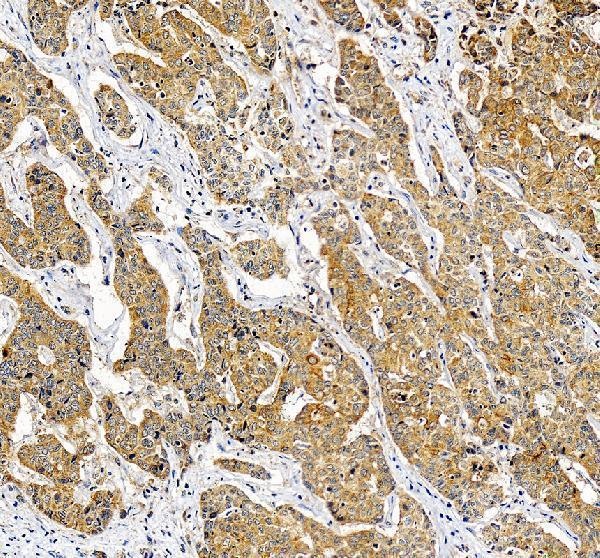
| Sample | Paraffin-embedded human liver cancer tissue; fixative not specified (datasheet A10068-2) |
| Fixation | Image fixative and duration unreported (datasheet A10068-2); verify before use. |
| Sectioning | 4–5 µm sections on charged slides (standard) |
| Deparaffinisation | Xylene, graded ethanol series to water (standard) |
| Antigen retrieval | Heat retrieval: EDTA pH 8.0 (datasheet A10068-2); 20 min, 95–100 °C (standard) |
| Peroxidase block | 3% H2O2, 10 min, room temperature (standard) |
| Blocking | 10% goat serum (datasheet A10068-2) |
| Primary antibody | Rabbit anti-RPS21, 2-5 μg/ml (datasheet A10068-2) |
| Primary incubation | Overnight at 4 °C (datasheet A10068-2) |
| Detection | HRP-conjugated secondary, DAB chromogen (datasheet A10068-2) |
| Counterstain | Hematoxylin, blue, dehydrate and mount (standard) |
| Expected result | RPS21-positive staining in glandular cells of adrenal gland (HPA tissue IHC: High). HPA tissue profile: Ubiquitous cytoplasmic expression. No signal in the no-primary control. |
RPS21 is a 40S ribosomal protein detected on cytosolic polysomes and in ribosomes associated with rough endoplasmic reticulum (UniProt P63220). Expect predominantly cytoplasmic staining in many cell types, including breast glandular cells and cortical neurons (HPA tissue IHC: High). HPA describes the tissue pattern as ubiquitous cytoplasmic expression, but rates its reliability Approved, with medium agreement between staining and RNA data and external verification pending (HPA tissue IHC). RPS21 has no transmembrane segment (UniProt P63220 topology).
| Cytoplasmic chromogenic signal is strong in breast glandular cells or cortical neurons (HPA tissue IHC: High). | This fits the reported compartment and known positive cell types (UniProt P63220; HPA tissue IHC). Compare staining within the tissue: HPA also reports High signal in lymph-node germinal center cells and cerebellar Purkinje cells (HPA tissue IHC). Intensity alone does not establish antibody specificity (HPA antibody validation: IHC Approved; external verification pending). |
| Signal is predominantly nuclear, or outlines the plasma membrane while cytoplasm is unstained. | That distribution conflicts with the reported cytosolic and rough-ER association and the HPA cytoplasmic IHC profile (UniProt P63220; HPA tissue IHC). Check morphology, counterstain, detection controls, and a known positive tissue before interpreting it as RPS21; a single unusual compartment cannot establish a new localization. |
| Adipocytes, skeletal myocytes, or smooth-muscle cells stain strongly. | HPA reports RPS21 as Not detected in those cell types (HPA tissue IHC). Strong staining there raises concern for cross-reactivity or endogenous chromogenic activity (general IHC practice). Compare a matched negative-reagent control and inspect whether the color follows tissue structures rather than cell cytoplasm; HPA's Not detected call does not prove absolute protein absence. |
| Brown color spreads across stroma, tissue edges, or empty areas without clear cellular boundaries. | A diffuse deposit is hard to reconcile with the reported cytoplasmic pattern (HPA tissue IHC). Uneven detection or insufficient washing can create background (general IHC practice). Judge the result against a matched negative-reagent control and a positive tissue section, then score only signal that can be assigned to intact cells. |
| Breast glandular cells or lymph-node germinal center cells show no convincing signal (HPA tissue IHC: High). | A blank known positive section makes a negative study section uninterpretable (general IHC practice). Verify tissue preservation, retrieval, primary antibody application, and chromogen performance with run controls. HPA's IHC Approved rating has medium staining–RNA consistency and pending external verification, so investigate the assay before treating absence as biology (HPA tissue IHC). |
| Compartment and epitope context | RPS21 is a 40S subunit component on cytosolic polysomes and rough-ER-associated ribosomes; it has no transmembrane segment (UniProt P63220). Expect cellular cytoplasmic staining, while routine chromogenic IHC may not resolve cytosol from ER-associated ribosomes (general IHC practice). |
| Tissue-dependent contrast | HPA reports High staining in breast glandular cells and lymph-node germinal center cells, Low staining in kidney tubule cells, and Not detected in adipocytes (HPA tissue IHC). Choose a reported High cell population as a run control and use the Low and Not detected calls as comparisons, not absolute expression limits. |
| Antibody evidence | The listed antibody HPA003371 is IHC Approved, while HPA describes medium consistency with RNA expression and pending external verification (HPA antibody validation; HPA tissue IHC). Treat the predicted pattern as a reference for evaluation, not proof that every colored cell is specific. |
| Maturation and isoform annotation | UniProt lists one chain spanning residues 1–83, no signal peptide or propeptide, and no annotated isoforms (UniProt P63220). The supplied record therefore gives no processing or alternate-isoform pattern to explain compartment shifts; these annotations do not specify retrieval conditions. |
| IF/ICC Q&A: where should signal appear? | HPA reports ER localization as approved and cytosol localization as supported in ICC-IF; its listed antibody has ICC Supported status (HPA subcellular; HPA antibody validation). Interpret IF/ICC localization on its dedicated guide page: those image-based compartment calls do not set a paraffin IHC protocol. |
| Situation | Likely cause | Next action |
|---|---|---|
| Known High tissue is blank while its morphology is intact (HPA tissue IHC: breast glandular cells High). | A failed staining step or weak detection is possible (general IHC practice); HPA staining levels do not establish RPS21 fixation sensitivity. | Check run controls, primary antibody addition, detection reagents, chromogen, and the locally validated retrieval workflow (general IHC practice). Repeat with a reported High cell population before scoring other sections negative (HPA tissue IHC). |
| All compartments and tissue structures appear brown, including areas without intact cells. | Diffuse background may arise from inadequate washing or excess detection signal (general IHC practice), rather than the HPA cytoplasmic pattern (HPA tissue IHC). | Inspect the negative-reagent control, wash and detection steps, and staining time; compare intact cells with adjacent acellular areas (general IHC practice). |
| Unexpected strong color appears in adipocytes or skeletal myocytes (HPA tissue IHC: Not detected). | Cross-reactivity or endogenous chromogenic activity is possible (general IHC practice). HPA's Not detected designation is an observation, not a universal absence claim (HPA tissue IHC). | Run a matched negative-reagent control, examine the intracellular distribution, and compare a reported High tissue in the same staining run (general IHC practice; HPA tissue IHC). |
| Only nuclei stain, with no corresponding cytoplasmic signal. | A nuclear-only result conflicts with cytosolic and rough-ER-associated localization (UniProt P63220) and HPA's cytoplasmic IHC profile (HPA tissue IHC). | Confirm nuclear boundaries with the counterstain, check control sections and detection artifacts, then repeat before assigning the nuclear color to RPS21 (general IHC practice). |
| Low-level cells look negative after the assay detects a reported High control. | Weak signal may fall below the scoring threshold; HPA reports Low staining in kidney tubule cells and prostate glandular cells (HPA tissue IHC). | Record cell-specific intensity and the assay's detection threshold; avoid converting a Low reference call into a required strong-positive control (HPA tissue IHC; general IHC practice). |
| Replicate sections give inconsistent cytoplasmic staining. | Variation in section handling or staining steps can affect chromogenic IHC (general IHC practice). HPA reports medium staining–RNA consistency and pending external verification, without target-specific fixation-effect data (HPA tissue IHC). | Compare matched sections and run controls, document retrieval and detection settings, and reproduce the result before drawing a biological conclusion (general IHC practice). |



Comprehensive Human Protein Atlas IHC scoring per tissue (reliability: Approved — Medium consistency between antibody staining and RNA expression data. Pending external verification.). Rows are taken directly from the HPA tissue chart — click any row's HPA link to view the source.
| Tissue | Cell type | Level | Evidence | Source |
|---|---|---|---|---|
| Adrenal gland | Glandular cells | High | Protein (IHC) | HPA → |
| Breast | Glandular cells | High | Protein (IHC) | HPA → |
| Caudate | Neuronal cells | High | Protein (IHC) | HPA → |
| Cerebellum | Purkinje cells | High | Protein (IHC) | HPA → |
| Cerebral cortex | Neuronal cells | High | Protein (IHC) | HPA → |
Troubleshoot RPS21 staining by checking retrieval, cytoplasmic localisation and cell type before comparing signal intensity across paraffin sections.
Two anti-RPS21 antibodies have human paraffin-section IHC images (catalog IHC captions). A10068-2 also has an IF image of HeLa cells (catalog IF caption); both list Human, Mouse and Rat reactivity (catalog applications).
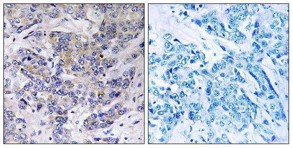
A10068-2 will render with IHC of a human liver cancer paraffin section and has IF data from HeLa cells (A10068-2 IHC and IF captions). A10068 will render with IHC of a human breast carcinoma paraffin section and lists IF/ICC among its applications, though no IF image is supplied (A10068 IHC caption; catalog applications and IF image list).
Which to pick: For tissue IHC, choose A10068-2 if its documented EDTA retrieval and 2 μg/ml overnight incubation fit your workflow (A10068-2 IHC caption); A10068 is another paraffin-section option, with peptide-blocked human breast carcinoma staining and a listed 1:100–1:300 IHC range (A10068 IHC caption; catalog dilution). For IF/ICC, choose A10068-2 because its HeLa-cell IF image documents staining at 5 μg/ml (A10068-2 IF caption); A10068 lists IF/ICC and is described as polyclonal, but has no IF image in this payload (A10068 catalog applications, dilution_raw and IF image list). Both list Human, Mouse and Rat reactivity, while the supplied IHC images show human samples only; neither IHC caption reports a fixative (catalog reactivity; A10068 and A10068-2 IHC captions).