This website uses cookies to ensure you get the best experience on our website.


- Table of Contents
Plan RPS24 paraffin-section IHC around the cytoplasmic staining observed across several tissues (HPA tissue IHC). This guide covers fixation, staining controls and interpretation in light of the reported mismatch between antibody staining and RNA expression (HPA tissue IHC).

Expected localisation, validated protocols, controls and antibodies — the at-a-glance facts below, then the full design guide.
| Expected localisation | Cytoplasm (HPA tissue IHC); nucleus and nucleolus (UniProt) | |
| Staining pattern | Cytoplasmic staining in several tissues (HPA tissue IHC) | |
| Antigen retrieval | Citrate pH 6.0 HIER, heat-mediated (datasheet A05730) | |
| Positive control | Breast+4 more · see all | |
| Negative control | Adipose tissue+4 more · see all |
| Fixation | Keep paraffin-section fixation consistent across samples (standard IHC practice; not target-specific) | |
| Caveat | Antibody staining has low consistency with RNA data (HPA tissue IHC) | |
| Regulation | Higher in tissues rich in proliferating cells (UniProt) | |
| Isoform / epitope | 4 isoforms; epitope coverage is unknown (UniProt) |
The catalog antibody’s IHC-P protocol (datasheet A05730) is accompanied by published RPS24 IHC protocols for liver cancer and prostate tissue (PMC9820840; PMC5634644).
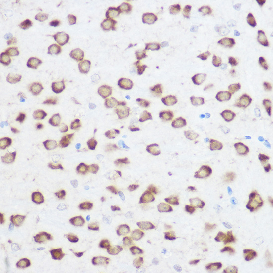
| Sample | Paraffin-embedded rat brain tissue; fixative not specified (datasheet A05730) |
| Fixation | Image fixative and duration unreported (datasheet A05730); verify before use. |
| Sectioning | 4–5 µm sections on charged slides (standard) |
| Deparaffinisation | Xylene, graded ethanol series to water (standard) |
| Antigen retrieval | Heat retrieval: Citrate pH 6.0 (datasheet A05730); 20 min, 95–100 °C (standard) |
| Peroxidase block | 3% H2O2, 10 min, room temperature (standard) |
| Blocking | 10% normal serum of the secondary host, 30 min, room temperature (standard) |
| Primary antibody | Rabbit anti-RPS24, 1:50-1:200 (datasheet A05730) |
| Primary incubation | Overnight at 4 °C (standard) |
| Detection | HRP-polymer secondary, DAB chromogen 5–10 min (standard) |
| Counterstain | Hematoxylin, blue, dehydrate and mount (standard) |
| Expected result | RPS24-positive staining in glandular cells of breast (HPA tissue IHC: Medium). HPA tissue profile: Cytoplasmic expression in several tissues. No signal in the no-primary control. |
RPS24 is a cytoplasmic ribosomal protein that is also reported in the nucleus and nucleolus (UniProt P62847: location and function). In paraffin-section IHC, expect mainly cytoplasmic staining in several tissues, including glandular cells in breast, duodenum, endometrium, and small intestine (HPA: tissue IHC). RPS24 has no transmembrane segment (UniProt P62847: topology). HPA rates its tissue IHC staining Approved but reports low consistency with RNA expression (HPA: tissue IHC reliability).
| Cytoplasmic staining in breast or duodenal glandular cells. | This matches HPA's Medium staining in those cell types and its broader cytoplasmic IHC profile (HPA: tissue IHC). Judge the pattern in the named cells rather than expecting every cell in the section to stain equally; HPA reports low tissue RNA specificity and low consistency between staining and RNA data (HPA: tissue IHC). |
| Predominantly nuclear, nucleolar, or sharply membrane-bound chromogen, with little cytoplasmic staining. | UniProt includes nuclear and nucleolar RPS24, so nuclear signal alone does not prove an artifact (UniProt P62847: location). A pattern dominated by these compartments conflicts with HPA's cytoplasmic tissue IHC profile; a membrane outline also lacks support from RPS24 topology (HPA: tissue IHC; UniProt P62847: topology). Recheck controls before assigning either pattern to RPS24. |
| Strong staining in a cell population that HPA reports as Not detected. | For example, HPA reports Not detected in adipocytes and heart cardiomyocytes (HPA: tissue IHC). Unexpected staining may reflect antibody cross-reactivity or endogenous detection activity, both general IHC possibilities. A Not detected HPA result describes that assay's observation; it does not establish that the cell population can never contain RPS24. |
| Diffuse color over cells and surrounding tissue, with no clear intracellular pattern. | This is difficult to score as RPS24 because the reported tissue IHC pattern is cytoplasmic (HPA: tissue IHC). In general IHC practice, nonspecific antibody binding, incomplete blocking, or residual detection-reagent activity can produce background. Compare a no-primary control and review the staining distribution before increasing the assigned score. |
| No signal in breast or pancreatic glandular cells selected as a positive comparator. | HPA reports Medium staining in breast glandular cells and pancreatic exocrine glandular cells (HPA: tissue IHC). An absent signal in the selected cells makes the run difficult to interpret; check section identity, primary-antibody use, retrieval conditions, and detection controls as general IHC troubleshooting steps. HPA's Approved rating does not guarantee staining in every preparation (HPA: tissue IHC reliability). |
| Compartment and application | HPA tissue IHC describes cytoplasmic expression, whereas HPA ICC-IF places signal in the endoplasmic reticulum and cytosol; UniProt also records nuclear and nucleolar localisation (HPA: tissue IHC and subcellular; UniProt P62847: location). Interpret paraffin-section chromogen against the tissue IHC pattern, since the ICC-IF result comes from a different application. |
| Choice of comparison tissue | HPA reports Medium staining in several glandular populations, Low staining in skin keratinocytes, and Not detected in adipocytes, bone-marrow hematopoietic cells, and heart cardiomyocytes (HPA: tissue IHC). These observed categories provide comparisons, with HPA's stated low staining-to-RNA consistency limiting predictions for an unlisted sample (HPA: tissue IHC reliability). |
| Antibody evidence | HPA003364 has Approved IHC status, while HPA073508 has no supplied IHC rating; both have Enhanced ICC status (HPA: antibody validation). ICC enhancement therefore cannot be treated as Enhanced IHC evidence. Use an antibody's IHC evidence when interpreting paraffin sections (HPA: antibody validation). |
| Isoforms and epitope coverage | UniProt lists four RPS24 isoforms and modified residues including N-acetylmethionine at position 1 and phosphothreonine at position 9 (UniProt P62847: isoforms and modified residues). The payload gives no antibody epitope, so isoform coverage or modification-dependent staining cannot be predicted; these annotations do not establish a retrieval or fixation effect. |
| Retrieval and detection conditions | Antigen retrieval and blocking are general paraffin-section IHC variables that can affect interpretability (general IHC practice). No supplied UniProt or HPA evidence establishes RPS24-specific fixation sensitivity or an optimal retrieval condition. Record the conditions used and interpret a failed run with its controls before attributing it to target biology. |
| Situation | Likely cause | Next action |
|---|---|---|
| The expected cytoplasmic signal is absent in a selected Medium-staining glandular comparator. | A failed staining step or unsuitable run conditions are possible general IHC explanations; HPA's Medium category is an observed tissue result, not a guarantee for each section (HPA: tissue IHC). | Confirm the comparator cells are present, review primary-antibody and detection steps, and assess the run's controls before calling the test sample negative (general IHC practice). |
| Chromogen appears mainly in nuclei or along cell borders. | A nucleus-dominant result diverges from HPA's cytoplasmic tissue pattern, though UniProt also lists nuclear and nucleolar RPS24; a membrane outline is unsupported by the recorded topology (HPA: tissue IHC; UniProt P62847: location and topology). | Compare compartments in the positive comparator and inspect a no-primary control; report a reproducible discordant pattern separately from the expected IHC result (general IHC practice). |
| Adipocytes or cardiomyocytes stain more strongly than the chosen positive comparator. | HPA reports Not detected in adipocytes and cardiomyocytes, making cross-reactivity or endogenous detection activity possibilities to investigate (HPA: tissue IHC; general IHC practice). | Examine a no-primary control for detection-related color, then compare the suspect cells and comparator under the same staining conditions (general IHC practice). |
| Diffuse background obscures glandular cell boundaries. | Nonspecific binding or residual detection activity can obscure intracellular staining in chromogenic IHC (general IHC practice). The resulting diffuse pattern cannot be matched confidently to HPA's cytoplasmic profile (HPA: tissue IHC). | Check the no-primary control and review blocking, washing, and detection steps; score only cells whose staining can be localized reliably (general IHC practice). |
| Bone marrow appears negative despite a biological expectation of RPS24 expression. | UniProt describes higher levels in tissues with many proliferating cells, including bone marrow, while HPA reports Not detected in bone-marrow hematopoietic cells by tissue IHC (UniProt P62847: tissue specificity; HPA: tissue IHC). | Record the source-specific discrepancy. Use an HPA Medium-staining tissue as the staining comparator and avoid treating bone marrow as a guaranteed positive IHC control (HPA: tissue IHC). |
| Why does an ICC-IF image emphasize endoplasmic reticulum or cytosol when tissue IHC looks cytoplasmic? | HPA reports enhanced endoplasmic-reticulum and cytosol localisation for ICC-IF, while its paraffin tissue IHC summary says cytoplasmic expression (HPA: subcellular and tissue IHC). | Interpret each result within its application; use the cytoplasmic tissue IHC profile for this slide and the separate IF/ICC guide for fluorescence interpretation (HPA: tissue IHC and subcellular). |
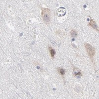


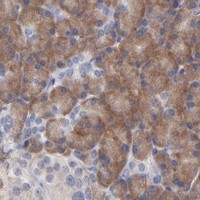


Comprehensive Human Protein Atlas IHC scoring per tissue (reliability: Approved — Low consistency between antibody staining and RNA expression data.). Rows are taken directly from the HPA tissue chart — click any row's HPA link to view the source.
| Tissue | Cell type | Level | Evidence | Source |
|---|---|---|---|---|
| Breast | Glandular cells | Medium | Protein (IHC) | HPA → |
| Cerebellum | Purkinje cells | Medium | Protein (IHC) | HPA → |
| Duodenum | Glandular cells | Medium | Protein (IHC) | HPA → |
| Endometrium | Glandular cells | Medium | Protein (IHC) | HPA → |
| Pancreas | Exocrine glandular cells | Medium | Protein (IHC) | HPA → |
| Tissue | Cell type | Level | Evidence | Source |
|---|---|---|---|---|
| Adipose tissue | Adipocytes | Not detected | Protein (IHC) | HPA → |
| Adrenal gland | Glandular cells | Not detected | Protein (IHC) | HPA → |
| Bone marrow | Hematopoietic cells | Not detected | Protein (IHC) | HPA → |
| Bronchus | Respiratory epithelial cells | Not detected | Protein (IHC) | HPA → |
| Epididymis | Glandular cells | Not detected | Protein (IHC) | HPA → |
Troubleshoot RPS24 staining by checking retrieval, compartment, cell type, and controls before comparing signal across sections.
A05730 has real IHC data from paraffin-embedded rat brain (catalog IHC image caption). Human, mouse, and rat reactivity is listed (catalog reactivity); no IF data are shown (catalog IF images).
A05730 is listed for IHC and has an image of paraffin-embedded rat brain stained at 1:200 after citrate retrieval (catalog applications; catalog IHC image caption). Human, mouse, and rat are listed as reactive species, while the image documents rat tissue only (catalog reactivity; catalog IHC image caption).
Which to pick: For tissue IHC, choose A05730: it is a rabbit polyclonal antibody listed for IHC at 1:50–1:200, with a paraffin-embedded rat brain example (catalog host and dilution; catalog IHC image caption). Its listed human, mouse, and rat reactivity makes it the cross-species candidate, though the supplied IHC image shows rat tissue only (catalog reactivity; catalog IHC image caption). No IF/ICC application or image is supplied for A05730, so an IF/ICC choice cannot be supported here; the IHC caption does not report the fixative (catalog applications; catalog IF images; catalog IHC image caption).