This website uses cookies to ensure you get the best experience on our website.


- Table of Contents
Plan chromogenic RPS26 IHC around cytoplasmic staining in several tissues (HPA tissue IHC). Use the catalog antibody’s paraffin-section protocol (datasheet A06454), and interpret tissue differences cautiously because HPA rates its staining reliability as uncertain (HPA tissue IHC).

Expected localisation, validated protocols, controls and antibodies — the at-a-glance facts below, then the full design guide.
| Expected localisation | Cytoplasmic staining in several tissues (HPA tissue IHC) | |
| Staining pattern | Cytoplasmic staining in glandular cells and cholangiocytes (HPA tissue IHC) | |
| Antigen retrieval | EDTA pH 8.0 HIER, heat-mediated (datasheet A06454) | |
| Positive control | Gallbladder+4 more · see all | |
| Negative control | Adipose tissue+4 more · see all |
| Fixation | Keep fixation consistent across sections (standard IHC practice; not target-specific); optimize as needed. | |
| Caveat | Tissue staining has low consistency with RNA expression (HPA tissue IHC) | |
| Regulation | Low tissue specificity; no regulator annotated (HPA tissue IHC; UniProt) | |
| Isoform / epitope | 0 isoforms annotated; chain spans residues 2–115 (UniProt) |
The catalog antibody IHC-P protocol (datasheet A06454) is paired with one published mouse embryo IHC protocol (PMC5333091).
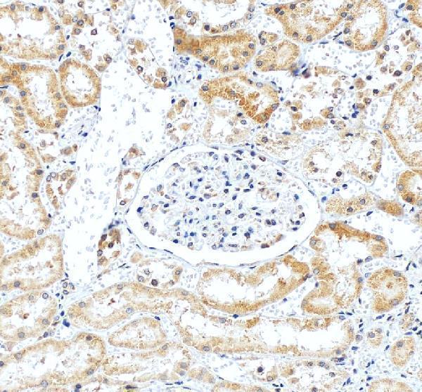
| Sample | Paraffin-embedded human kidney tissue; fixative not specified (datasheet A06454) |
| Fixation | Image fixative and duration unreported (datasheet A06454); verify before use. |
| Sectioning | 4–5 µm sections on charged slides (standard) |
| Deparaffinisation | Xylene, graded ethanol series to water (standard) |
| Antigen retrieval | Heat retrieval: EDTA pH 8.0 (datasheet A06454); 20 min, 95–100 °C (standard) |
| Peroxidase block | 3% H2O2, 10 min, room temperature (standard) |
| Blocking | 10% goat serum (datasheet A06454) |
| Primary antibody | Rabbit anti-RPS26, 1:50 recommended; image 2 μg/ml (datasheet A06454) |
| Primary incubation | Overnight at 4 °C (datasheet A06454) |
| Detection | HRP-conjugated secondary, DAB chromogen (datasheet A06454) |
| Counterstain | Hematoxylin, blue, dehydrate and mount (standard) |
| Expected result | RPS26-positive staining in glandular cells of gallbladder (HPA tissue IHC: High). HPA tissue profile: Cytoplasmic expression in several tissues. No signal in the no-primary control. |
RPS26 is a 40S ribosomal protein detected on cytosolic polysomes and in ribosomes associated with rough endoplasmic reticulum; it has no transmembrane segment (UniProt P62854). In paraffin-section IHC, expect cytoplasmic staining, including in gallbladder glandular cells, liver cholangiocytes and pancreatic exocrine glandular cells (HPA: High in each). Treat intensity comparisons cautiously: HPA rates tissue IHC reliability Uncertain because staining and RNA expression have low consistency (HPA: tissue IHC).
| Cytoplasmic staining in gallbladder glandular cells, cholangiocytes or pancreatic exocrine glandular cells. | This matches the reported High IHC staining in those cell types (HPA: tissue IHC). Cytoplasmic localisation also fits RPS26 on cytosolic polysomes and rough-ER-associated ribosomes (UniProt P62854). Judge the pattern by cell type as well as intensity because the tissue IHC assessment is Uncertain (HPA: tissue IHC). |
| Strong chromogenic signal confined to nuclei, with little cytoplasmic staining. | A nuclear-only IHC pattern does not match the reported cytoplasmic tissue profile (HPA: tissue IHC). Check morphology, counterstain and detection controls before interpreting it as RPS26 (general IHC practice). Nucleolar signal is an additional ICC-IF location, so that IF finding alone does not validate nuclear-only chromogenic IHC (HPA: subcellular). |
| Strong staining in adipocytes, cardiomyocytes or cells in kidney glomeruli. | Those cell types were Not detected in the supplied tissue IHC profile (HPA: tissue IHC). Unexpected staining could reflect cross-reactivity or endogenous detection activity (general IHC practice); inspect controls and tissue morphology. These HPA observations are comparison points, not proof that every specimen of those tissues lacks RPS26 (HPA: IHC reliability Uncertain). |
| Uniform colour over cells, stroma and empty spaces, without a recognisable cytoplasmic pattern. | Diffuse staining obscures the cell-specific cytoplasmic profile reported for RPS26 (HPA: tissue IHC). Assess background with a primary-omission control and review blocking, washing and detection chemistry (general IHC practice). Background intensity alone cannot be scored as target expression when cellular boundaries and localisation are unclear (general IHC practice). |
| No stain in gallbladder glandular cells, cholangiocytes or pancreatic exocrine glandular cells. | Absence in these reported High groups warrants a technical check before calling the sample negative (HPA: tissue IHC). Verify that the cells are present and assess the primary antibody and detection controls (general IHC practice). The HPA tissue pattern is Uncertain, so a single negative section cannot resolve expression on its own (HPA: tissue IHC). |
| Ribosomal location | RPS26 is part of the 40S subunit and occurs on cytosolic polysomes and rough-ER-associated ribosomes (UniProt P62854). Interpret an intracellular cytoplasmic pattern in light of this distribution; the record reports no transmembrane segment (UniProt P62854). |
| Strength of tissue evidence | HPA calls the IHC tissue profile Uncertain because antibody staining has low consistency with RNA expression; RNA shows low tissue specificity (HPA: tissue IHC). Use listed High and Not detected groups as observed comparators, rather than absolute expression rules (HPA: tissue IHC). |
| IF/ICC: where is signal expected? | Mainly endoplasmic reticulum and cytosol, with additional nucleolar localisation (HPA: subcellular ICC-IF). This is an IF localisation reference, not an IHC protocol: HPA043961 is ICC Approved but IHC Uncertain, while HPA055803 is ICC Approved with no IHC status listed (HPA: antibodies). |
| Situation | Likely cause | Next action |
|---|---|---|
| Expected cytoplasmic stain is absent in a reported High group (HPA: tissue IHC). | Missing target cells or an unsuccessful staining run are possible (general IHC practice). | Confirm cell identity and section quality, then review primary-antibody and detection controls before interpreting absence (general IHC practice; HPA: IHC reliability Uncertain). |
| Colour appears across the whole section, including spaces without cells. | Nonspecific background or detection chemistry may obscure localisation (general IHC practice). | Compare a primary-omission control and review blocking and washing; score only interpretable, cell-associated cytoplasmic signal (general IHC practice; HPA: cytoplasmic tissue profile). |
| Nuclear-only chromogen dominates the section. | The pattern conflicts with HPA's cytoplasmic tissue profile; its cause is unresolved (HPA: tissue IHC). | Check counterstain and detection controls, then reassess whether the stain follows cytoplasm in identifiable cells (general IHC practice; HPA: tissue IHC). |
| A reported Not detected cell group stains strongly (HPA: tissue IHC). | Cross-reactivity or endogenous detection activity is possible (general IHC practice). | Check primary-omission and detection controls and compare with a reported High cell group; avoid treating one discordant specimen as definitive (general IHC practice; HPA: IHC reliability Uncertain). |
| Intensity differs between two listed tissue groups. | The supplied profile spans High, Medium, Low and Not detected observations, and its IHC reliability is Uncertain (HPA: tissue IHC). | Record cell type, compartment and intensity separately; compare equivalent cells under the same scoring criteria before drawing an expression conclusion (general IHC practice; HPA: tissue IHC). |
| An IF image shows nucleolar signal alongside cytosol or ER signal. | Nucleoli are an additional ICC-IF location; ER and cytosol are the main locations (HPA: subcellular ICC-IF). | Interpret the IF compartment pattern using the separate IF/ICC guide; do not use it to override the cytoplasmic paraffin IHC profile (HPA: subcellular ICC-IF; HPA: tissue IHC). |


Comprehensive Human Protein Atlas IHC scoring per tissue (reliability: Uncertain — Low consistency between antibody staining and RNA expression data.). Rows are taken directly from the HPA tissue chart — click any row's HPA link to view the source.
| Tissue | Cell type | Level | Evidence | Source |
|---|---|---|---|---|
| Gallbladder | Glandular cells | High | Protein (IHC) | HPA → |
| Liver | Cholangiocytes | High | Protein (IHC) | HPA → |
| Pancreas | Exocrine glandular cells | High | Protein (IHC) | HPA → |
| Appendix | Glandular cells | Medium | Protein (IHC) | HPA → |
| Breast | Glandular cells | Medium | Protein (IHC) | HPA → |
| Tissue | Cell type | Level | Evidence | Source |
|---|---|---|---|---|
| Adipose tissue | Adipocytes | Not detected | Protein (IHC) | HPA → |
| Adrenal gland | Glandular cells | Not detected | Protein (IHC) | HPA → |
| Epididymis | Glandular cells | Not detected | Protein (IHC) | HPA → |
| Heart muscle | Cardiomyocytes | Not detected | Protein (IHC) | HPA → |
| Kidney | Cells in glomeruli | Not detected | Protein (IHC) | HPA → |
Troubleshoot RPS26 staining in paraffin sections by checking retrieval, cytoplasmic localisation, assay controls, and cell-specific scoring (datasheet A06454; UniProt P62854; HPA tissue IHC).
A06454 has real IHC data from a human paraffin-embedded kidney section (A06454 image caption). IF/ICC is listed as an application; human, mouse and rat are listed as reactive species (A06454 catalog).
A06454 will render with its own IHC figure showing RPS26 staining in a human paraffin-embedded kidney section (A06454 image caption). The catalog lists IHC, IF and ICC applications and human, mouse and rat reactivity; it provides no IF image (A06454 catalog).
Which to pick: Choose A06454 for paraffin-section IHC: its figure documents heat retrieval in EDTA at pH 8.0, overnight primary incubation at 4°C and chromogenic detection in human kidney (A06454 image caption). A06454 is also the listed choice for IF/ICC and for evaluating mouse or rat samples because it is polyclonal and those applications and species appear in its catalog entry; the supplied figure demonstrates human IHC only (A06454 catalog; A06454 image caption). The IHC caption does not report the tissue fixative (A06454 image caption).