This website uses cookies to ensure you get the best experience on our website.


- Table of Contents
Plan RRAD staining in paraffin sections using the observed cytoplasmic pattern and its uncertain reliability (HPA tissue IHC). Start the IHC-validated antibody at 1:100–1:300 (datasheet), then compare test sections with matched controls (standard IHC practice).

Expected localisation, validated protocols, controls and antibodies — the at-a-glance facts below, then the full design guide.
| Expected localisation | Predominantly cytoplasmic; nuclear in a few tissues (HPA tissue IHC) | |
| Staining pattern | General cytoplasmic staining, with some nuclear staining (HPA tissue IHC) | |
| Antigen retrieval | Citrate pH 6.0 HIER, 95–98 °C, 20 min (rule: cytoplasmic / membrane antigen) | |
| Positive control | Bronchus+4 more · see all | |
| Negative control | Adipose tissue+3 more · see all |
| Fixation | Matched tissue-IHC evidence does not establish the fixation claim. Validate the specimen-specific method before use. (selected-SKU IHC image A30715) | |
| Caveat | Staining has low consistency with RNA expression (HPA tissue IHC) | |
| Regulation | Higher in type II diabetic muscle (UniProt) | |
| Isoform / epitope | One 1–308 chain; no isoforms or transmembrane segment (UniProt) |
The catalog antibody’s IHC-P protocol is accompanied by two published RRAD IHC protocols (PMC6923381; PMC10024404).
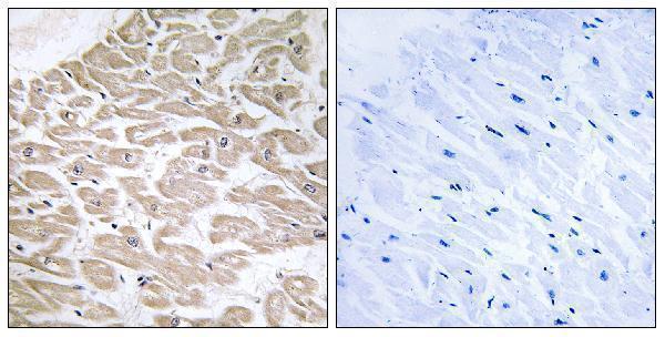
| Sample | Paraffin-embedded human heart tissue; fixative not specified (datasheet A30715) |
| Fixation | Image fixative and duration unreported (datasheet A30715); verify before use. |
| Sectioning | 4–5 µm sections on charged slides (standard) |
| Deparaffinisation | Xylene, graded ethanol series to water (standard) |
| Antigen retrieval | Heat-induced epitope retrieval in citrate buffer, pH 6.0, 20 min at 95–98 °C (standard rule: cytoplasmic / membrane antigen) |
| Peroxidase block | 3% H2O2, 10 min, room temperature (standard) |
| Blocking | 10% normal serum of the secondary host, 30 min, room temperature (standard) |
| Primary antibody | Rabbit anti-RRAD, 1:100 - 1:300 (datasheet A30715) |
| Primary incubation | Overnight at 4 °C (standard) |
| Detection | HRP-polymer secondary, DAB chromogen 5–10 min (standard) |
| Counterstain | Hematoxylin, blue, dehydrate and mount (standard) |
| Expected result | RRAD-positive staining in respiratory epithelial cells of bronchus (HPA tissue IHC: High). HPA tissue profile: General cytoplasmic expression with additional nuclear expression in a few tissues. No signal in the no-primary control. |
RRAD is annotated at the cell membrane without a transmembrane segment (UniProt P55042 topology). In tissue IHC, expect predominantly cytoplasmic staining in the cell populations HPA reports as positive, including respiratory epithelial cells and placental decidual cells (HPA: tissue IHC). Interpret the pattern cautiously: HPA rates tissue IHC reliability “Uncertain” because staining and RNA expression show low consistency (HPA: tissue IHC).
| Distinct cytoplasmic staining in bronchial respiratory epithelial cells or placental decidual cells. | These are reported High staining populations (HPA: tissue IHC). Cell-associated signal with visible unstained structures supports an interpretable IHC result; HPA’s “Uncertain” reliability still limits confidence that every stained cell contains RRAD (HPA: tissue IHC). |
| Predominantly nuclear staining, with little cytoplasmic or membrane-associated signal. | Recheck this pattern before assigning it to RRAD: HPA describes general cytoplasmic tissue staining and nuclear staining in only a few tissues (HPA: tissue IHC). Nucleoplasmic localisation is reported in ICC/IF, so a nuclear component alone does not prove artefact (HPA: subcellular ICC/IF). |
| Strong staining in a cell population reported as “Not detected,” such as adipocytes. | This conflicts with the reported adipocyte IHC result (HPA: adipose tissue IHC). Consider antibody cross-reactivity or endogenous chromogenic activity, then compare cell identity and staining against controls (general IHC practice). The HPA tissue assay is rated “Uncertain” (HPA: tissue IHC). |
| Diffuse colour across tissue and empty spaces, with poor separation between cells. | This is difficult to score as cell-specific RRAD staining. Evaluate background from detection reagents, endogenous activity, or excess antibody using appropriate control sections (general IHC practice). The reported tissue pattern is cellular and generally cytoplasmic (HPA: tissue IHC). |
| No staining in a section expected to contain bronchial respiratory epithelial cells. | HPA reports High staining in those cells, making them a useful comparator, though not an independently validated positive control (HPA: bronchus IHC; HPA: tissue IHC reliability “Uncertain”). Check tissue preservation, retrieval, antibody application, and chromogenic detection as general IHC workflow steps. |
| Tissue and cell selection | HPA reports High staining in bronchial and nasopharyngeal respiratory epithelium, oral squamous epithelium, fallopian tube glandular cells, and placental decidual cells (HPA: tissue IHC). Specify the cell population when scoring; a whole-section score can hide that distinction (general IHC practice). |
| Compartment and topology | UniProt annotates RRAD at the cell membrane and lists no transmembrane segment (UniProt P55042 topology). HPA describes general cytoplasmic tissue staining (HPA: tissue IHC). Evaluate compartment alongside tissue morphology; a crisp membrane outline is not required by the reported IHC pattern. |
| Strength of tissue evidence | HPA assigns “Uncertain” tissue IHC reliability because antibody staining and RNA expression have low consistency (HPA: tissue IHC). Its listed antibody, HPA041755, is “Uncertain” for IHC and “Approved” for ICC (HPA: antibody validation). Treat tissue positives as reported observations, not definitive specificity proof. |
| Expression and assay disagreement | UniProt reports greatest abundance in heart and detection in adipose tissue (UniProt P55042 tissue specificity). HPA reports group-enriched heart and skeletal muscle RNA, but adipocytes as “Not detected” by tissue IHC (HPA: tissue IHC). Avoid using abundance or RNA alone to predict a slide’s staining intensity. |
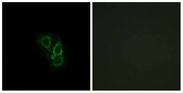
| IF/ICC Q: Where should RRAD appear? | A: Mainly at the plasma membrane, with additional nucleoplasmic, Golgi, and flagellar centriole localisation (HPA: subcellular ICC/IF). HPA rates the listed antibody “Approved” for ICC, while its IHC rating is “Uncertain” (HPA: antibody validation); interpret each assay against its own reported pattern. |
| Situation | Likely cause | Next action |
|---|---|---|
| Expected positive epithelium has little or no chromogenic signal. | The tissue may lack the intended cell population, or a general IHC workflow step may have failed. Bronchial respiratory epithelial cells are reported High (HPA: bronchus IHC). | Confirm the cells on the counterstain, then review section condition, retrieval, antibody incubation, and detection with a working control (general IHC practice). Do not infer RRAD-specific retrieval sensitivity from the HPA result. |
| Colour is strongest in nuclei throughout the section. | A widespread nuclear-only pattern exceeds HPA’s description of nuclear expression in a few tissues (HPA: tissue IHC). Nucleoplasm is an additional ICC/IF location (HPA: subcellular ICC/IF). | Compare nuclei and cytoplasm within identified cell populations; inspect negative controls and scoring criteria before calling the IHC pattern positive (general IHC practice). |
| Adipocytes stain strongly while the expected positive population is weak. | HPA reports adipocytes as “Not detected” by IHC, although UniProt reports RRAD detection in adipose tissue (HPA: adipose tissue IHC; UniProt P55042 tissue specificity). | Verify cell identity and compare controlled sections. Report the assay disagreement rather than treating either source as proof that the strong adipocyte stain is specific. |
| Brown colour is diffuse, including spaces without cells. | Background from antibody or detection reagents, or endogenous chromogenic activity, can obscure localisation (general IHC practice). | Inspect no-primary and detection controls, confirm blocking and washing, and score only signal tied to identifiable cells (general IHC practice). |
| A membrane outline is faint despite cytoplasmic staining in reported positive cells. | HPA’s tissue IHC profile is generally cytoplasmic, whereas its ICC/IF profile is mainly plasma membrane (HPA: tissue IHC; HPA: subcellular ICC/IF). | Score the IHC slide against the tissue pattern and cell identity. Record membrane accentuation if visible; do not require an ICC/IF-like outline to accept tissue staining. |
| Replicate sections disagree in staining intensity or location. | The HPA tissue IHC assessment itself is “Uncertain” because staining and RNA data have low consistency (HPA: tissue IHC). Routine section or detection variation may also contribute (general IHC practice). | Compare the same identified cell population across sections, include appropriate controls, and document the disagreement before drawing a biological conclusion (general IHC practice). |


Comprehensive Human Protein Atlas IHC scoring per tissue (reliability: Uncertain — Low consistency between antibody staining and RNA expression data.). Rows are taken directly from the HPA tissue chart — click any row's HPA link to view the source.
| Tissue | Cell type | Level | Evidence | Source |
|---|---|---|---|---|
| Bronchus | Respiratory epithelial cells | High | Protein (IHC) | HPA → |
| Cerebral cortex | Neuropil | High | Protein (IHC) | HPA → |
| Fallopian tube | Glandular cells | High | Protein (IHC) | HPA → |
| Nasopharynx | Respiratory epithelial cells | High | Protein (IHC) | HPA → |
| Oral mucosa | Squamous epithelial cells | High | Protein (IHC) | HPA → |
| Tissue | Cell type | Level | Evidence | Source |
|---|---|---|---|---|
| Adipose tissue | Adipocytes | Not detected | Protein (IHC) | HPA → |
| Caudate | Glial cells | Not detected | Protein (IHC) | HPA → |
| Seminal vesicle | Glandular cells | Not detected | Protein (IHC) | HPA → |
| Spleen | Cells in red pulp | Not detected | Protein (IHC) | HPA → |
Troubleshoot RRAD staining in paraffin sections by checking retrieval, controls, cellular distribution, and scoring before interpreting chromogenic signal.
A30715 has IHC data from paraffin-embedded human heart tissue and IF data from A549 cells (catalog image captions). Its listed reactivity covers human, mouse, and rat (catalog).
A30715 will render with an IHC image of paraffin-embedded human heart tissue, including a peptide-blocked comparison (catalog IHC caption). A30715 also has an IF image of A549 cells; IHC, IF, and ICC are listed applications, and human, mouse, and rat are listed species (catalog IF caption; catalog).
Which to pick: For tissue IHC, choose A30715: its own image shows paraffin-embedded human heart tissue, but the fixative is unreported (catalog IHC caption). For IF/ICC, A30715 lists both applications and shows IF in A549 cells (catalog applications; catalog IF caption). For work across species, A30715 lists human, mouse, and rat reactivity and is polyclonal; the supplied IHC image documents human tissue only (catalog reactivity; catalog dilution record; catalog IHC caption).