RTCA / RNA 3'-terminal phosphate cyclase · IHC design guide

Design Immunohistochemistry for RTCA

Plan RTCA staining in paraffin sections using the IHC-validated antibody at 1:100–1:300 (datasheet: A30754). Compare glandular cells in gallbladder or prostate, where staining is high, and interpret the pattern cautiously because tissue staining awaits external verification (HPA tissue IHC).

Evidence assembled Oct 2026 · For research use; verify linked source records and product datasheet before use
Immunohistochemistry protocol sheet for RTCA (IHC for RTCA): expected localisation Nuclear and cytoplasmic staining (HPA tissue IHC), antibody A30754, validated IHC image, and IHC protocol steps
Printable RTCA IHC protocol sheet — expected localisation Nuclear and cytoplasmic staining (HPA tissue IHC), antibody A30754, controls and protocol steps. Open the full RTCA IHC guide →

RTCA Immunohistochemistry Experimental Design Guide

Expected localisation, validated protocols, controls and antibodies — the at-a-glance facts below, then the full design guide.

Must know before staining
Expected localisation Nuclear and cytoplasmic staining (HPA tissue IHC)
Staining pattern Nuclear and cytoplasmic signal across most tissues (HPA tissue IHC)
Antigen retrieval Tris-EDTA pH 9.0 HIER, 95–98 °C, 20 min (rule: nuclear antigen)
Positive control ⓘ Gallbladder+4 more · see all
Negative control ⓘ Adipose tissue+4 more · see all
Important caveats
Reasons your staining may differ from the expected pattern.
Fixation Keep fixation consistent across sections. (standard IHC practice; not target-specific)
Caveat Tissue staining awaits external verification (HPA tissue IHC)
Regulation No staining-linked regulator reported (UniProt)
Isoform / epitope 2 isoforms; epitope coverage is unreported (UniProt)
Section 1

Recommended RTCA IHC & IF Protocols

The catalog antibody’s IHC-P protocol is paired with one published RTCA immunohistochemistry method (PMC4446171: IHC methods).

Recommended immunohistochemistry (IHC-P) protocol parameters
SampleParaffin-embedded human breast carcinoma tissue; fixative not specified (datasheet A30754)
FixationImage fixative and duration unreported (datasheet A30754); verify before use.
Sectioning4–5 µm sections on charged slides (standard)
DeparaffinisationXylene, graded ethanol series to water (standard)
Antigen retrievalHeat-induced epitope retrieval in Tris-EDTA buffer, pH 9.0, 20 min at 95–98 °C (standard rule: nuclear antigen)
Peroxidase block3% H2O2, 10 min, room temperature (standard)
Blocking10% normal serum of the secondary host, 30 min, room temperature (standard)
Primary antibodyRabbit anti-RTCA, 1:100 - 1:300 (datasheet A30754)
Primary incubationOvernight at 4 °C (standard)
DetectionHRP-polymer secondary, DAB chromogen 5–10 min (standard)
CounterstainHematoxylin, blue, dehydrate and mount (standard)
Expected resultRTCA-positive staining in glandular cells of gallbladder (HPA tissue IHC: High). HPA tissue profile: Nuclear and cytoplasmic expression in most tissues. No signal in the no-primary control.
💡Decision noteFor paraffin sections, start with Tris-EDTA pH 9.0 retrieval at 95–98 °C for 20 min (page retrieval rule: nuclear antigen; UniProt: nucleoplasm).
Section 2

What Is the Expected RTCA Staining Pattern?

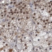
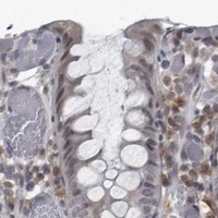
RTCA is assigned to the nucleus and nucleoplasm (UniProt O00442), with no transmembrane segment (UniProt O00442 topology). In tissue IHC, HPA reports nuclear and cytoplasmic staining across most tissues, including high staining in gallbladder and prostate glandular cells and testicular Leydig cells (HPA tissue IHC). Treat those patterns as provisional: HPA rates its tissue IHC reliability Uncertain because antibody staining and RNA expression show only medium consistency (HPA tissue IHC).

What am I looking at on my slide?
Gallbladder or prostate glandular cells, or testicular Leydig cells, show conspicuous nuclear staining, with possible cytoplasmic staining (HPA tissue IHC).This fits HPA's high staining in those cells and its broader nuclear and cytoplasmic tissue pattern (HPA tissue IHC). Nucleoplasmic localisation provides a second compartment expectation (UniProt O00442; HPA ICC-IF), although neither source establishes that every positive cell must stain equally.
Staining is confined to cell membranes or extracellular material, while nuclei in reference-positive cells remain unstained.A membrane-only or extracellular pattern conflicts with the nucleoplasmic assignment (UniProt O00442; HPA ICC-IF) and reported tissue staining (HPA tissue IHC). Review morphology and controls before calling it an artefact; the supplied sources do not identify its cause.
Strong staining appears mainly in a cell population listed as not detected, such as adipocytes or cardiomyocytes (HPA tissue IHC).That distribution conflicts with HPA's observations for those specific cells (HPA tissue IHC). Consider cross-reactivity or endogenous detection activity, then compare a reference-positive tissue and detection controls (general IHC practice). An HPA not-detected call is not proof of biological absence.
A diffuse chromogenic haze obscures nuclei and cell boundaries throughout the section.This cannot be scored reliably against the reported nuclear and cytoplasmic distribution (HPA tissue IHC). Excess detection background or nonspecific antibody binding is possible; inspect the no-primary control and reassess blocking, washing and detection conditions (general IHC practice).
No convincing signal appears in gallbladder or prostate glandular cells, or testicular Leydig cells.These are HPA high reference populations (HPA tissue IHC), so check tissue identity, antibody performance, retrieval and detection controls (general IHC practice). Absence in one section is inconclusive because HPA rates tissue IHC Uncertain (HPA tissue IHC).
💡Expected RTCA appearanceA persuasive positive shows conspicuous nuclear staining, potentially accompanied by cytoplasmic staining, in HPA high glandular or Leydig reference cells; isolated membrane or extracellular staining is discordant (HPA tissue IHC; UniProt O00442; HPA ICC-IF).
How each factor affects the staining
Compartment and topologyRTCA is nucleoplasmic and has no transmembrane segment (UniProt O00442 topology; HPA ICC-IF). Score nuclear staining against cell morphology, while allowing the cytoplasmic component reported in tissue IHC (HPA tissue IHC).
Choice of reference tissueGallbladder and prostate glandular cells and testicular Leydig cells are HPA high examples; adrenal gland glandular cells and cortical or hippocampal neurons are medium examples (HPA tissue IHC). Match the expected intensity to the named cell population, not the whole section.
Tissue IHC confidenceHPA labels the tissue pattern Uncertain; its listed IHC assessments for HPA027982 and HPA028151 are also Uncertain (HPA tissue IHC; HPA antibody validation). Use these observations as comparison points, not definitive proof of antibody specificity.
IF/ICC Q: Does a fluorescent nuclear signal support the IHC interpretation?Yes, as compartment evidence: HPA reports enhanced nucleoplasmic ICC-IF localisation, with images from A-431, U-251MG, U2OS, Hep-G2 and SK-MEL-30 (HPA ICC-IF). That result does not upgrade HPA's Uncertain tissue IHC rating (HPA tissue IHC).
Isoforms and processingUniProt lists 2 isoforms, a single 1–366 chain, and no signal peptide or propeptide (UniProt O00442). The payload gives no antibody epitope or isoform coverage, so it cannot predict which isoform a given stain detects.
Antigen retrievalAssess retrieval using the IHC-validated antibody's documented conditions and a reference-positive tissue (general IHC practice; HPA tissue IHC for reference cells). Neither UniProt nor HPA supplies RTCA-specific retrieval requirements or evidence that fixation changes RTCA staining.
Why is my staining missing, weak or wrong?
SituationLikely causeNext action
Reference-positive glandular or Leydig cells lack signal (HPA tissue IHC).The run may have failed, or the sampled cells may not match the HPA reference population (general IHC practice). HPA's Uncertain IHC rating also limits certainty (HPA tissue IHC).Verify cell identity and run controls; review the antibody's documented retrieval, dilution and detection conditions without assuming an RTCA-specific fixation effect (general IHC practice).
Chromogen covers tissue broadly and obscures nuclei.Nonspecific binding or detection background may prevent comparison with the reported pattern (general IHC practice; HPA tissue IHC).Examine a no-primary control, then review blocking, washes and detection conditions; score RTCA only where cell boundaries and nuclei remain interpretable (general IHC practice).
Signal is membrane-only or extracellular.That location conflicts with nucleoplasmic RTCA and the absence of a transmembrane segment (UniProt O00442 topology; HPA ICC-IF).Check the cell outline and counterstain, compare a HPA high reference population, and inspect controls before attributing the signal to RTCA (HPA tissue IHC; general IHC practice).
A listed not-detected population stains strongly, such as adipocytes (HPA tissue IHC).Cross-reactivity or endogenous detection activity is possible (general IHC practice); HPA's not-detected category is an observation, not a biological exclusion (HPA tissue IHC).Compare staining with the no-primary control and a HPA reference-positive population. Report the discrepancy by cell type rather than treating the entire tissue as uniformly positive (general IHC practice; HPA tissue IHC).
Cytoplasmic staining accompanies nuclear staining.HPA reports both nuclear and cytoplasmic tissue expression, while UniProt and HPA ICC-IF emphasise nucleoplasm (HPA tissue IHC; UniProt O00442; HPA ICC-IF).Record nuclear and cytoplasmic compartments separately, with intensity and cell type. Do not reject the cytoplasmic component solely because ICC-IF shows enhanced nucleoplasmic localisation (HPA tissue IHC; HPA ICC-IF).
IF/ICC looks nucleoplasmic, but tissue IHC is ambiguous.The assays have different HPA evidence: ICC-IF localisation is enhanced, whereas tissue IHC reliability is Uncertain (HPA ICC-IF; HPA tissue IHC).Use IF/ICC as a compartment cross-check and interpret the chromogenic section using its own cell-level controls and HPA tissue references; do not transfer an IF/ICC validation label to IHC (HPA ICC-IF; HPA tissue IHC; general IHC practice).

Sample controls for RTCA IHC & IF

🧪Run gallbladder first and assess its glandular cells for nuclear staining (HPA: High in gallbladder glandular cells; UniProt O00442: nucleoplasm). Use adipose tissue as the negative comparator (HPA: Not detected in adipocytes); on the gallbladder slide, internal negative cells should lack nuclear chromogen above background, although no specific negative cell population is identified in the supplied HPA rows (HPA: gallbladder glandular cells High).
Positive control tissue: Gallbladder (Glandular cells, HPA High)
Negative control tissue: Adipose tissue (HPA Not detected)
ICC-IF cell lines (HPA subcellular resource): HPA ICC-IF images show RTCA in A-431, U-251MG, U2OS, Hep-G2, SK-MEL-30, with annotated localisation: Nucleoplasm (enhanced) (HPA subcellular).
Technical controls: Include a no-primary, secondary-only control and an isotype control matched to the primary antibody’s host species and clonality (standard IHC practice). Use RTCA knockout material or a peptide block as a biological specificity control (selected A30754 tissue-IHC caption: peptide-blocked image), and check endogenous peroxidase background on the gallbladder slide before interpreting chromogenic staining (standard IHC practice).
⚠️Feasibility: A target-specific fixation window and retrieval dependency are unreported in the supplied evidence; the selected A30754 paraffin-section caption does not state its fixative (selected A30754 tissue-IHC caption). The evidence does not establish whether frozen sections or IF would be easier than paraffin IHC for RTCA (selected A30754 tissue-IHC caption; HPA: ICC-IF images). Assess gallbladder sections for glandular background staining and endogenous peroxidase signal during scoring (HPA: High in gallbladder glandular cells; standard IHC practice).

HPA tissue IHC evidence for RTCA

Comprehensive Human Protein Atlas IHC scoring per tissue (reliability: Uncertain — Medium consistency between antibody staining and RNA expression data. Pending external verification.). Rows are taken directly from the HPA tissue chart — click any row's HPA link to view the source.

Positive expression · recommended positive controls

TissueCell typeLevelEvidenceSource
Gallbladder Glandular cells High Protein (IHC) HPA →
Prostate Glandular cells High Protein (IHC) HPA →
Testis Leydig cells High Protein (IHC) HPA →
Adrenal gland Glandular cells Medium Protein (IHC) HPA →
Bone marrow Hematopoietic cells Medium Protein (IHC) HPA →

Undetected expression · recommended negative controls

TissueCell typeLevelEvidenceSource
Adipose tissue Adipocytes Not detected Protein (IHC) HPA →
Epididymis Glandular cells Not detected Protein (IHC) HPA →
Heart muscle Cardiomyocytes Not detected Protein (IHC) HPA →
Lung Alveolar cells Not detected Protein (IHC) HPA →
Skeletal muscle Myocytes Not detected Protein (IHC) HPA →
Section 3

Advanced RTCA IHC Tips

Troubleshoot RTCA staining in paraffin sections using the page retrieval setting, catalog antibody image, and reported nuclear and tissue expression patterns.

How should I retrieve RTCA antigen when nuclear staining is weak?
Use Tris-EDTA pH 9.0 heat-induced retrieval at 95–98 °C for 20 min (page retrieval setting: nuclear antigen). Compare retrieved and unretrieved sections from the same block, keeping antibody incubation and chromogen development identical so retrieval remains the tested variable (standard IHC practice). Examine nucleoplasmic staining first, because that is the annotated location, while recording cytoplasmic staining separately because tissue IHC has reported both patterns (UniProt O00442; HPA tissue IHC). If staining remains weak, test an alternative retrieval condition on adjacent sections only as a fallback, and assess tissue integrity alongside signal (standard IHC practice).
Could fixation explain weak RTCA staining in my paraffin sections?
RTCA-specific sensitivity to fixation is unknown from the supplied evidence; the catalog image identifies paraffin-embedded human breast carcinoma but does not state its fixative (A30754 caption). Record the actual fixative, fixation duration, processing history, and block age for each specimen before comparing staining across runs (standard IHC practice). If archival material stains weakly, compare blocks with documented processing histories using the same retrieval, antibody incubation, and chromogen development conditions (standard IHC practice). Interpret any difference as an association with specimen handling until matched material isolates fixation itself; neither the tissue expression profile nor RTCA topology establishes a fixation effect (standard IHC practice; HPA tissue IHC; UniProt O00442).
What subcellular pattern should I expect for RTCA in chromogenic IHC?
Prioritize nucleoplasmic staining when judging whether a cellular signal matches the annotated RTCA location (UniProt O00442; HPA subcellular). Record nuclear and cytoplasmic intensity separately, because the tissue IHC profile reports both compartments in most tissues and has uncertain reliability (HPA tissue IHC). RTCA has no annotated transmembrane segment or signal peptide, so a crisp plasma-membrane-only pattern warrants scrutiny rather than a membrane-localisation claim (UniProt O00442 topology and processing). Use a counterstain that leaves nuclei readable, then inspect the same cell population at high magnification before assigning compartment scores (standard IHC practice). Compare compartment patterns across serial sections and controls before interpreting an unexpected signal biologically (standard IHC practice).
Can this antibody distinguish RTCA isoforms in tissue sections?
RTCA has 2 listed isoforms, but the supplied catalog caption does not map the antibody epitope or establish isoform selectivity (UniProt O00442 isoforms; A30754 caption). Treat staining as RTCA-associated signal rather than an isoform-specific readout unless the antibody's immunogen and both isoform sequences establish what it recognizes (standard IHC interpretation). The caption shows staining in paraffin-embedded human breast carcinoma tissue and a synthesized-peptide blocking image, which supports an epitope-competition check without identifying that epitope here (A30754 caption). Run the peptide-blocked condition beside an unblocked section under identical chromogenic conditions, then assess whether the relevant cellular signal falls (A30754 caption; standard IHC practice).
How can I check RTCA localisation by IF alongside the IHC result?
Use IF as a separate localisation check: nucleoplasmic signal is reported in cell imaging, while tissue IHC reports nuclear and cytoplasmic expression (HPA subcellular; HPA tissue IHC). For a tissue comparison, multiplex RTCA with a marker identifying the expected cell population, such as neuronal cells in cerebral cortex, and include a nuclear counterstain (HPA: Medium in cerebral-cortex neuronal cells; standard IF practice). Choose spectrally separated fluorophores and inspect unstained tissue autofluorescence before assigning a weak RTCA channel, especially in pigmented or intrinsically fluorescent regions (standard IF practice). Because RTCA lacks a transmembrane segment and is annotated in the nucleoplasm, validate permeabilisation sufficient for nuclear access without assuming an extracellular epitope (UniProt O00442 topology and localisation; standard IF practice).
How do I separate RTCA signal from chromogenic background?
Inspect a no-primary control for detection-system signal and an isotype control where appropriate, using the same section processing and chromogen exposure (standard IHC practice). Quench endogenous peroxidase before peroxidase-based DAB detection, and check whether apparent staining follows tissue deposits, damaged edges, or necrotic areas (standard IHC practice). Optimize blocking, antibody concentration, washing, and DAB development one variable at a time; the supplied caption gives no antibody dilution to copy (A30754 caption; standard IHC practice). A peptide-blocked companion image is available for the selected antibody, so use loss of cellular staining with its synthesized peptide as supporting specificity evidence rather than treating every brown deposit as RTCA (A30754 caption; standard IHC interpretation).
How should I score RTCA staining across specimens? ⚠ ANSWER MARKED FOR VERIFICATION
Define the scoring compartment before analysis: report nucleoplasmic and cytoplasmic staining separately because their evidence differs (UniProt O00442 localisation; HPA tissue IHC). For each annotated cell population, record the percentage of positive cells and intensity categories, then calculate an H-score if the study needs a combined measure (standard IHC quantification). Normalize positive counts to the number of eligible cells, or stained-cell density to sampled tissue area in mm², while excluding folds and necrosis by a preset rule (standard IHC quantification). Keep retrieval, chromogen development, imaging, and thresholds consistent across specimens; document the cell type because reported RTCA levels vary by tissue population (standard IHC practice; HPA tissue IHC).
When is an apparent RTCA-positive cell likely an artefact?
A credible result identifies a defined cell population and a reproducible intracellular pattern, with nucleoplasmic staining consistent with the annotated location (UniProt O00442; standard IHC interpretation). Cytoplasmic staining alone needs caution because tissue IHC reports it but rates its overall evidence uncertain, pending external verification (HPA tissue IHC). Check whether the positive cells match the sampled population: glandular cells are reported high in gallbladder and prostate, whereas lung alveolar cells are reported not detected (HPA tissue IHC). Discount staining confined to section edges, necrosis, or deposits, and use a no-primary control to identify endogenous enzyme or detection background before calling a biological positive (standard IHC practice).
Boster reagents

Best RTCA / RNA 3'-terminal phosphate cyclase IHC Antibodies

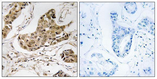
The catalog shows RTCA IHC in paraffin-embedded human breast carcinoma and IF in PC-3 cells; listed antibody reactivity covers human, mouse and rat (A30754 and A07570-1 image captions; catalog reactivity).

Real IHC data Immunohistochemistry analysis of paraffin-embedded human breast carcinoma tissue, using RTCD1 Antibody. The picture on the right is blocked with the synthesized peptide.
Anti-RTCD1 Antibody
Cat # A30754
Real IF data IF analysis of RTCD1/RTCA using anti-RTCD1/RTCA antibody (A07570-1). RTCD1/RTCA was detected in immunocytochemical section of PC-3 cells. Enzyme antigen retrieval was performed using IHC enzyme antigen retrieval reagent (AR0022) for 15 mins. The cells were blocked with 10% goat serum. And then incubated with 5μg/mL rabbit anti-RTCD1/RTCA Antibody (A07570-1) overnight at 4°C. DyLight®488 Conjugated Goat Anti-Rabbit IgG (BA1127) was used as secondary antibody at 1:100 dilution and incubated for 30 minutes at 37°C. The section was counterstained with DAPI. Visualize using a fluorescence microscope and filter sets appropriate for the label used.
Anti-RTCD1/RTCA Antibody ®
Cat # A07570-1

A30754 lists IHC and IF applications, with an IHC image from paraffin-embedded human breast carcinoma; its listed reactivity is human, mouse and rat (A30754 image caption; catalog applications/reactivity). A07570-1 lists human IF/ICC, with an IF image from PC-3 cells; IHC is not a listed application (A07570-1 image caption; catalog applications/reactivity).

Which to pick: For tissue IHC, choose A30754: its listed dilution is 1:100–1:300, and its own image shows paraffin-embedded human breast carcinoma; the fixative is unreported (A30754 catalog dilution; image caption). For human IF/ICC, choose A07570-1: its PC-3 IF image uses 5 μg/mL, and its listed applications include IF and ICC (A07570-1 image caption; catalog applications). For mouse or rat work, A30754 is the listed cross-species option and is polyclonal; its displayed IHC example is human tissue only (A30754 catalog reactivity; dilution_raw; image caption).

Each figure is that product's own IHC / IF validation image from its datasheet.

References

  1. UniProt Consortium. UniProt entry O00442 (RTCA_HUMAN, RNA 3'-terminal phosphate cyclase).
  2. Human Protein Atlas. RTCA tissue IHC expression (reliability: Uncertain).
  3. Human Protein Atlas. RTCA subcellular location (ICC-IF): Localized to the nucleoplasm..
  4. Human Protein Atlas. RTCA antibody validation summary (4 antibodies).
  5. PD-L1 intrinsically promotes the proliferation of breast cancer cells through the SKP2-p27/p21 axis. Cancer cell international 2024 — PMC11084005.
  6. Upregulated expression of miR-4443 and miR-4488 in drug resistant melanomas promotes migratory and invasive phenotypes through downregulation of intermediate filament nestin. Journal of experimental & clinical cancer research : CR 2023 — PMC10680267.
  7. KLF14 directly downregulates the expression of GPX4 to exert antitumor effects by promoting ferroptosis in cervical cancer. Journal of translational medicine 2024 — PMC11465826.
  8. Regulation of axon regeneration by the RNA repair and splicing pathway. Nature neuroscience 2015 — PMC4446171.
  9. PubMed PMID:9184239 — UniProt-cited evidence.
  10. PubMed PMID:16710414 — UniProt-cited evidence.
  11. PubMed PMID:15489334 — UniProt-cited evidence.