This website uses cookies to ensure you get the best experience on our website.


- Table of Contents
Plan RTL6 chromogenic IHC on paraffin sections with the catalog antibody at 1:100–1:300 (datasheet). Compare kidney tubule staining, reported as high, with adipocytes, reported as not detected (HPA tissue IHC).

Expected localisation, validated protocols, controls and antibodies — the at-a-glance facts below, then the full design guide.
| Expected localisation | Cytoplasmic staining in most tissues (HPA tissue IHC) | |
| Staining pattern | Cytoplasmic signal across most tissues (HPA tissue IHC) | |
| Antigen retrieval | Citrate pH 6.0 HIER, 95–98 °C, 20 min (rule: cytoplasmic / membrane antigen) | |
| Positive control | Adrenal gland+4 more · see all | |
| Negative control | Adipose tissue+1 more · see all |
| Fixation | Keep fixation consistent across sections (standard IHC practice; not target-specific) | |
| Caveat | Staining has low consistency with RNA expression (HPA tissue IHC) | |
| Regulation | No expression regulation annotated (UniProt) | |
| Isoform / epitope | No isoforms; one 1–239 chain (UniProt) |
The catalog antibody’s IHC-P protocol (datasheet) is accompanied by one published tissue microarray IHC protocol (PMC13048935).
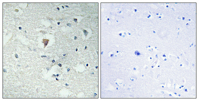
| Sample | Paraffin-embedded Human brain tissue; fixative not specified (datasheet A31816) |
| Fixation | Image fixative and duration unreported (datasheet A31816); verify before use. |
| Sectioning | 4–5 µm sections on charged slides (standard) |
| Deparaffinisation | Xylene, graded ethanol series to water (standard) |
| Antigen retrieval | Heat-induced epitope retrieval in citrate buffer, pH 6.0, 20 min at 95–98 °C (standard rule: cytoplasmic / membrane antigen) |
| Peroxidase block | 3% H2O2, 10 min, room temperature (standard) |
| Blocking | 10% normal serum of the secondary host, 30 min, room temperature (standard) |
| Primary antibody | Rabbit anti-RTL6, 1:100-1:300 (datasheet A31816) |
| Primary incubation | Overnight at 4 °C (standard) |
| Detection | HRP-polymer secondary, DAB chromogen 5–10 min (standard) |
| Counterstain | Hematoxylin, blue, dehydrate and mount (standard) |
| Expected result | RTL6-positive staining in glandular cells of adrenal gland (HPA tissue IHC: High). HPA tissue profile: Cytoplasmic expression in most tissues. No signal in the no-primary control. |
For paraffin-section IHC, expect predominantly cytoplasmic RTL6 staining in many tissues, with high staining reported in kidney tubule cells, cerebellar Purkinje cells and several other defined cell populations (HPA tissue IHC). RTL6 has no annotated transmembrane segment or subcellular location (UniProt Q6ICC9). Treat the tissue pattern as a guide rather than proof of specificity: HPA rates its IHC evidence Approved but reports low consistency between antibody staining and RNA expression (HPA tissue IHC).
| Distinct cytoplasmic chromogen in kidney tubule cells or cerebellar Purkinje cells, with surrounding structures still distinguishable. | This matches the reported compartment and two high-staining cell populations (HPA tissue IHC). Score the fraction and intensity of stained cells within each population; a convincing positive result retains cellular boundaries. HPA's Approved rating supports using this pattern as a reference, while its low antibody–RNA consistency limits how confidently staining alone identifies RTL6 (HPA tissue IHC). |
| Predominantly nuclear or membrane-rim staining in an IHC section, with little discernible cytoplasmic signal. | Recheck interpretation and controls before calling this RTL6: the tissue IHC profile is cytoplasmic (HPA tissue IHC), and UniProt annotates no transmembrane segment (UniProt Q6ICC9 topology). Nuclear speckles are approved in HPA ICC-IF, so a nuclear signal alone does not prove an artefact; the assay-specific discrepancy needs investigation (HPA ICC-IF). |
| Strong staining in adipocytes or soft-tissue fibroblasts, especially when expected positive cells are faint. | Those populations were reported as not detected (HPA tissue IHC). Consider cross-reactivity or endogenous chromogenic detection activity as possible explanations, then examine negative and detection controls. A negative HPA observation is a comparator, not proof that every specimen of that tissue must be blank (HPA tissue IHC). |
| Similar diffuse colour across cells, extracellular areas and tissue edges, without a defined cytoplasmic pattern. | Treat this as background until controls establish otherwise. Check whether the same colour appears without primary antibody; if so, investigate the detection system, blocking and washing as general IHC troubleshooting steps. The diffuse distribution does not resemble HPA's predominantly cytoplasmic tissue profile (HPA tissue IHC). |
| No signal in an adequately preserved kidney-tubule or Purkinje-cell positive-control section. | Both cell populations are reported as High (HPA tissue IHC). First check that the expected cells are present, then review the catalog antibody's IHC-P instructions and detection controls. Absence of signal raises a technical or specimen question; it cannot establish biological absence from one failed section. |
| Tissue and cell selection | HPA reports High staining in adrenal, seminal-vesicle and stomach glandular cells; kidney tubules; Purkinje and cortical neuronal cells; lymph-node germinal-center cells; and late spermatids (HPA tissue IHC). Its low-tissue-specificity RNA classification means tissue identity alone is a weak specificity test (HPA tissue IHC). |
| Antibody evidence | HPA005697 is rated Approved for IHC, while the tissue profile notes low consistency between antibody staining and RNA data (HPA antibodies; HPA tissue IHC). Interpret morphology together with controls and avoid treating the staining score as an independently verified measure of RTL6 abundance. |
| Topology and processing | UniProt lists a single 1–239 chain, no signal peptide, no propeptide, no transmembrane segment and no annotated glycosylation sites (UniProt Q6ICC9). These annotations do not identify the antibody epitope or predict antigen-retrieval conditions. |
| Antigen retrieval | Use the catalog antibody's IHC-P instructions to choose initial retrieval conditions; the supplied HPA and UniProt records give no RTL6-specific retrieval condition or fixation-sensitivity result. If optimisation is needed, compare conditions with the same positive tissue and controls as general IHC practice. |
| IF/ICC Q: Should its nuclear speckles change the IHC call? | A: HPA ICC-IF places RTL6 mainly in nuclear speckles and additionally in cytosol, whereas HPA tissue IHC describes cytoplasmic staining (HPA ICC-IF; HPA tissue IHC). Interpret each assay against its own evidence; a compartment difference merits validation rather than an automatic positive or negative call. |
| Situation | Likely cause | Next action |
|---|---|---|
| The positive-control kidney section is blank. | The sampled section may lack identifiable tubules, or the IHC staining workflow may have failed; kidney tubule cells are High in the reference profile (HPA tissue IHC). | Confirm tubules on the counterstain, inspect the detection control and follow the catalog antibody's IHC-P instructions. Repeat with a suitable positive section before interpreting study samples; do not assign an RTL6-negative score from the failed run. |
| Every tissue shows similar diffuse colour. | Background from detection reagents, inadequate blocking or insufficient washing is plausible in chromogenic IHC; this appearance differs from the defined cytoplasmic HPA profile (HPA tissue IHC). | Compare a no-primary control and examine whether colour follows cells or tissue edges. Review blocking, wash steps and detection chemistry as general IHC practice, then rescore only when cell-specific signal can be separated from background. |
| Adipocytes or fibroblasts stain strongly. | These cells are reported as Not detected (HPA tissue IHC). Cross-reactivity or endogenous detection activity is possible, although HPA's observation cannot exclude genuine differences in another specimen. | Check staining in a no-primary control, examine morphology and compare with an HPA High population processed in the same run (HPA tissue IHC). If the discordance persists, report it as unresolved rather than assigning RTL6 positivity. |
| The only convincing IHC signal is nuclear. | The result conflicts with HPA's cytoplasmic tissue IHC profile, although HPA ICC-IF independently reports approved nuclear-speckle localisation (HPA tissue IHC; HPA ICC-IF). | Verify that nuclei and cytoplasm are correctly identified with the counterstain, inspect background controls and document the compartment separately. Do not transfer the ICC-IF localisation directly into an IHC scoring rule. |
| Low signal appears in prostate or colon glandular cells. | Low staining is reported for those glandular populations (HPA tissue IHC); a weak result there does not by itself show assay failure. | Judge run performance in a clearly represented High population, such as kidney tubules or stomach glandular cells (HPA tissue IHC). Score the low population separately, using the same background threshold across sections. |
| Changing retrieval gives a different staining score. | The supplied records do not establish an RTL6-specific retrieval response or fixation effect; technical conditions can change chromogenic IHC appearance as a general practice consideration. | Compare matched sections, positive tissue and no-primary controls under the tested conditions. Record the chosen condition and its control performance without claiming that HPA tissue levels or UniProt topology explain the retrieval difference. |
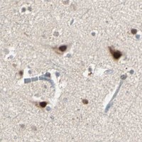

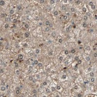
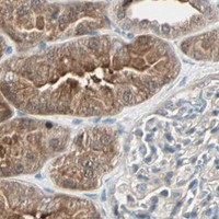


Comprehensive Human Protein Atlas IHC scoring per tissue (reliability: Approved — Low consistency between antibody staining and RNA expression data.). Rows are taken directly from the HPA tissue chart — click any row's HPA link to view the source.
| Tissue | Cell type | Level | Evidence | Source |
|---|---|---|---|---|
| Adrenal gland | Glandular cells | High | Protein (IHC) | HPA → |
| Cerebellum | Purkinje cells | High | Protein (IHC) | HPA → |
| Cerebral cortex | Neuronal cells | High | Protein (IHC) | HPA → |
| Kidney | Cells in tubules | High | Protein (IHC) | HPA → |
| Lymph node | Germinal center cells | High | Protein (IHC) | HPA → |
Use RTL6 compartment and cell-type patterns to assess paraffin-section staining, while treating antibody performance and fixation sensitivity as separate validation questions.
A31816 has a human brain paraffin-section IHC image (catalog IHC caption), lists IF as an application, and lists Human and Mouse reactivity (catalog applications and reactivity).
A31816 will render with an IHC image of paraffin-embedded human brain at 1:100 (catalog IHC caption). IF is listed as an application, but no IF image is supplied (catalog applications and image alts).
Which to pick: For tissue IHC, choose A31816: its image shows paraffin-embedded human brain, and the listed IHC dilution is 1:100–1:300 (catalog IHC caption; datasheet: IHC dilution). For IF, A31816 lists a 1:50 dilution; ICC validation and an IF image are unreported (catalog applications, dilution and image alts). A31816 lists Human and Mouse reactivity, but the supplied IHC image shows Human only; the tissue fixative and antibody clonality are unreported (catalog reactivity, IHC caption and antibody fields).