This website uses cookies to ensure you get the best experience on our website.
- Table of Contents
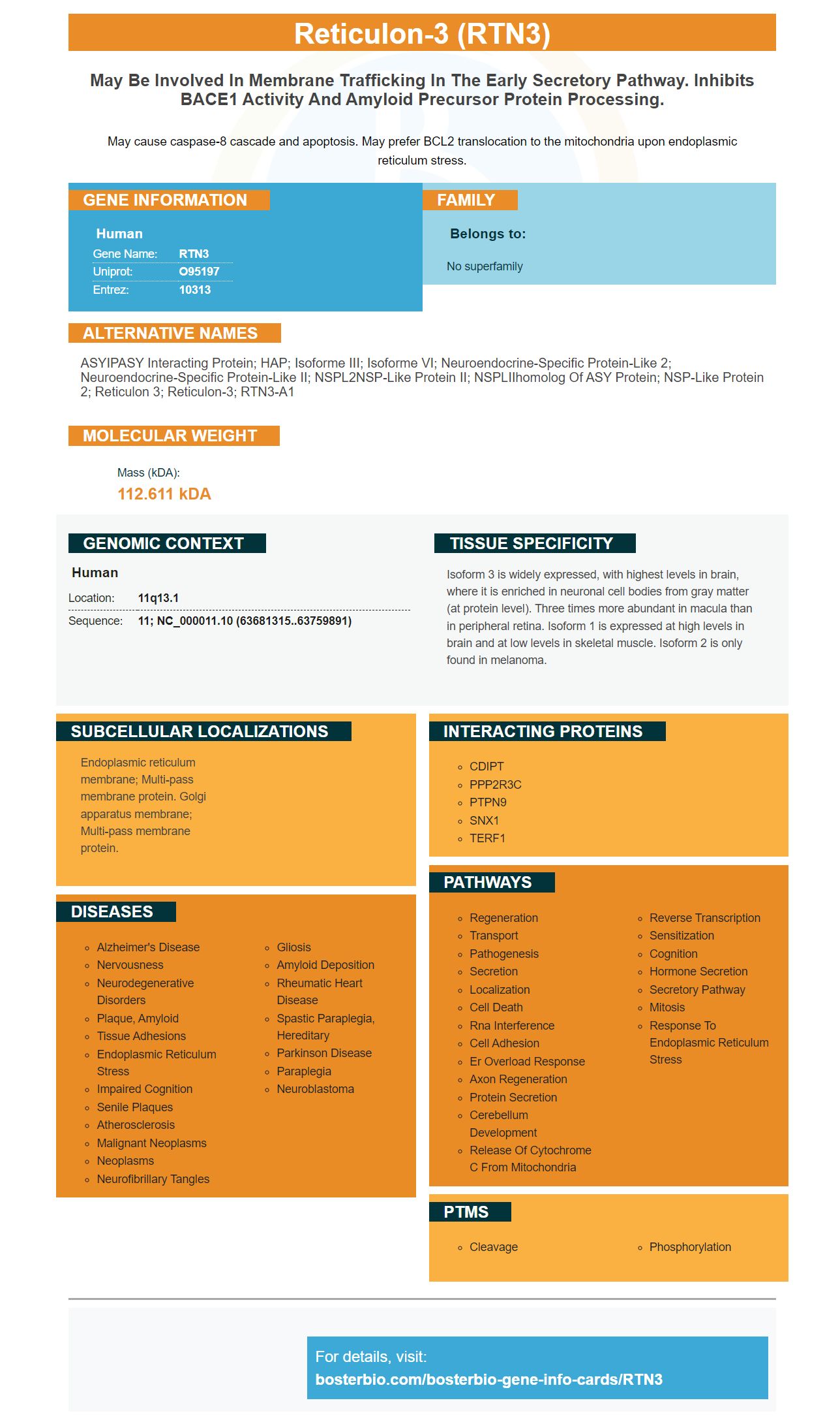
Facts about Reticulon-3.
May cause caspase-8 cascade and apoptosis. May prefer BCL2 translocation to the mitochondria upon endoplasmic reticulum stress.
Human | |
---|---|
Gene Name: | RTN3 |
Uniprot: | O95197 |
Entrez: | 10313 |
Belongs to: |
---|
No superfamily |
ASYIPASY interacting protein; HAP; isoforme III; isoforme VI; Neuroendocrine-specific protein-like 2; Neuroendocrine-specific protein-like II; NSPL2NSP-like protein II; NSPLIIhomolog of ASY protein; NSP-like protein 2; reticulon 3; reticulon-3; RTN3-A1
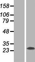
Mass (kDA):
112.611 kDA
Human | |
---|---|
Location: | 11q13.1 |
Sequence: | 11; NC_000011.10 (63681315..63759891) |
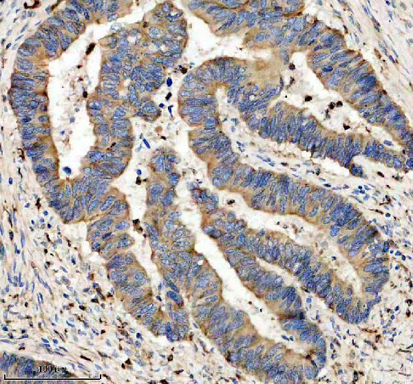
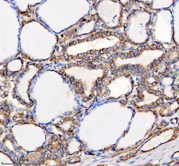
Isoform 3 is widely expressed, with highest levels in brain, where it is enriched in neuronal cell bodies from gray matter (at protein level). Three times more abundant in macula than in peripheral retina. Isoform 1 is expressed at high levels in brain and at low levels in skeletal muscle. Isoform 2 is only found in melanoma.
Endoplasmic reticulum membrane; Multi-pass membrane protein. Golgi apparatus membrane; Multi-pass membrane protein.
The Boster Bio guide is a fantastic source for optimizing experiments made using this marker. It includes information on how to control the experiment, creating the best conditions, and optimizing your results. Every researcher faces issues during experiments, but by using the correct controls you can eliminate the majority of causes of error. Use optimization tips and troubleshooting guides to determine the source of the error and fix it quickly.
The function of PGRMC1 in the clearance of ER-phages of pro-hormones that are mutant is not fully understood. PGRMC1 is known to target LMW cargoes within the ER. The function of PGRMC1 could be an ideal therapeutic target for disorders that are caused by misfolding of prohormone peptides. Molecular analysis of PGRMC1 identified that it plays a crucial role in ER-phagic clearance.
Both mutant POMC (and mutant proinsulin are oligomerized) in the ER during misfolding. They capture their wild-type counterparts leading to a decrease in secretion. PGRMC1 promotes the degradation of small oligomers. The proinsulin turnover of mutants is affected by PGRMC1's genetic and pharmacological deactivation. In the presence of POMC that is mutant and proinsulin, proinsulin that is mutant cannot be eliminated by ER-phagy. Mutant POMC and proinsulin trafficking that is wild type could be caused by PGRMC1 for diseases characterized by ER proteins retention.
In addition to promoting mutant prohormone degrading, PGRMC1 also facilitates ER exit and anterograde transport of C28F-POMC. PGRMC1 loss is not a factor in the quality control of anterograde ER import. It is therefore possible that a role for PGRMC1 in ER-phagic clearance mutant prohormones has not yet been identified.
The ER-phagic elimination is a multi-step process that involves the breakdown of misfolded protein in the ER. This process involves the re-admission of misfolded proteins from the cytosol through a receptor embedded in the ER membrane. The misfolded protein cargo is transported to the lysosomal degradation machinery after physical engagement. In addition, the protein aggregation process ensures that are targets of ER-phagy are lysosomal.
Autophagic removal of mutant proinsulin is a key factor in tumorigenesis. Autophagy is an adaptive response to ER stress, and ER-phagy is one of the mechanisms used to alleviate this stress. The prohormone ER-phagic clearing mutant is an essential component of the process of developing tumors. It regulates the growth of tumors as well as metastasis and long-term survival.
A16P-mutant proinsulin also accumulates in heterogeneous proinsulin complexes. In cells that lack the PGRMC1-RTN3 targeting complex, it accumulates in the cytoplasm. Additionally, in cells deficient in the PGRMC1 protein, ER-phagy is impaired in the presence of proinsulin that is mutant. While the mechanisms that govern ER-phagic clearance are still not fully understood, it is clear that it is an important adaptive regulatory mechanism in the pathogenesis of disc degeneration in the intervertebral region.
We focused our attention on a particular protein called PGRMC1 to better to understand the role played by RTN3 in ER-phagy. This protein is associated with the RTN3 marker in ER-phagy and targets LMW cargos. It has a transmembrane space that is distinct. This protein is vital in the degradation of POMCs with mutants by ER-phagy targeting complex.
Our studies also demonstrated that RTN3 is part of a binding complex which targets ER-phagy cells to misfolded mutant prohomones. This complex is topologically located within the cytosolic ER membrane leaflet and has to be in contact with a genuine ER transmembrane receiver in order to activate. We identified this protein by attaching 3xFLAG-GFP to the N-terminus of RTN3C. This construct was functionally expressed in the cell line during our research and was used as a control. We also tested the negative control with Sec61b, a transmembrane protein that plays no part in ER-phagy.
RTN3 is inserted into the cytosolic leaflet of the ER membrane, and requires additional machinery to capture luminally localized cargoes. In our research we have discovered that RTN3 is able to recruit misfolded prohormones to ER-phagy, which rids of massive fibrils. The RTN3-interacting component of the ER membrane may also recruit misfolded prohormones.
ER-phagy involves several ER membrane proteins. The ER-phagy-targeting complex is located at the ER-cytosol interface , and allows the accumulation of luminally localized cargo to be degraded. Six ER membrane proteins are involved in distinct ER–phagy targeting complexes in mammals. The ER-phagy targeting complexes have the cytosolic LC3-interacting region (RC3L) that is anchored within the phagophore membrane. However, it isn't yet understood if ER-phagy components interact with cargo located within the ER lumen.
The function of RTN3 in the ER tubular network is not clear. The long isoform of RTN3 is restricted to ER tubules and preferentially attracts LC3B, a major marker of autophagosomes. These results have implications for the biology of the ER tubular network as well as ER turnover. However, more research is needed to understand the role of RTN3 in the ER tubular network.
In the 1960s, Nirenberg and Matthaei first demonstrated in vitro translation of proteins using cell extracts. Cell-free protein synthesis has become a key component of molecular biology. It has important applications in functional genomics and proteomics. Cell-free protein synthesis is an important instrument in many research projects in biology, and it could be the most powerful high-throughput technology for these fields.
To produce functional protein from a cell extract the RTN3 marker was used. GST-tagged members of the ATG8 protein family were used for affinity pull-down studies. Mono-ubiquitin binds to RTN3L, RTN3S, but not to LC3B or GABARAP–L.
The ER-phagy receptor RTN3 (a new member of the RTN family) is an essential component in selective ER tube degrading. It enhances local concentrations through creating oligomerization, which leads to the disintegration of ER tubules via lysosomes. To understand the function of RTN3 in autophagy. The receptor's huge amino-terminal domain is comprised of multiple LIR domains.
RTN3's role in ER structure is not fully understood. It is believed that RTN3L promotes ER tubular fragmentation, but other reticulons and short forms don't show this effect. These differences should be addressed by using electron microscopy or gradient analysis. This study reveals how vital RTN3 plays in the ER. Download the study here.
During autophagy the RTN3 protein is found in ER tubules. RTN3L is an ER-phagy-receptor that functions independently of FAM134B. It is required for autophagy to break down ER membranes. This interaction is dependent on functional autophagy's central autophagy machinery that RTN3 is able to inhibit. This study clarifies the role of RTN3 in ER phagy.
Numerous studies have revealed that chemotherapy drugs are more effective by inhibiting the process of ER-phagy. This process enhances ER function and is linked to improved survival of tumor cells. It also plays a role in metastasis, tumor cell invasion and drug resistance. The ability to inhibit ER-phagy may increase tumor cell sensitivity and prevent the migration of tumor cells.
The function of RTN3 is closely tied to its amino terminal domain. The amino termini permit an array of protein sizes, ranging from several to more than a thousand amino acids. However, the importance of individual RTN isoforms has been undervalued. Although several reports have highlighted the unique functions of RTN1 and RTN4, very little is known about RTN4 or RTN3S.
A study of the human ER revealed that inhibition of RTN3 caused the accumulation of FAM134B during 4PBA pretreatment under starvation. Fam134b knockdown did not affect the efficacy of ERphagy or result in an alteration in ER structure. The ER structure is complex and RTN3 is composed of six LIR motifs. These are involved in the precise shaping of the ER and biochemical functions. There could be other resident proteins that regulate turnover in different ER subdomains.
Stimulants like caffeine and fatty acids, as well as glucose and fatty acids can stimulate ER-phagy. It is also believed to affect the ER's diverse physiological functions, which include protein modification, transport, lipid synthesis, and Ca2+ storage. Its inhibition is an attractive treatment option for a variety of cancers. This is the most recent breakthrough in the field of cancer research.
Palmitate protects hypothalamic cells from stress-induced starvation. It inhibits ER-phagy by inhibiting ER-phagy. Palmitate inhibits ER-phagy. It also helps protect the hypothalamic cells of the CNS from deficiency in nutrients. It is highly selective and inhibits ER-phagy, so it is able to be used in a variety of ways.
Palmitate increased mRFP LC3 and LC3II levels. Baf A1 increased ER-phagy but did not affect the formation of autophagosomes , or the degrading of ER compartments. Both compounds inhibit autophagy however, their impact on ER-phagy is contingent upon their doses. Palmitate also hinders the formation of autophagosomes.
PMID: 10331947 by Moreira E.F., et al. Cloning of a novel member of the reticulon gene family (RTN3): gene structure and chromosomal localization to 11q13.
PMID: 12811824 by Qi B., et al. Pro-apoptotic ASY/Nogo-B protein associates with ASYIP.