This website uses cookies to ensure you get the best experience on our website.


- Table of Contents
Plan chromogenic RUVBL1 IHC around variable cytoplasmic staining, with high signal in bronchial and fallopian tube ciliated cells and testis pachytene spermatocytes (HPA tissue IHC). Start with the catalog antibody’s 2–5 μg/ml IHC range (datasheet A02049-2), then score staining by cell type.

Expected localisation, validated protocols, controls and antibodies — the at-a-glance facts below, then the full design guide.
| Expected localisation | Cytoplasmic tissue stain (HPA tissue IHC); mainly nuclear by annotation (UniProt) | |
| Staining pattern | Variable cytoplasmic staining in most tissues (HPA tissue IHC) | |
| Antigen retrieval | EDTA pH 8.0 HIER, heat-mediated (datasheet A02049-2) | |
| Positive control | Bronchus+4 more · see all | |
| Negative control | Adipose tissue+4 more · see all |
| Fixation | Matched tissue-IHC evidence does not establish the fixation claim. Validate the specimen-specific method before use. (selected-SKU IHC image A02049-2) | |
| Caveat | Skeletal myocytes lack detectable IHC despite high tissue expression (HPA tissue IHC; UniProt tissue specificity) | |
| Regulation | High in testis pachytene spermatocytes (HPA tissue IHC) | |
| Isoform / epitope | 2 isoforms; epitope coverage is unspecified (UniProt) |
The catalog antibody has an IHC-P protocol (A02049-2 datasheet). The published options below report RUVBL1 staining in paraffin-embedded cancer tissues (PMC9899616; PMC11011037; PMC9569266).
| Sample | Paraffin-embedded human colorectal adenocarcinoma tissue; fixative not specified (datasheet A02049-2) |
| Fixation | Image fixative and duration unreported (datasheet A02049-2); verify before use. |
| Sectioning | 4–5 µm sections on charged slides (standard) |
| Deparaffinisation | Xylene, graded ethanol series to water (standard) |
| Antigen retrieval | Heat retrieval: EDTA pH 8.0 (datasheet A02049-2); 20 min, 95–100 °C (standard) |
| Peroxidase block | 3% H2O2, 10 min, room temperature (standard) |
| Blocking | 10% goat serum (datasheet A02049-2) |
| Primary antibody | Rabbit anti-RUVBL1, 2-5 μg/ml (datasheet A02049-2) |
| Primary incubation | Overnight at 4 °C (datasheet A02049-2) |
| Detection | HRP-conjugated secondary, DAB chromogen (datasheet A02049-2) |
| Counterstain | Hematoxylin, blue, dehydrate and mount (standard) |
| Expected result | RUVBL1-positive staining in ciliated cells (cell body) of bronchus (HPA tissue IHC: High). HPA tissue profile: Cytoplasmic expression at variable levels in most tissues. No signal in the no-primary control. |
In paraffin-section IHC, expect variable cytoplasmic RUVBL1 staining across most tissues, including strong staining in bronchial and fallopian-tube ciliated cells and testis pachytene spermatocytes (HPA: tissue IHC, Enhanced reliability). Nuclear staining is biologically plausible because RUVBL1 is mainly nuclear, with cytoplasmic localization also reported; it has no transmembrane segment (UniProt Q9Y265: subcellular location and topology).
| Clear cytoplasmic staining in bronchial or fallopian-tube ciliated cell bodies, or in testis pachytene spermatocytes (HPA: High in each cell population). | This matches the reported strong-cell examples for tissue IHC. Compare staining within the named cell population, since the HPA profile describes variable cytoplasmic expression across most tissues rather than uniform staining of every cell (HPA: tissue IHC profile). |
| A section shows nuclear staining with little of the expected cytoplasmic pattern (HPA: tissue IHC profile). | Assess tissue identity, morphology and controls before calling it an artefact: nuclear RUVBL1 is plausible (UniProt Q9Y265: mainly nuclear), and ICC-IF supports nucleoplasm and nuclear speckles (HPA: subcellular). Those observations do not establish that a nuclear-only paraffin IHC pattern is validated. |
| Strong staining appears mainly in adipocytes or skeletal-muscle myocytes (HPA: Not detected in these cells). | This conflicts with the supplied HPA tissue IHC observations and warrants a specificity check; cross-reactivity or endogenous chromogenic detection activity are possible explanations, not diagnoses. UniProt reports high skeletal-muscle expression, so the IHC result alone cannot settle the discrepancy (UniProt Q9Y265: tissue specificity). |
| Diffuse color covers cells and extracellular spaces, obscuring compartment boundaries. | Treat the slide as difficult to interpret until background is controlled. Review the no-primary control for detection-system signal, then optimize blocking, washes and antibody concentration as general IHC practice; diffuse color alone cannot establish RUVBL1 localization (standard IHC practice). |
| No signal appears in bronchial or fallopian-tube ciliated cells, or testis pachytene spermatocytes (HPA: High in each cell population). | A negative result in these reference populations raises a run-level or specimen-level question before it supports absence of RUVBL1. Confirm that the named cells are present and evaluate the positive control and detection steps (standard IHC practice; HPA: tissue IHC). |
| Which compartment should IHC scoring use? | Use the observed paraffin IHC profile as the primary comparison: cytoplasmic expression at variable levels in most tissues (HPA: tissue IHC). UniProt also places RUVBL1 mainly in the nucleus and partly in the cytoplasm, so compartment alone is insufficient to judge specificity (UniProt Q9Y265: subcellular location). |
| Does IF/ICC predict the paraffin IHC appearance? | IF/ICC supports nucleoplasm and nuclear speckles, with cytosol and basal body as additional sites (HPA: subcellular ICC-IF). This is useful context for a nuclear signal, but it is a different assay from the cytoplasmic tissue IHC profile and does not supply a paraffin IHC protocol. |
| How strong is the tissue-pattern evidence? | HPA labels tissue IHC reliability Enhanced while describing medium consistency between antibody staining and RNA expression; both listed antibodies have Enhanced IHC validation (HPA: tissue IHC reliability and antibody validation). Use the named cell observations as references, while allowing the reported variation between tissues. |
| Can an HPA-undetected cell serve as an absolute negative? | No signal was detected in the listed adipocytes, skeletal-muscle myocytes and several other named populations under the reported tissue IHC observations (HPA: tissue IHC). That observation is useful for comparison, but it does not prove biological absence; UniProt reports high skeletal-muscle expression (UniProt Q9Y265: tissue specificity). |
| Do protein features establish a fixation adjustment? | UniProt lists two isoforms and no transmembrane segment, signal peptide or propeptide (UniProt Q9Y265: isoforms, topology and processing). The supplied sources give no antibody epitope or target-specific fixation-sensitivity result, so they do not justify an isoform-specific score or a RUVBL1-specific retrieval setting. |
| Situation | Likely cause | Next action |
|---|---|---|
| Known high-staining reference cells are blank (HPA: High in bronchial and fallopian-tube ciliated cells and testis pachytene spermatocytes). | The named cells may be absent from the section, or a staining step may have failed; the slide alone cannot distinguish these possibilities (standard IHC practice). | Confirm the cells on the counterstained section, check a concurrent positive control, then review antibody dilution, retrieval and detection against the assay's validated procedure (standard IHC practice). |
| Nuclear staining dominates a paraffin section. | Nuclear localization is plausible (UniProt Q9Y265: mainly nuclear; HPA: ICC-IF nucleoplasm and speckles), though HPA's tissue IHC summary emphasizes cytoplasm. | Record nuclear and cytoplasmic signal separately, compare the named reference cells and controls, and avoid assigning a validated nuclear-only IHC pattern from ICC-IF data (HPA: tissue IHC and subcellular). |
| An HPA-undetected cell population stains strongly (HPA: tissue IHC). | Cross-reactivity or endogenous chromogenic activity is possible; differences between tissue IHC and expression evidence may also matter for skeletal muscle (UniProt Q9Y265: tissue specificity). | Check morphology and a no-primary control, then compare another validated antibody where available; report the disagreement rather than forcing a negative interpretation (standard IHC practice; HPA: antibody validation). |
| Brown color appears broadly outside recognizable cells. | Background from the detection system, insufficient blocking or excessive antibody concentration can obscure localization (standard chromogenic IHC practice). | Inspect the no-primary control; review endogenous-activity blocking, reagent concentration and washes, then rescore only where cell boundaries and compartments remain readable (standard IHC practice). |
| Signal is patchy across one section. | HPA reports variable tissue staining, but the supplied sources do not establish a RUVBL1-specific fixation effect (HPA: tissue IHC profile). | First compare the same named cell type across intact areas and inspect section quality; evaluate processing and retrieval as general assay checks without attributing the patchiness to RUVBL1-specific epitope masking (standard IHC practice). |
| An IF/ICC image and a paraffin IHC slide appear to disagree. | The supplied observations emphasize different compartments: nuclear sites in ICC-IF and variable cytoplasmic staining in tissue IHC (HPA: subcellular and tissue IHC). | Interpret each result against its own assay and cell context; use controls to assess the IHC slide and do not transfer an IF/ICC appearance or workflow into paraffin IHC scoring (HPA: subcellular and tissue IHC; standard IHC practice). |
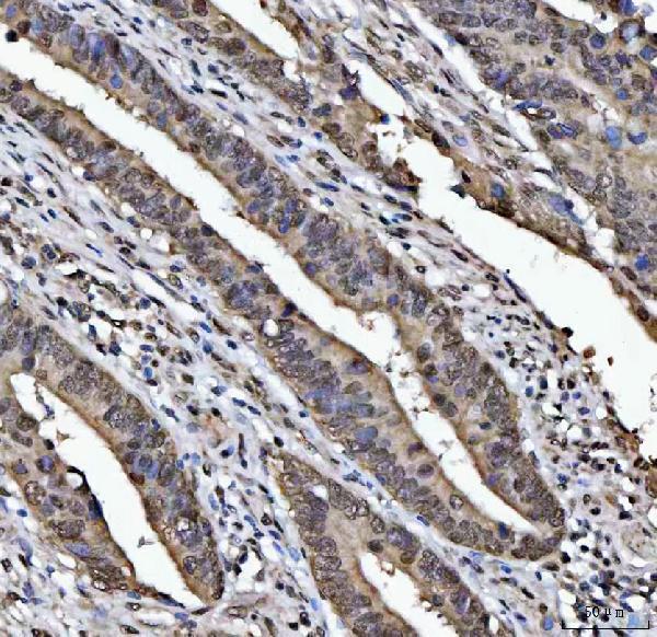
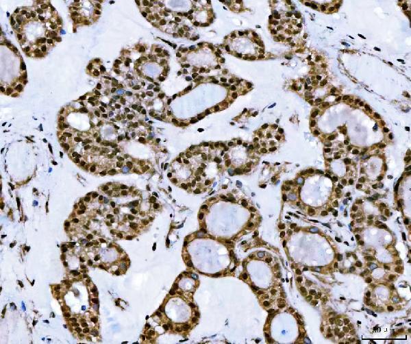
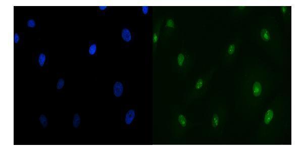
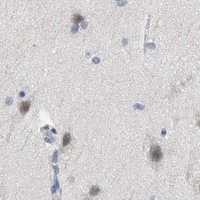

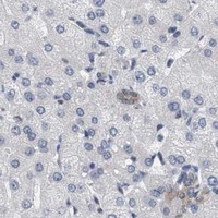
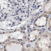


Comprehensive Human Protein Atlas IHC scoring per tissue (reliability: Enhanced — Medium consistency between antibody staining and RNA expression data.). Rows are taken directly from the HPA tissue chart — click any row's HPA link to view the source.
| Tissue | Cell type | Level | Evidence | Source |
|---|---|---|---|---|
| Bronchus | Ciliated cells (cell body) | High | Protein (IHC) | HPA → |
| Fallopian tube | Ciliated cells (cell body) | High | Protein (IHC) | HPA → |
| Testis | Pachytene spermatocytes | High | Protein (IHC) | HPA → |
| Appendix | Glandular cells | Medium | Protein (IHC) | HPA → |
| Breast | Glandular cells | Medium | Protein (IHC) | HPA → |
| Tissue | Cell type | Level | Evidence | Source |
|---|---|---|---|---|
| Adipose tissue | Adipocytes | Not detected | Protein (IHC) | HPA → |
| Caudate | Glial cells | Not detected | Protein (IHC) | HPA → |
| Cerebellum | Cells in granular layer | Not detected | Protein (IHC) | HPA → |
| Parathyroid gland | Glandular cells | Not detected | Protein (IHC) | HPA → |
| Salivary gland | Glandular cells | Not detected | Protein (IHC) | HPA → |
Troubleshoot RUVBL1 staining in paraffin sections by checking retrieval, compartment pattern, controls, and scoring before interpreting chromogenic signal.
A02049-2 has RUVBL1 IHC images from human colorectal, thyroid and lung cancers and spleen (catalog: IHC captions), plus IF images from A549 cells and human colon cancer tissue (catalog: IF captions).
A02049-2 shows staining in paraffin sections of human colorectal adenocarcinoma, thyroid cancer, lung adenocarcinoma and spleen (catalog: IHC captions). Its IF images show A549 cells and a paraffin section of human colon cancer (catalog: IF captions).
Which to pick: For tissue IHC, choose A02049-2: its paraffin-section images use EDTA retrieval at pH 8.0 and 2 μg/ml primary antibody; the fixative is unreported (catalog: IHC captions). For IF/ICC, the same SKU has A549-cell and human colon-section images at 5 μg/ml (catalog: IF captions). For cross-species work, A02049-2 lists Human, Monkey, Mouse and Rat reactivity, although its shown tissue images are human; it is rabbit-hosted, and clonality is unreported (catalog: reactivity; catalog: image captions; catalog: host and clone).