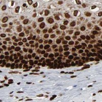
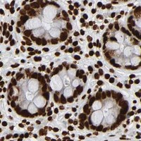
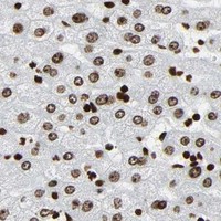
RXRA / Retinoic acid receptor RXR-alpha · IHC design guide

Design Immunohistochemistry for RXRA

Plan chromogenic RXRA IHC around widespread nuclear tissue staining (HPA tissue IHC). This guide covers fixation consistency and nuclear scoring (standard IHC practice), using the IHC-validated antibody at 0.5–1 μg/mL (datasheet A01299-1).

Evidence assembled Oct 2026 · For research use; verify linked source records and product datasheet before use
Immunohistochemistry protocol sheet for RXRA (IHC for RXRA): expected localisation Nuclear staining in tissue IHC (HPA tissue IHC), antibody A01299-1, validated IHC image, and IHC protocol steps
Printable RXRA IHC protocol sheet — expected localisation Nuclear staining in tissue IHC (HPA tissue IHC), antibody A01299-1, controls and protocol steps. Open the full RXRA IHC guide →

RXRA Immunohistochemistry Experimental Design Guide

Expected localisation, validated protocols, controls and antibodies — the at-a-glance facts below, then the full design guide.

Must know before staining
Expected localisation Nuclear staining in tissue IHC (HPA tissue IHC)
Staining pattern Widespread nuclear staining across tissue cell types (HPA tissue IHC)
Antigen retrieval Citrate pH 6 HIER, heat-mediated (datasheet A01299-1)
Positive control ⓘ Adrenal gland+4 more · see all
Negative control ⓘ None in HPA (detected in all 45 tissues); use no-primary + isotype controls
Important caveats
Reasons your staining may differ from the expected pattern.
Fixation Keep fixation consistent across sections (standard IHC practice; not target-specific); Matched tissue-IHC evidence does not establish the fixation claim. Validate the specimen-specific method before use. (selected-SKU IHC image A01299-1)
Caveat NR4A1 interaction can redirect RXRA to mitochondria (UniProt)
Regulation Vitamin D3 enhances nuclear localisation (UniProt)
Isoform / epitope Two isoforms; antibody epitope and isoform coverage unspecified (UniProt; datasheet A01299-1)
Section 1

Recommended RXRA IHC & IF Protocols

Compare the catalog antibody’s citrate pH 6 retrieval protocol (datasheet A01299-1) with two published human tissue IHC protocols (PMC12995935; PMC13513811).

Recommended immunohistochemistry (IHC-P) protocol parameters
SampleParaffin-embedded mouse liver tissue; fixative not specified (datasheet A01299-1)
FixationImage fixative and duration unreported (datasheet A01299-1); verify before use.
Sectioning4–5 µm sections on charged slides (standard)
DeparaffinisationXylene, graded ethanol series to water (standard)
Antigen retrievalHeat retrieval: Citrate pH 6, 20 min (datasheet A01299-1)
Peroxidase block3% H2O2, 10 min, room temperature (standard)
Blocking10% goat serum (datasheet A01299-1)
Primary antibodyRabbit anti-RXRA, 0.5-1μg/ml (datasheet A01299-1)
Primary incubationOvernight at 4 °C (datasheet A01299-1)
DetectionStreptavidin-biotin complex (SABC), DAB chromogen (datasheet A01299-1)
CounterstainHematoxylin, blue, dehydrate and mount (standard)
Expected resultRXRA-positive staining in glandular cells of adrenal gland (HPA tissue IHC: High). HPA tissue profile: Ubiquitous nuclear expression. No signal in the no-primary control.
💡Decision noteStart with citrate pH 6 heat retrieval for the catalog antibody (datasheet A01299-1); the published protocols use citrate retrieval but do not state its pH (PMC12995935; PMC13513811).
Section 2

What Is the Expected RXRA Staining Pattern?

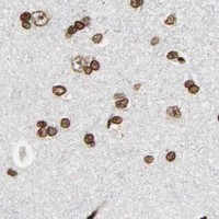
In paraffin-section IHC, expect predominantly nuclear RXRA staining across many cell types (HPA: ubiquitous nuclear expression; reliability Supported, with medium antibody–RNA consistency and external verification pending). High staining has been observed in glandular, hematopoietic, respiratory epithelial and glial cells, among others (HPA: tissue IHC). RXRA has no transmembrane segment, although cytoplasmic and mitochondrial localization is also reported (UniProt P19793: topology and subcellular location).

What am I looking at on my slide?
Clear nuclear chromogen in the expected cells, with interpretable counterstained nuclei.This fits the principal tissue pattern (HPA: ubiquitous nuclear expression). Stronger staining is plausible in the listed High groups, including adrenal glandular cells and bone-marrow hematopoietic cells (HPA: tissue IHC). Score the cell type and nuclear fraction; intensity alone does not establish specificity.
Predominantly cytoplasmic staining with little or no nuclear staining.This departs from the principal tissue-IHC pattern (HPA: ubiquitous nuclear expression). RXRA can also occur in cytoplasm or mitochondria (UniProt P19793: subcellular location), so compartment alone cannot prove artefact. Review the nuclear counterstain and controls before calling the signal RXRA.
Signal concentrates in an unexpected cell population while the expected nuclei are unstained.Consider antibody cross-reactivity or endogenous detection activity. The comparison is contextual: HPA reports broad nuclear expression and lists no negative tissue here (HPA: tissue IHC). An unlisted cell type is therefore not automatically RXRA-negative; check morphology and detection controls.
Brown haze covers nuclei, cytoplasm and extracellular spaces without crisp cell boundaries.Treat this as background until controls and morphology support a cellular pattern. It does not resemble the principal nuclear profile (HPA: tissue IHC). Uneven blocking, residual detection activity or excess chromogen are general IHC possibilities, not documented RXRA-specific effects.
No nuclear signal appears in a section containing a listed High population.Check the run before inferring absent RXRA. Examples include bronchial respiratory epithelial cells and caudate glial cells (HPA: High in those cell types). A single blank section cannot resolve failed staining from biological variation; HPA rates the tissue profile Supported, with external verification pending.
💡Expected RXRA appearanceCall a convincing positive when distinct nuclear chromogen is present in the relevant cells, especially a listed High population; diffuse acellular color or dominant cytoplasmic haze alone is unconvincing (HPA: tissue IHC; UniProt P19793: subcellular location).
How each factor affects the staining
Cell context and localizationThe nuclear fraction can vary: vitamin D3, VDR interaction and pulsatile shear stress have been linked to increased nuclear localization, while NR4A1 interaction can drive mitochondrial translocation (UniProt P19793: subcellular location). These observations do not predict the result in every paraffin tissue.
Choice of comparison tissueUse a listed High population as a practical positive comparator, such as adrenal glandular or bronchial respiratory epithelial cells (HPA: tissue IHC). HPA also describes ubiquitous nuclear expression and provides no negative tissue in this payload, so avoid treating another tissue as a proven negative.
Antibody evidenceCAB004565 and CAB005352 each have Supported IHC status (HPA: antibodies). The tissue profile has medium antibody–RNA consistency and awaits external verification (HPA: reliability). Concordant cell morphology and controls still matter when interpreting either antibody.
Isoforms and epitopeTwo isoforms and multiple modified residues are recorded (UniProt P19793: isoforms and modifications). No antibody epitope is supplied, so their effects on either antibody’s staining cannot be predicted. RXRA has no signal peptide or propeptide listed (UniProt P19793: processing).
IF/ICC Q&AWhat should an IF/ICC signal look like? Mainly nucleoplasmic, with additional Golgi localization in the HPA cell images; A-431 and U2OS images are listed (HPA: subcellular ICC-IF). This is cell-based evidence, separate from the paraffin-section IHC pattern.
Why is my staining missing, weak or wrong?
SituationLikely causeNext action
A listed High population is blank.The run or antibody may have failed, or the sampled cells may differ; the slide alone cannot distinguish these.Check section identity, nuclear morphology, detection controls and the antibody’s IHC-P instructions. Reassess against a listed High population (HPA: tissue IHC). Do not infer RXRA absence from one blank field.
Nuclear staining is faint or patchy.Biological variation or uneven staining may contribute; no RXRA-specific fixation sensitivity is reported in these sources.Compare like cell types across the section and a positive comparator (HPA: tissue IHC). Check general IHC retrieval and detection steps against the antibody instructions without assuming a target-specific fixation effect.
Diffuse brown background obscures nuclei.Residual endogenous detection activity, incomplete blocking or excess chromogen are general IHC possibilities.Inspect reagent and no-primary controls; review blocking, washing and development under the detection system’s instructions. Only score signal with clear cellular boundaries and a readable counterstain.
Cytoplasmic color dominates the field.It may reflect background or context-dependent RXRA localization; cytoplasm and mitochondrion are reported locations (UniProt P19793: subcellular location).Check whether convincing nuclei remain in expected cells (HPA: ubiquitous nuclear expression). Compare control sections and avoid calling cytoplasmic color alone a standard positive IHC pattern.
Only unexpected cells or acellular material stain.Cross-reactivity or endogenous detection activity is possible; an unlisted cell type is not a validated negative (HPA: ubiquitous nuclear expression).Confirm cell identity by morphology, inspect no-primary controls and compare a listed High population (HPA: tissue IHC). Treat extracellular deposits as background unless separately validated.
Two antibodies give different cellular patterns.Each has Supported IHC status, but the overall tissue profile has only medium antibody–RNA consistency (HPA: antibodies and reliability).Compare matched sections, controls and the same cell populations before interpretation. Record the disagreement; neither result alone establishes a new RXRA tissue pattern.

Sample controls for RXRA IHC & IF

🧪Start with bone marrow and expect staining in hematopoietic cells (HPA: High in bone-marrow hematopoietic cells). HPA detects RXRA in all 45 scored tissues, so there is no designated negative tissue; no-primary and isotype slides provide background controls, while unstained cells on the positive slide are only an internal background reference, not a validated RXRA-negative cell type (HPA: no negative rows; HPA: RXRA detected in all 45 tissues).
Positive control tissue: Adrenal gland (Glandular cells, HPA High)
Negative control tissue: None in HPA: RXRA is detected in all 45 scored tissues. Use a no-primary (secondary-only) and an isotype control instead.
ICC-IF cell lines (HPA subcellular resource): HPA ICC-IF images show RXRA in A-431, U2OS, with annotated localisation: Nucleoplasm (supported) (HPA subcellular).
Technical controls: Run a no-primary, secondary-only slide and a concentration-matched rabbit IgG isotype control alongside the rabbit primary (A01299-1 caption: rabbit primary). Use RXRA-knockout tissue, if available, as a biological specificity control; for chromogenic staining of bone marrow, quench endogenous peroxidase and check for residual signal on the no-primary slide (HPA: High in bone-marrow hematopoietic cells; standard IHC practice).
⚠️Feasibility: The A01299-1 mouse-liver paraffin-section caption reports citrate retrieval at pH 6 for 20 minutes, but does not report a fixative; a target-specific fixation window and fixation effect are therefore unreported (A01299-1 caption). This is a retrieval condition to test, not evidence that retrieval is universally required; the supplied evidence does not establish whether frozen sections or IF are easier, although HPA provides ICC-IF images in A-431 and U2OS (A01299-1 caption; HPA subcellular). In bone marrow, endogenous peroxidase can produce chromogenic background in hematopoietic cells (HPA: High in bone-marrow hematopoietic cells; standard IHC practice).

HPA tissue IHC evidence for RXRA

Comprehensive Human Protein Atlas IHC scoring per tissue (reliability: Supported — Medium consistency between antibody staining and RNA expression data. Pending external verification.). Rows are taken directly from the HPA tissue chart — click any row's HPA link to view the source.

Positive expression · recommended positive controls

TissueCell typeLevelEvidenceSource
Adrenal gland Glandular cells High Protein (IHC) HPA →
Appendix Glandular cells High Protein (IHC) HPA →
Bone marrow Hematopoietic cells High Protein (IHC) HPA →
Breast Adipocytes High Protein (IHC) HPA →
Bronchus Respiratory epithelial cells High Protein (IHC) HPA →

Undetected expression · recommended negative controls

TissueCell typeLevelEvidenceSource
None in HPA: RXRA is detected in all 45 scored tissues. Use a no-primary (secondary-only) and an isotype control instead.
Section 3

Advanced RXRA IHC Tips

Troubleshoot RXRA staining in paraffin sections by checking retrieval, nuclear localisation, antibody specificity and scoring controls.

What retrieval conditions should I start with for RXRA in paraffin sections?
Start with heat-mediated retrieval in citrate buffer at pH 6 for 20 minutes (datasheet A01299-1). The selected mouse liver image used those conditions before overnight incubation with 1 μg/ml catalog antibody at 4°C (datasheet A01299-1). If nuclear staining is weak, compare a modestly adjusted heating time on matched sections while keeping staining and development conditions constant (standard IHC practice). Include a no-primary control to distinguish newly exposed tissue background from RXRA signal (standard IHC practice). Record the retrieval conditions for each comparison, since changes in nuclear intensity alone cannot establish improved antibody specificity (standard IHC practice).
Could fixation explain weak or patchy RXRA staining?
Target-specific sensitivity of RXRA staining to fixation is unknown from the supplied evidence; the selected paraffin-section caption does not state a fixative (datasheet A01299-1). Document the actual fixative and fixation duration for each specimen before comparing nuclear signal across sections (standard IHC practice). Use similarly processed sections when adjusting retrieval, antibody concentration or development time, because differences in processing can confound those comparisons (standard IHC practice). Assess tissue preservation and staining together, and avoid treating a brighter section as evidence of a better fixation condition by itself (standard IHC practice). The supported HPA nuclear pattern provides a localisation reference, not a fixation comparison (HPA: tissue IHC, Supported).
How should I assess nuclear versus cytoplasmic RXRA staining?
Score nuclear staining separately: HPA describes ubiquitous nuclear tissue expression, while its cell imaging places RXRA mainly in the nucleoplasm (HPA: tissue IHC; HPA: subcellular). Cytoplasmic or mitochondrial signal is biologically possible, but depends on context in the UniProt record (UniProt P19793: subcellular location). Check whether nonnuclear chromogen follows intact cell boundaries and recurs in matched sections before assigning it to RXRA (standard IHC practice). Vitamin D3 and VDR are associated with enhanced nuclear localisation, and NR4A1 interaction with mitochondrial translocation (UniProt P19793: subcellular location). Report compartment-specific results; a total staining score would conceal a shift between nucleus and cytoplasm (standard IHC practice).
Can this stain distinguish RXRA isoforms or modified epitopes?
RXRA has 2 listed isoforms, but the supplied antibody caption does not map its epitope to either one (UniProt P19793: isoforms; datasheet A01299-1). Its ligand-binding domain spans residues 227–458, and phosphorylation is reported at several earlier residues, including 21 and 27 (UniProt P19793: domains and modified residues). Neither feature establishes which molecular form this antibody detects in paraffin sections (datasheet A01299-1; standard IHC interpretation). If an isoform or modification is the research endpoint, obtain epitope mapping and validate discrimination with appropriate controls before assigning stained cells to that form (standard IHC practice). Report ordinary chromogenic staining as RXRA immunoreactivity until that validation is available (standard IHC practice).
How can I compare RXRA IHC with multiplex immunofluorescence?
Use a marker of the cell type expected in the chosen section and assess RXRA within those marked cells, since HPA reports nuclear signal across many tissues (HPA: tissue IHC; standard IF practice). Choose fluorophores after measuring the section's autofluorescence, and reserve a clearly separated channel for the nuclear RXRA signal (standard IF practice). RXRA has no transmembrane segment and is mainly nucleoplasmic in HPA cell imaging, so permeabilise sufficiently for an intracellular nuclear epitope (UniProt P19793: topology; HPA: subcellular; standard IF practice). Optimise that permeabilisation with no-primary and single-stain controls, because the IHC caption supplies no IF conditions (datasheet A01299-1; standard IF practice). Compare compartment and cell identity across methods, rather than equating fluorescence intensity with DAB intensity (standard IHC/IF practice).
What should I check when DAB background obscures RXRA nuclei?
First inspect a no-primary section and the counterstain to locate background independent of the catalog antibody (standard IHC practice). The selected protocol used 10% goat serum blocking, a biotinylated secondary antibody, a streptavidin–biotin complex and DAB (datasheet A01299-1). Include a peroxidase block and check for tissue-dependent signal from the biotin-based detection system when staining appears outside cell boundaries (standard chromogenic IHC practice). If background persists, compare shorter DAB development or a lower primary concentration on matched sections, preserving a known nuclear reference (standard IHC practice; HPA: ubiquitous nuclear expression). Evaluate section edges separately because drying and uneven reagent coverage can concentrate chromogen there (standard IHC practice).
How should I quantify RXRA staining across paraffin sections? ⚠ ANSWER MARKED FOR VERIFICATION
Define the cell population and score nuclear RXRA before reviewing group labels, using the same threshold and imaging settings across sections (standard IHC practice; HPA: ubiquitous nuclear expression). An H-score combines the percentage of cells at each nuclear intensity; percentage-positive cells is simpler when intensity grading is unreliable (standard IHC practice). For spatial comparisons, report positive-cell density per mm² of viable analysed tissue and exclude folds, necrosis and section edges by preset rules (standard IHC practice). Normalise counts to the number of eligible cells or analysed area, and compare like compartments and cell populations across specimens (standard IHC practice). Report cytoplasmic staining separately if it is a study endpoint (UniProt P19793: subcellular location; standard IHC practice).
How can I distinguish true RXRA positivity from staining artefacts?
A reproducible nuclear pattern in intact cells is consistent with the HPA tissue profile, but staining alone does not establish antibody specificity (HPA: ubiquitous nuclear expression; standard IHC practice). Check whether positive cells match the annotated tissue compartment: HPA reports high signal in adrenal glandular cells and bone-marrow hematopoietic cells, among others (HPA: tissue IHC). Investigate diffuse extracellular DAB, edge-heavy staining, necrotic areas and signal retained in a no-primary section as potential artefacts (standard IHC practice). Peroxidase-related background can mimic positivity in chromogenic detection, so interpret it with the detection controls (standard IHC practice). Cytoplasmic or mitochondrial staining warrants separate validation because those locations are context-dependent in the UniProt record (UniProt P19793: subcellular location).
Boster reagents

Best RXRA / Retinoic acid receptor RXR-alpha IHC Antibodies

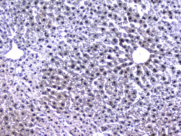
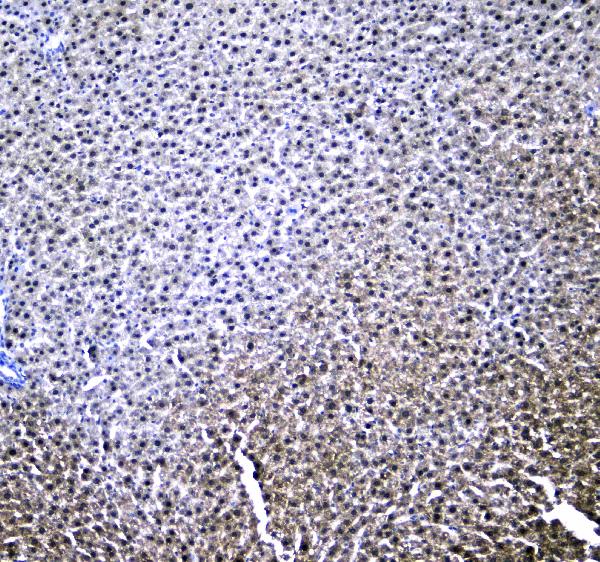
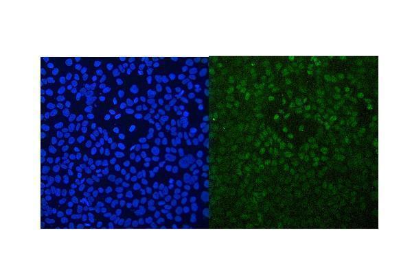
A01299-1 has IHC images of paraffin sections from mouse and rat liver and human liver and liver cancer tissue, plus an IF image of A431 cells (catalog image captions).

Real IHC data IHC analysis of RXRA using anti-RXRA antibody (A01299-1). RXRA was detected in paraffin-embedded section of mouse liver tissue. Heat mediated antigen retrieval was performed in citrate buffer (pH6, epitope retrieval solution) for 20 mins. The tissue section was blocked with 10% goat serum. The tissue section was then incubated with 1μg/ml rabbit anti-RXRA Antibody (A01299-1) overnight at 4°C. Biotinylated goat anti-rabbit IgG was used as secondary antibody and incubated for 30 minutes at 37°C. The tissue section was developed using Strepavidin-Biotin-Complex (SABC)(Catalog # SA1022) with DAB as the chromogen.
Anti-Retinoid X Receptor alpha/RXRA Antibody ®
Cat # A01299-1

A01299-1 is the only SKU that will render as an IHC card; its IHC captions document paraffin sections from mouse and rat liver and human liver and liver cancer tissue (catalog IHC image captions). Its IF caption documents A431 cells, and the catalog lists human, mouse and rat reactivity (catalog IF image caption; catalog reactivity).

Which to pick: Choose A01299-1 for tissue IHC: its own captions document paraffin sections across human, mouse and rat; the fixative is unreported (A01299-1 IHC image captions). For IF/ICC, A01299-1 has an A431 IF image, while rabbit monoclonal M01299-2, clone 20R23, lists ICC/IF without an IF image in the payload (A01299-1 IF image caption; M01299-2 catalog clone and applications). For cross-species IHC, A01299-1 has images across all three listed species; M01299-2 lists the same reactivity but does not list IHC as an application (A01299-1 IHC image captions and catalog reactivity; M01299-2 catalog reactivity and applications).

Each figure is that product's own IHC / IF validation image from its datasheet.

References

  1. UniProt Consortium. UniProt entry P19793 (RXRA_HUMAN, Retinoic acid receptor RXR-alpha).
  2. Human Protein Atlas. RXRA tissue IHC expression (reliability: Supported).
  3. Human Protein Atlas. RXRA subcellular location (ICC-IF): Mainly localized to the nucleoplasm. In addition localized to the Golgi apparatus..
  4. Human Protein Atlas. RXRA antibody validation summary (2 antibodies).
  5. TEAD4 and RXRA Regulate the Function of Nucleus Pulposus Cells in Intervertebral Disc Degeneration Via the TNF-α/NF-κB Pathway: An Integrated Analysis of Single-Cell RNA-Seq, Bulk RNA-Seq, and In Vitro Validation. Applied biochemistry and biotechnology 2026 — PMC12995935.
  6. Gaseous Air Pollutant Exposure and Atherosclerosis: A Systematic Study Integrating Multi-Omics and Network Toxicology. International journal of molecular sciences 2026 — PMC13513811.
  7. PubMed PMID:2159111 — UniProt-cited evidence.
  8. PubMed PMID:18619963 — UniProt-cited evidence.
  9. PubMed PMID:14702039 — UniProt-cited evidence.