This website uses cookies to ensure you get the best experience on our website.


- Table of Contents
Plan chromogenic RYBP IHC using placental trophoblasts as a high staining reference (HPA tissue IHC). Score nuclear and cytoplasmic signal (HPA tissue IHC), and consider 5 μg/mL as the catalog antibody’s IHC starting concentration (datasheet: A04316).

Expected localisation, validated protocols, controls and antibodies — the at-a-glance facts below, then the full design guide.
| Expected localisation | Nuclear and cytoplasmic staining (HPA tissue IHC) | |
| Staining pattern | Placental trophoblasts show nuclear and cytoplasmic staining (HPA tissue IHC) | |
| Antigen retrieval | Tris-EDTA pH 9.0 HIER, 95–98 °C, 20 min (rule: nuclear antigen) | |
| Positive control | Placenta+4 more · see all | |
| Negative control | Adipose tissue+4 more · see all |
| Fixation | Keep fixation consistent across sections (standard IHC practice; not target-specific) | |
| Caveat | Antibody staining and RNA show low consistency (HPA tissue IHC) | |
| Regulation | Reduced in breast cancer tissue (UniProt) | |
| Isoform / epitope | One full-length chain; no annotated isoforms (UniProt) |
The catalog antibody protocol is followed by published RYBP IHC methods for colorectal cancer, hepatocellular carcinoma, and mouse embryo sections (PMC10546375; PMC5244986; PMC11638158).
| Sample | Tissue sections; selected-image fixative not specified (standard IHC workflow) |
| Fixation | Image fixative and duration unreported (datasheet A04316); verify before use. |
| Sectioning | 4–5 µm sections on charged slides (standard) |
| Deparaffinisation | Xylene, graded ethanol series to water (standard) |
| Antigen retrieval | Heat-induced epitope retrieval in Tris-EDTA buffer, pH 9.0, 20 min at 95–98 °C (standard rule: nuclear antigen) |
| Peroxidase block | 3% H2O2, 10 min, room temperature (standard) |
| Blocking | 10% normal serum of the secondary host, 30 min, room temperature (standard) |
| Primary antibody | Rabbit anti-RYBP, 5 μg/mL (datasheet A04316) |
| Primary incubation | Overnight at 4 °C (standard) |
| Detection | HRP-polymer secondary, DAB chromogen 5–10 min (standard) |
| Counterstain | Hematoxylin, blue, dehydrate and mount (standard) |
| Expected result | RYBP-positive staining in trophoblastic cells of placenta (HPA tissue IHC: High). HPA tissue profile: Nuclear and cytoplasmic expression in several tissues, mainly in placenta. No signal in the no-primary control. |
RYBP is primarily nuclear, with nucleoplasmic localization and possible puncta; cytoplasmic staining is also reported (UniProt Q8N488, some puncta inferred by similarity; HPA tissue IHC; HPA ICC-IF). In paraffin sections, expect the clearest staining in placental trophoblastic cells, where HPA reports high staining (HPA tissue IHC). HPA rates its IHC evidence Approved but notes low consistency between antibody staining and RNA expression (HPA tissue IHC). RYBP has no transmembrane segment (UniProt Q8N488 topology).
| Placental trophoblastic cells show strong nuclear staining, with some cytoplasmic signal. | This fits the high trophoblast staining and nuclear plus cytoplasmic tissue profile (HPA tissue IHC). A predominantly nuclear result also fits UniProt Q8N488; compare compartments within the same cells rather than treating every cytoplasmic signal as unexpected. |
| Fine nuclear puncta appear within otherwise positive cells. | Puncta may fit the proposed Polycomb group body pattern, but that UniProt annotation is inferred by similarity (UniProt Q8N488). The supported ICC-IF location is nucleoplasm (HPA ICC-IF); puncta are therefore possible, not a required acceptance criterion for chromogenic IHC. |
| Signal is predominantly membranous or confined to extracellular material. | That distribution conflicts with the reported nuclear and cytoplasmic localization and the absence of a transmembrane segment (HPA tissue IHC; UniProt Q8N488 topology). Check section morphology and detection controls before calling it RYBP-specific staining (standard IHC practice). |
| Cells reported as unstained show signal as strong as placental trophoblasts. | For example, HPA reports adipocytes as not detected and placental trophoblasts as high (HPA tissue IHC). Unexpected staining raises possible cross-reactivity or endogenous detection activity; HPA's Approved rating and RNA–staining inconsistency warrant cautious interpretation (HPA tissue IHC; standard IHC practice). |
| Color spreads across stroma, empty spaces, and cells without a clear intracellular pattern. | Diffuse deposition lacks the reported nuclear and cytoplasmic cellular distribution (HPA tissue IHC). Consider nonspecific binding, incomplete blocking or washing, or chromogen development before scoring cells as positive (standard IHC practice). |
| Tissue and cell selection | Placental trophoblastic cells are a high-staining reference; squamous epithelial cells in esophagus, oral mucosa, tonsil and vagina are medium (HPA tissue IHC). Compare the named cell population, since a tissue-wide score can conceal differences among its cells (standard IHC practice). |
| Compartment and topology | UniProt places RYBP primarily in the nucleus and also lists cytoplasm; HPA tissue IHC reports both compartments (UniProt Q8N488; HPA tissue IHC). No transmembrane segment is annotated, so a membrane-only pattern needs control review (UniProt Q8N488 topology). |
| Strength of staining evidence | HPA calls tissue IHC Approved while reporting low consistency between antibody staining and RNA expression; the listed HPA053357 antibody is IHC Approved (HPA tissue IHC; HPA antibodies). Treat the tissue images as observed patterns, not proof that every positive cell has confirmed target-specific signal. |
| Target processing | UniProt annotates one chain spanning residues 1–228, with no signal peptide, propeptide or isoforms in this record (UniProt Q8N488). These annotations offer no basis for expecting a secreted compartment or a distinct isoform-specific staining pattern; the antibody epitope is unspecified. |
| IF/ICC Q: where should RYBP appear? | A: The supported HPA ICC-IF location is the nucleoplasm; its listed images include A-431, RT-4, U2OS and NIH 3T3 (HPA ICC-IF). IF/ICC has a separate guide; its localization evidence can inform compartment checks here without serving as an IHC protocol. |
| Situation | Likely cause | Next action |
|---|---|---|
| Placental trophoblasts show no convincing signal. | The expected high-staining reference is missing (HPA tissue IHC); a failed staining run or unsuitable assay conditions are possibilities (standard IHC practice). | Confirm that trophoblasts are present and intact, then review the validated IHC procedure, antibody preparation, detection reagents and a concurrent positive section (standard IHC practice). No RYBP-specific fixation sensitivity is supplied. |
| Only diffuse background appears. | Diffuse color does not match the reported cellular nuclear and cytoplasmic pattern (HPA tissue IHC); nonspecific binding or excess detection signal may contribute (standard IHC practice). | Check primary-omission and detection controls, blocking, washes and chromogen development; then reassess whether staining resolves to individual cells (standard IHC practice). |
| Signal is mainly at cell membranes. | RYBP lacks an annotated transmembrane segment and is reported in nuclear and cytoplasmic compartments (UniProt Q8N488 topology; HPA tissue IHC). | Inspect morphology and control sections, and score nuclear and cytoplasmic staining separately. Investigate persistent membrane signal as a possible assay artefact (standard IHC practice). |
| A reported unstained cell population scores strongly positive. | HPA reports adipocytes and several other specified cell populations as not detected, but its IHC evidence has low RNA–staining consistency (HPA tissue IHC). Cross-reactivity or endogenous detection activity is possible (standard IHC practice). | Verify the exact cell type and compare it with a concurrent placental section; review primary-omission and endogenous-activity controls before assigning RYBP positivity (HPA tissue IHC; standard IHC practice). |
| Placental staining is weak while surrounding background is high. | The expected high trophoblast signal has poor contrast (HPA tissue IHC). Background or insufficient effective detection may prevent a reliable cell-level call (standard IHC practice). | Review tissue preservation, assay controls, antibody dilution and detection development under the validated IHC procedure (standard IHC practice). The supplied sources give no RYBP-specific retrieval or fixation-response rule. |
| Puncta are absent despite clear nuclear staining. | Puncta are described as likely Polycomb group bodies by similarity, whereas HPA supports nucleoplasmic localization (UniProt Q8N488; HPA ICC-IF). | Assess the overall cellular and compartment pattern against the positive section; do not reject an otherwise convincing IHC result solely because discrete puncta are unresolved (HPA tissue IHC; standard IHC practice). |


Comprehensive Human Protein Atlas IHC scoring per tissue (reliability: Approved — Low consistency between antibody staining and RNA expression data.). Rows are taken directly from the HPA tissue chart — click any row's HPA link to view the source.
| Tissue | Cell type | Level | Evidence | Source |
|---|---|---|---|---|
| Placenta | Trophoblastic cells | High | Protein (IHC) | HPA → |
| Esophagus | Squamous epithelial cells | Medium | Protein (IHC) | HPA → |
| Oral mucosa | Squamous epithelial cells | Medium | Protein (IHC) | HPA → |
| Testis | Sertoli cells | Medium | Protein (IHC) | HPA → |
| Tonsil | Squamous epithelial cells | Medium | Protein (IHC) | HPA → |
| Tissue | Cell type | Level | Evidence | Source |
|---|---|---|---|---|
| Adipose tissue | Adipocytes | Not detected | Protein (IHC) | HPA → |
| Adrenal gland | Glandular cells | Not detected | Protein (IHC) | HPA → |
| Appendix | Glandular cells | Not detected | Protein (IHC) | HPA → |
| Bone marrow | Hematopoietic cells | Not detected | Protein (IHC) | HPA → |
| Caudate | Glial cells | Not detected | Protein (IHC) | HPA → |
Troubleshoot RYBP chromogenic IHC in paraffin sections by checking retrieval, compartment-specific staining, controls, and cell-level scoring before interpreting signal.
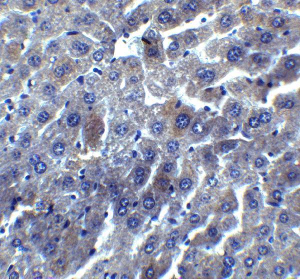
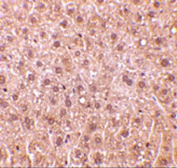
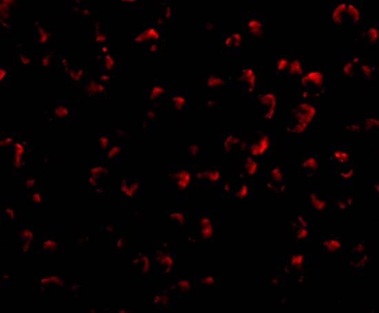
The IHC-validated antibody A04316 has mouse liver IHC images and IF images from A549 cells and mouse liver (catalog: A04316 image captions). Its listed reactivity is human, mouse and rat (catalog: A04316 reactivity).
A04316 is the SKU with a card and its own IHC figure, showing mouse liver stained at 5 μg/mL (catalog: A04316 IHC image caption). Its IF images show A549 cells and mouse liver at 20 μg/mL (catalog: A04316 IF image captions).
Which to pick: Choose A04316 for paraffin section IHC: IHC-P is listed, and its own IHC image shows mouse liver staining at 5 μg/mL; the fixative is unreported (catalog: A04316 applications and IHC image caption). For IF/ICC work, A04316 has IF images in A549 cells and mouse liver, while M04316-3 lists ICC/IF but has no supplied IF image (catalog: A04316 IF image captions; M04316-3 applications and image captions). Both list human, mouse and rat reactivity; M04316-3 is monoclonal, and A04316 has documented mouse liver IHC and IF images (catalog: reactivity, clone and image captions).