This website uses cookies to ensure you get the best experience on our website.
- Table of Contents
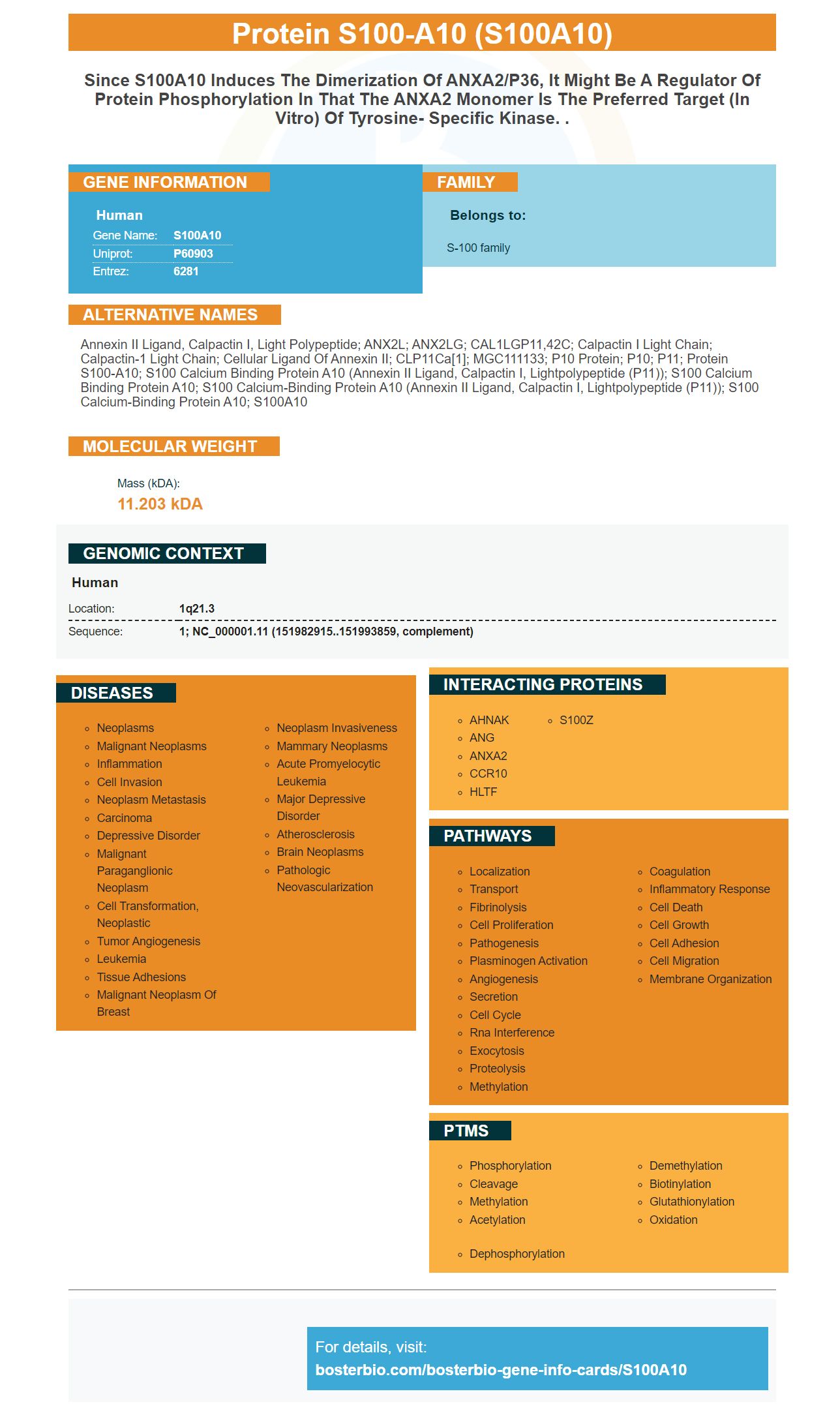
Facts about Protein S100-A10.
Human | |
---|---|
Gene Name: | S100A10 |
Uniprot: | P60903 |
Entrez: | 6281 |
Belongs to: |
---|
S-100 family |
annexin II ligand, calpactin I, light polypeptide; ANX2L; ANX2LG; CAL1LGP11,42C; Calpactin I light chain; Calpactin-1 light chain; Cellular ligand of annexin II; CLP11Ca[1]; MGC111133; p10 protein; p10; P11; protein S100-A10; S100 calcium binding protein A10 (annexin II ligand, calpactin I, lightpolypeptide (p11)); S100 calcium binding protein A10; S100 calcium-binding protein A10 (annexin II ligand, calpactin I, lightpolypeptide (p11)); S100 calcium-binding protein A10; S100A10
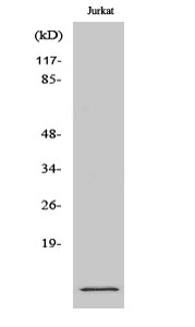
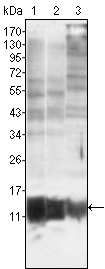
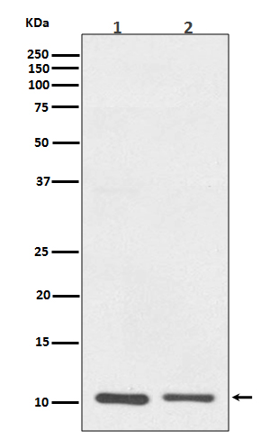
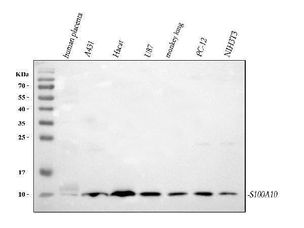
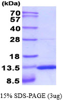
Mass (kDA):
11.203 kDA
Human | |
---|---|
Location: | 1q21.3 |
Sequence: | 1; NC_000001.11 (151982915..151993859, complement) |
It is important to determine what the S100A10 marker's function is and where it is located in order to make the most of it. The S100A10 protein is produced by E.coli and contains a 1-97aa-sequence. It can be stored at +2degC- +8degC up to one week, or -20degC- -80degC long-term storage. It is strongly advised to avoid repeated freeze-thaw cycles. Scientists who use the protein to conduct their experiments can earn credit by sharing their findings with other scientists. Every scientist in the world can benefit from the rewards.
The monoclonal antibody Anti-Bax (Apoptoses marker) from the Boster Bio catalog reacts to Human, Mouse, and Rat Bax. This antibody has been tested for a variety of assays. It reacts with the human Bax's N-terminus. It was developed to target the N-terminus of human Bax.
In the studies, xenograft tumor cells from mice that were expressing tumors were pulverized through a 100mm cell strainer. The cells were then lysed using hard pipetting. The instructions of the manufacturer were followed after the antibodies were applied. The results revealed that the OE group had a higher rate of apoptosis than the other two groups. Western Analyses of blots showed that the OE group had lower expression of Bcl-2 PCNA and an increase in expression of caspase-3. The expression of the pro-apoptotic molecule GAPDH was not affected.
This antibody is able to bind and inhibit FAS which is a vital marker for apoptosis. The antibody also increased CC3 levels in tumor cells and inhibited the proliferation marker Ki67 in the same tumors. In addition, it dramatically reduced the amount of Ki67, a proliferative marker. The results presented here show that the Anti-Bax (Apoptosis Marker) monoclonal antibody made by Boster Bio can be useful in a variety of ways.
In addition to targeting tumor antigens and immune checkpoints the immune checkpoints have also demonstrated promising clinical results against different malignancies. These antibodies are expensive and can have serious side effects, such as resistance. Monoclonal antibodies can be expensive and difficult to produce in large quantities. Certain patients might not be able to tolerate these antibodies. However, active immunization may be a great alternative in many situations and, in some instances patients can benefit from both.
Anti-S100A10 antibody recognizes S100A10 protein in perfusion-fixed sections of the mouse kidney tissue. It shows no cross-reactivity the rhS100B antibody or the rhS100A10 antibody. The general protocols for this antibody may be found in the Technical Information section of our website. This antibody is suitable for use in Western Blots as well as Direct ELISA.
S100A10 is a calcium binding protein is found most often in mast cells. It is an heterotetrameric compound consisting of two S100A10 subunits and two Annexin A2 subunits. It is located on the cell surface and intracellularly within the plasma membrane. It plays a role in the production of plasmins by cells as well as invasiveness.
The S100A10 marker is stable at temperatures of room temperature, which makes it suitable for biophysical research. The protein is stable at all temperatures. The purified protein will permit researchers to perform biophysical experiments and investigate the mechanisms of membrane binding. These experiments can also be used to determine the conditions that alter binding and cause a loss of function. These biophysical studies in the future will assist researchers in determining the role of S100A10.
The S100A10 protein has been expressed in E.coli cells and then removed using standard chromatography methods. S100 proteins belong to the EF-hand calcium binding protein family and play a variety of roles, including regulating the expression of specific proteins. S100A10 is involved in a variety of processes throughout the body, such as protein phosphorylation, enzymatic activity, and Ca2+ homeostaemia. S100 proteins also have roles in the cytoskeleton as well as transcription factors. They are involved in the detection of colorectal cancer.
The S100A10 marker is part of the S100 family. It is mostly found in mast cells, and is located in heterotetrameric structures that include two annexin A2 subunits as well as one S100A10. The protein is present in the plasma membrane as well as on the extracellular surface of cells. It is a key regulator for cellular processes including the production and utilization of plasminogen.
The cell's cytoplasm has the S100A10 protein. It forms a heterotetramer with the annexin A2. It is in contact with the actin-cytoskeleton. It regulates neurotransmitter receptors as well as neuron-specific ion channelslike NaV1.8, ASIC1, TASK1, and TASK1. It also modulates the activity and invasion of macrophages by releasing receptor-like signals.
S100A10 was detected in paraffin embedded sections of human kidney cancer tissue by using a goat anti-human polyclonal antibody. The staining was done at 4°C and counterstained with the hematoxylin. These results suggest that the S100A10 marker plays an important role in the emergence and development of aggressive tumor symptoms.
HESCs with the S100A10 knockout showed a decrease in cell migration. HESCs as well as HEECs treated with 8-Br-cAMP demonstrated a decrease in surface area colonized by the. The knockdown slowed down the cell movement of both types of cells. They also reduced the amount of cellular debris on their surfaces. This is in line with previous studies. It is important to know that S100A10 knockout cells do not have a significant increase in the size of their colonies when exposed to 8-Br-cAMP.
PMID: 1831433 by Kube E., et al. Primary structure of human, chicken, and Xenopus laevis p11, a cellular ligand of the Src-kinase substrate, annexin II.
PMID: 1533380 by Harder T., et al. Cloning and characterization of the human gene encoding p11: structural similarity to other members of the S-100 gene family.