This website uses cookies to ensure you get the best experience on our website.


- Table of Contents
S100A14 is cytoplasmic (UniProt), while tissue IHC highlights squamous epithelial cells and sometimes membrane staining (HPA tissue IHC). This guide pairs human paraffin-section IHC at 2–5 µg/ml (datasheet A06301-1) with cell-specific controls selected from the tissue profile (HPA tissue IHC).

Expected localisation, validated protocols, controls and antibodies — the at-a-glance facts below, then the full design guide.
| Expected localisation | Cytoplasmic (UniProt); membranous in some tissues (HPA tissue IHC) | |
| Staining pattern | Squamous epithelial staining; membranous in some tissues (HPA tissue IHC) | |
| Antigen retrieval | EDTA pH 8.0 HIER, heat-mediated (datasheet A06301-1) | |
| Positive control | Appendix+4 more · see all | |
| Negative control | Adipose tissue+4 more · see all |
| Fixation | Keep fixation conditions consistent across sections. (standard IHC practice; not target-specific) | |
| Caveat | Whole-tissue scoring may obscure endocrine-cell staining (HPA tissue IHC) | |
| Regulation | Expression varies by cell type (HPA tissue IHC) | |
| Isoform / epitope | One 1–104 chain, no isoforms; cytoplasmic epitope expected (UniProt) |
The catalog antibody has a datasheet IHC protocol; four published S100A14 chromogenic IHC protocols provide tissue-specific examples (datasheet A06301-1; PMC4348405; PMC6026614; PMC5550849; PMC4116497).
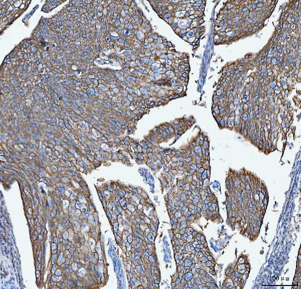
| Sample | Paraffin-embedded human cervica squamous carcinoma tissue; fixative not specified (datasheet A06301-1) |
| Fixation | Image fixative and duration unreported (datasheet A06301-1); verify before use. |
| Sectioning | 4–5 µm sections on charged slides (standard) |
| Deparaffinisation | Xylene, graded ethanol series to water (standard) |
| Antigen retrieval | Heat retrieval: EDTA pH 8.0 (datasheet A06301-1); 20 min, 95–100 °C (standard) |
| Peroxidase block | 3% H2O2, 10 min, room temperature (standard) |
| Blocking | 10% goat serum (datasheet A06301-1) |
| Primary antibody | Rabbit anti-S100A14, 2-5 µg/ml (datasheet A06301-1) |
| Primary incubation | Overnight at 4 °C (datasheet A06301-1) |
| Detection | HRP-conjugated secondary, DAB chromogen (datasheet A06301-1) |
| Counterstain | Hematoxylin, blue, dehydrate and mount (standard) |
| Expected result | S100A14-positive staining in endocrine cells of appendix (HPA tissue IHC: High). HPA tissue profile: Expression in squamous epithelial cells with additional membranous expression in some tissues. No signal in the no-primary control. |
In paraffin-section IHC, expect S100A14 mainly in squamous epithelial cells, with additional membranous staining in some tissues (HPA tissue IHC: Enhanced). Cytoplasmic staining is consistent with its reported location (UniProt Q9HCY8: Cytoplasm). S100A14 has no transmembrane segment, so membrane-associated staining should be judged against the tissue pattern, not treated as evidence of a membrane-spanning protein (UniProt Q9HCY8: topology; HPA tissue IHC: profile).
| Strong staining in esophageal or cervical squamous epithelial cells, with cytoplasmic and possibly membranous signal. | This fits the expected cell type and reported high staining in both tissues (HPA tissue IHC: Esophagus and Cervix, High). Score signal within the squamous cells; the surrounding tissue is not expected to stain uniformly on that basis. |
| Strong staining is confined to scattered endocrine cells in colon or appendix rather than every epithelial cell. | This can be a correct positive result: HPA assigns High staining specifically to endocrine cells in colon and appendix (HPA tissue IHC: Colon and Appendix, High). Check cell identity before calling the section weak or patchy. |
| Signal appears only in extracellular material or stroma, without staining in the expected cells. | That distribution does not match the reported cytoplasmic location or the HPA cell-specific IHC profile (UniProt Q9HCY8: Cytoplasm; HPA tissue IHC: profile). Review morphology and controls before attributing it to S100A14. |
| Broad staining appears in a cell type reported as undetected, or colour develops on the negative control. | Suspect nonspecific binding or endogenous detection activity and compare controls (general IHC practice). HPA reports adipocytes and bronchial respiratory epithelial cells as Not detected; that finding is specific to those cells (HPA tissue IHC). |
| A known-positive squamous epithelium has no visible signal while the tissue and counterstain remain interpretable. | Treat this as a possible assay failure before concluding that S100A14 is absent: esophageal and cervical squamous cells are High in HPA IHC (HPA tissue IHC). Recheck antibody use, detection and controls (general IHC practice). |
| Choice of positive tissue and cells | Use the named cells when assessing a control: squamous epithelial cells in esophagus or cervix, basal-layer cells in skin, or endocrine cells in colon are High (HPA tissue IHC). Whole-tissue averages can hide these restricted patterns (general IHC interpretation). |
| Compartment and topology | UniProt places S100A14 in the cytoplasm and reports no transmembrane segment (UniProt Q9HCY8). HPA also observes membrane-associated staining in some tissues (HPA tissue IHC: profile); assess that signal alongside the expected cell type. |
| IHC evidence and protocol scope | HPA rates tissue IHC reliability Enhanced and lists HPA027613 as IHC Enhanced (HPA tissue IHC; HPA antibodies). These ratings support pattern interpretation; neither supplies a target-specific retrieval condition or fixation-sensitivity claim. |
| IF/ICC Q&A: Should its compartments match IHC exactly? | No exact match is established: ICC-IF reports nuclear bodies, plasma membrane, cell junctions and cytosol, while HPA027613 is ICC Uncertain (HPA subcellular; HPA antibodies). Interpret IF/ICC with its own controls; this IHC section supplies no IF/ICC protocol. |
| Situation | Likely cause | Next action |
|---|---|---|
| No staining in esophageal or cervical squamous epithelium. | The assay may have failed; those cells are reported High in tissue IHC (HPA tissue IHC). | Check section integrity, antibody application and detection with an appropriate positive control (general IHC practice). Avoid inferring target-specific fixation sensitivity from this result. |
| Colon looks mostly unstained despite a few strongly coloured cells. | The expected High result belongs to endocrine cells, not all colon cells (HPA tissue IHC: Colon, Endocrine cells). | Inspect the positive cells at higher magnification and score their distribution by cell type (general IHC interpretation). |
| Staining is concentrated in stroma or extracellular deposits. | That pattern conflicts with the reported cellular distribution (UniProt Q9HCY8: Cytoplasm; HPA tissue IHC: profile). | Compare the expected epithelial cells and a negative control; review nonspecific staining before scoring the deposits as positive (general IHC practice). |
| Diffuse colour obscures cell boundaries throughout the section. | Background may arise from nonspecific antibody binding or endogenous detection activity (general IHC practice). | Review reagent-only controls and blocking or detection steps, then assess whether a cell-specific pattern remains (general IHC practice). |
| Reported undetected cells stain as strongly as the intended positive cells. | The cell-level pattern is discordant with HPA for adipocytes or bronchial respiratory epithelial cells (HPA tissue IHC: Not detected). | Verify cell identity and compare negative controls before interpreting the signal as S100A14 (general IHC practice). Do not generalize those negative calls to every cell in either tissue. |
| Nuclear-body signal in IF/ICC seems inconsistent with the IHC slide. | HPA reports nuclear bodies in ICC-IF, but lists HPA027613 ICC validation as Uncertain (HPA subcellular; HPA antibodies). | Evaluate IF/ICC with its own controls and validation evidence; do not use its nuclear-body signal alone to overturn an IHC pattern (general assay interpretation). |
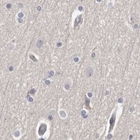
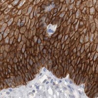

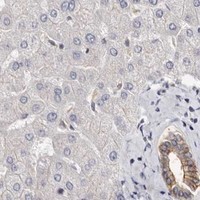


Comprehensive Human Protein Atlas IHC scoring per tissue (reliability: Enhanced — High consistency between antibody staining and RNA expression data.). Rows are taken directly from the HPA tissue chart — click any row's HPA link to view the source.
| Tissue | Cell type | Level | Evidence | Source |
|---|---|---|---|---|
| Appendix | Endocrine cells | High | Protein (IHC) | HPA → |
| Cervix | Squamous epithelial cells | High | Protein (IHC) | HPA → |
| Colon | Endocrine cells | High | Protein (IHC) | HPA → |
| Duodenum | Endocrine cells | High | Protein (IHC) | HPA → |
| Esophagus | Squamous epithelial cells | High | Protein (IHC) | HPA → |
| Tissue | Cell type | Level | Evidence | Source |
|---|---|---|---|---|
| Adipose tissue | Adipocytes | Not detected | Protein (IHC) | HPA → |
| Adrenal gland | Glandular cells | Not detected | Protein (IHC) | HPA → |
| Bone marrow | Hematopoietic cells | Not detected | Protein (IHC) | HPA → |
| Bronchus | Respiratory epithelial cells | Not detected | Protein (IHC) | HPA → |
| Caudate | Glial cells | Not detected | Protein (IHC) | HPA → |
Troubleshoot S100A14 chromogenic IHC in paraffin sections using the catalog antibody’s tissue result and the supplied protein localisation evidence.
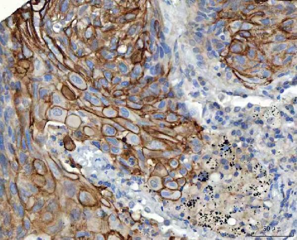
A06301-1 has real IHC data from paraffin sections of human cervical and lung squamous carcinomas (IHC image captions). Human is the only listed reactive species (catalog reactivity).
A06301-1 is listed for human IHC (catalog applications and reactivity). Its images show staining in paraffin sections of human cervical and lung squamous carcinomas (IHC image captions).
Which to pick: Choose A06301-1 for human paraffin-section IHC: it is a rabbit polyclonal antibody listed for IHC, with a 2–5 µg/ml catalog range (catalog antibody details and dilution); its images use 2 µg/ml after EDTA pH 8.0 heat retrieval (IHC image captions). The captions identify paraffin sections but do not report the fixative (IHC image captions). No SKU in this payload is listed for IF/ICC or another species, so there is no supported pick for either (catalog applications and reactivity).