This website uses cookies to ensure you get the best experience on our website.


- Table of Contents
Plan paraffin-section IHC around cytoplasmic and membranous staining in glandular and squamous epithelium (HPA tissue IHC). Use esophageal squamous epithelium as a high-staining reference and interpret nuclear localisation in light of calcium-dependent transport (HPA tissue IHC; UniProt).

Expected localisation, validated protocols, controls and antibodies — the at-a-glance facts below, then the full design guide.
| Expected localisation | Epithelial cytoplasm and membrane (HPA tissue IHC) | |
| Staining pattern | Glandular and squamous cells show cytoplasmic and membranous staining (HPA tissue IHC) | |
| Antigen retrieval | Tris-EDTA pH 9.0 HIER, 95–98 °C, 20 min (rule: nuclear antigen) | |
| Positive control | Esophagus+4 more · see all | |
| Negative control | Adipose tissue+4 more · see all |
| Fixation | Keep fixation consistent across sections. (standard IHC practice; not target-specific) | |
| Caveat | Calcium levels can shift nuclear and cytoplasmic localisation (UniProt) | |
| Regulation | Up-regulated in bladder and lung tumors (UniProt) | |
| Isoform / epitope | One 1–103 chain; no listed isoforms or signal peptide (UniProt) |
The catalog antibody’s IHC-P protocol is accompanied by four published S100A16 IHC methods (PMC8072221; PMC10572706; PMC5679695; PMC7039973).
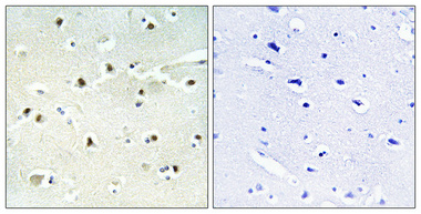
| Sample | Paraffin-embedded Human brain tissue; fixative not specified (datasheet A08032-1) |
| Fixation | Image fixative and duration unreported (datasheet A08032-1); verify before use. |
| Sectioning | 4–5 µm sections on charged slides (standard) |
| Deparaffinisation | Xylene, graded ethanol series to water (standard) |
| Antigen retrieval | Heat-induced epitope retrieval in Tris-EDTA buffer, pH 9.0, 20 min at 95–98 °C (standard rule: nuclear antigen) |
| Peroxidase block | 3% H2O2, 10 min, room temperature (standard) |
| Blocking | 10% normal serum of the secondary host, 30 min, room temperature (standard) |
| Primary antibody | Rabbit anti-S100A16, 1:100-1:300 (datasheet A08032-1) |
| Primary incubation | Overnight at 4 °C (standard) |
| Detection | HRP-polymer secondary, DAB chromogen 5–10 min (standard) |
| Counterstain | Hematoxylin, blue, dehydrate and mount (standard) |
| Expected result | S100A16-positive staining in squamous epithelial cells of esophagus (HPA tissue IHC: High). HPA tissue profile: Cytoplasmic and membranous expression in glandular and squamous epithelial cells. No signal in the no-primary control. |
S100A16 is an intracellular protein without a transmembrane segment (UniProt Q96FQ6 topology). In paraffin-section IHC, expect cytoplasmic and membranous staining in glandular and squamous epithelial cells, particularly strong staining in esophageal squamous epithelium (HPA: tissue IHC profile; esophagus High). HPA rates its tissue staining Enhanced, reflecting high consistency between antibody staining and RNA expression (HPA: tissue IHC reliability).
| Strong staining outlines or fills esophageal squamous epithelial cells. | This fits the expected positive pattern: esophageal squamous epithelial cells are High, and the broader tissue IHC profile is cytoplasmic and membranous (HPA: esophagus High; tissue IHC profile). Judge the distribution within the epithelium as well as the overall color intensity. |
| Staining is exclusively extracellular or confined to gland lumens, with little intracellular epithelial signal. | That distribution conflicts with the reported intracellular and membranous pattern (HPA: tissue IHC profile; UniProt Q96FQ6 subcellular location). Treat it as a possible staining artefact and inspect tissue morphology and controls before scoring it as S100A16; UniProt also reports nucleolar localization, so nuclear staining alone is not proof of artefact (UniProt Q96FQ6 subcellular location). |
| Strong staining appears mainly in alveolar cells while an esophageal positive control stains as expected. | HPA reports alveolar cells as Not detected and esophageal squamous epithelial cells as High (HPA: lung alveolar cells; esophagus squamous epithelial cells). Check for antibody cross-reactivity or endogenous chromogen-generating activity using appropriate controls (general IHC practice). Avoid inferring that all lung cell types are negative from the alveolar-cell entry. |
| Weak, diffuse color covers epithelium, stroma, and empty areas similarly. | A pattern without clear cellular boundaries cannot establish the reported cytoplasmic and membranous epithelial distribution (HPA: tissue IHC profile). Review the no-primary control, washes, blocking, and detection conditions as general IHC checks; diffuse color by itself does not identify a target-specific cause (general IHC practice). |
| No signal is visible in well-preserved esophageal squamous epithelium. | That is discordant with the High esophageal staining reported by HPA (HPA: esophagus squamous epithelial cells). Verify that the section contains the expected cells and that the detection run worked; then review antibody and retrieval conditions using the validated IHC procedure (general IHC practice). The supplied sources do not establish a S100A16-specific fixation effect. |
| Cell type and tissue context | HPA reports High staining in squamous epithelial cells of esophagus, tonsil, and vagina, and Medium staining in appendix glandular cells (HPA: tissue IHC). Compare like cells with like cells: an unstained alveolar cell is consistent with its Not detected entry, although this does not describe every lung cell (HPA: lung alveolar cells). |
| Compartment and topology | HPA describes cytoplasmic and membranous epithelial staining, while UniProt lists nucleus, nucleolus, and cytoplasm and reports no transmembrane segment (HPA: tissue IHC profile; UniProt Q96FQ6 subcellular location and topology). A membranous IHC appearance therefore does not establish that S100A16 spans the membrane. |
| Calcium-linked localization | UniProt reports primarily nucleolar localization, nucleolar exit and nucleocytoplasmic transport at high intracellular calcium, and nuclear accumulation at low calcium (UniProt Q96FQ6 subcellular location). This supports caution when interpreting a nuclear signal, but the supplied evidence does not predict a particular calcium state in a paraffin section. |
| Tissue evidence versus cell-level staining | UniProt describes S100A16 expression as ubiquitous and highly expressed in adipose tissue, whereas HPA lists adipocytes as Not detected by tissue IHC (UniProt Q96FQ6 tissue specificity; HPA: adipose adipocytes). Use the HPA cell-level result when choosing an IHC comparator; the sources do not resolve why these observations differ. |
| Antibody validation | HPA rates antibody HPA045841 Enhanced for IHC and describes high consistency between antibody staining and RNA expression (HPA: antibody validation; tissue IHC reliability). That supports the reported pattern but does not validate every antibody or make a control unnecessary for a new staining run (general IHC practice). |
| Q: What should IF/ICC show? | A: HPA reports mainly cytosolic signal, with additional plasma-membrane localization, in its ICC-IF summary (HPA: subcellular ICC-IF). This is localization context only; assess IF/ICC methods in its separate guide. |
| Situation | Likely cause | Next action |
|---|---|---|
| The esophageal positive control has no visible epithelial staining. | The result conflicts with HPA's High esophageal squamous-cell staining; the failed run alone cannot identify whether the section, antibody, retrieval, or detection is responsible (HPA: esophagus; general IHC practice). | Confirm the squamous epithelium is present, check the detection control, and repeat using the IHC-validated procedure's stated conditions (general IHC practice). |
| Brown color persists in a no-primary control. | The color cannot be attributed to primary-antibody binding; endogenous detection activity or detection-reagent background are possible explanations (general chromogenic IHC practice). | Review the detection system's endogenous-activity blocking step, reagent controls, and wash conditions before interpreting S100A16 staining (general IHC practice). |
| All tissue regions show a similar pale haze. | Indiscriminate color obscures the epithelial cytoplasmic and membranous pattern expected from HPA (HPA: tissue IHC profile); its exact source is undetermined. | Compare the no-primary control and review blocking, washes, and primary-antibody concentration under the validated procedure (general IHC practice). |
| Signal appears only over luminal material or outside cells. | An extracellular-only pattern conflicts with the reported cellular locations (HPA: tissue IHC profile; UniProt Q96FQ6 subcellular location). | Check section morphology and the no-primary control, then score only staining assignable to intact cells (general IHC practice). |
| Nuclei stain strongly, with little epithelial cytoplasmic signal. | HPA's tissue IHC profile emphasizes cytoplasmic and membranous staining, but UniProt reports nuclear and nucleolar localization (HPA: tissue IHC profile; UniProt Q96FQ6 subcellular location). | Compare a known-positive section and controls before deciding whether the nuclear pattern is biological or background (general IHC practice). |
| A presumed negative adipose section shows staining in cells other than adipocytes. | HPA's Not detected call applies specifically to adipocytes, not every cell in adipose tissue (HPA: adipose adipocytes). | Identify the stained cell type morphologically and compare the no-primary control before calling the section an invalid negative control (general IHC practice). |


Comprehensive Human Protein Atlas IHC scoring per tissue (reliability: Enhanced — High consistency between antibody staining and RNA expression data.). Rows are taken directly from the HPA tissue chart — click any row's HPA link to view the source.
| Tissue | Cell type | Level | Evidence | Source |
|---|---|---|---|---|
| Esophagus | Squamous epithelial cells | High | Protein (IHC) | HPA → |
| Fallopian tube | Ciliated cells (tip of cilia) | High | Protein (IHC) | HPA → |
| Skin | Cells in corneal layer | High | Protein (IHC) | HPA → |
| Tonsil | Squamous epithelial cells | High | Protein (IHC) | HPA → |
| Vagina | Squamous epithelial cells | High | Protein (IHC) | HPA → |
| Tissue | Cell type | Level | Evidence | Source |
|---|---|---|---|---|
| Adipose tissue | Adipocytes | Not detected | Protein (IHC) | HPA → |
| Adrenal gland | Glandular cells | Not detected | Protein (IHC) | HPA → |
| Bone marrow | Hematopoietic cells | Not detected | Protein (IHC) | HPA → |
| Epididymis | Glandular cells | Not detected | Protein (IHC) | HPA → |
| Heart muscle | Cardiomyocytes | Not detected | Protein (IHC) | HPA → |
Troubleshoot S100A16 staining in paraffin sections by checking retrieval, cellular pattern, controls and scoring before interpreting chromogenic signal.
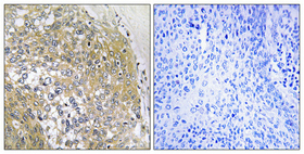
A08032-1 is listed for human and rat IHC and IF (catalog: applications and reactivity). Its supplied IHC images show paraffin-embedded human brain and cervix cancer (IHC image captions).
A08032-1 will render with human brain IHC at 1:100 on paraffin-embedded tissue (IHC image caption). Its second caption shows human cervix cancer IHC at 1:100 on paraffin-embedded tissue; IF is listed without a supplied IF image (IHC image caption; catalog: applications and IF image alts).
Which to pick: For tissue IHC, choose A08032-1 for paraffin sections based on its human brain and cervix cancer images; the fixative is unreported (IHC image captions). For IF, A08032-1 lists a 1:50 dilution, but the payload supplies no IF image or ICC validation (catalog: applications, IF dilution, IF image alts). For cross-species work, A08032-1 lists Human and Rat reactivity, while its supplied IHC images show Human tissue only (catalog: reactivity; IHC image captions).