This website uses cookies to ensure you get the best experience on our website.


- Table of Contents
Plan S100A2 chromogenic IHC in paraffin sections using the catalog antibody at 1:25 (datasheet M03698). Use bronchial respiratory or esophageal squamous epithelium as positive tissue and assess cytoplasmic and nuclear staining (HPA tissue IHC).

Expected localisation, validated protocols, controls and antibodies — the at-a-glance facts below, then the full design guide.
| Expected localisation | Cytoplasmic and nuclear staining in urothelium and epithelium (HPA tissue IHC) | |
| Staining pattern | Urothelial, respiratory and squamous cells: cytoplasm and nuclei (HPA tissue IHC) | |
| Antigen retrieval | Citrate pH 6 HIER, heat-mediated (datasheet M03698) | |
| Positive control | Bronchus+4 more · see all | |
| Negative control | Adipose tissue+4 more · see all |
| Fixation | Formaldehyde-fixed paraffin sections (selected-SKU IHC image M03698); keep fixation consistent (standard IHC practice; not target-specific) | |
| Caveat | Cell-type restriction may obscure staining in mixed tissue (HPA tissue IHC) | |
| Regulation | No expression regulator annotated (UniProt) | |
| Isoform / epitope | No annotated isoforms; one 1–98 chain with no processing (UniProt) |
The catalog antibody's citrate pH 6 IHC-P protocol (datasheet M03698) is followed by 3 published S100A2 IHC protocols (PMC3507318; PMC4324434; PMC4750537).
| Sample | Formaldehyde-fixed, paraffin-embedded human lung tissue (datasheet M03698) |
| Fixation | Image formalin-fixed; duration unreported (datasheet M03698); verify before use. |
| Sectioning | 4–5 µm sections on charged slides (standard) |
| Deparaffinisation | Xylene, graded ethanol series to water (standard) |
| Antigen retrieval | Heat retrieval: Citrate pH 6 (datasheet M03698); 20 min, 95–100 °C (standard) |
| Peroxidase block | 3% H2O2, 10 min, room temperature (standard) |
| Blocking | 10% normal serum of the secondary host, 30 min, room temperature (standard) |
| Primary antibody | Mouse monoclonal (clone 1592CT417.84.81) anti-S100A2, 1:25 (datasheet M03698) |
| Primary incubation | 1 hours at 37°C (datasheet M03698) |
| Detection | HRP-polymer secondary, DAB chromogen 5–10 min (standard) |
| Counterstain | Hematoxylin, blue, dehydrate and mount (standard) |
| Expected result | S100A2-positive staining in respiratory epithelial cells of bronchus (HPA tissue IHC: High). HPA tissue profile: Cytoplasmic and nuclear expression in urothelium, respiratory and squamous epithelium. No signal in the no-primary control. |
S100A2 should stain the cytoplasm and nuclei of selected epithelial cells, especially respiratory and squamous epithelium and urothelium (HPA tissue IHC: Enhanced; cytoplasmic and nuclear expression). Keratinocytes are among the epithelial cells reported to express it (UniProt P29034: tissue specificity). S100A2 has no transmembrane segment, so a membrane-only IHC pattern would need scrutiny (UniProt P29034: topology).
| Respiratory or squamous epithelial cells show cytoplasmic and nuclear staining, with nearby cell types less conspicuous. | This fits the reported distribution and compartments (HPA tissue IHC: cytoplasmic and nuclear expression in respiratory and squamous epithelium). Bronchial respiratory and esophageal squamous epithelial cells are useful positive references because each is reported at a High level (HPA tissue IHC: Bronchus and Esophagus, High). |
| Staining is confined to cell borders, with little or no cytoplasmic or nuclear signal. | A membrane-only pattern does not match the predominant tissue IHC pattern (HPA tissue IHC: cytoplasmic and nuclear expression). Plasma membrane is an additional ICC-IF location, so some peripheral signal is possible; assess the overall pattern before rejecting it (HPA subcellular: additional plasma membrane location, approved). |
| Strong signal appears in cells scored as negative, such as adipocytes or bone marrow hematopoietic cells. | Those cell types are reported as Not detected (HPA tissue IHC: adipose adipocytes and bone marrow hematopoietic cells). Check cell identity, cross-reactivity, and endogenous chromogenic activity with appropriate controls (general IHC practice). A negative score for breast adipocytes must not be applied to all breast epithelial cells (HPA tissue IHC: breast adipocytes, Not detected; UniProt P29034: normal mammary epithelial subset). |
| A uniform haze covers epithelium, stroma, and empty areas of the section. | That distribution does not provide interpretable cell-specific localization (general IHC practice). Check the no-primary control and detection reagents, then reassess blocking, washing, and antibody concentration under the assay's validated conditions (general IHC practice). |
| Known-positive bronchial respiratory or esophageal squamous epithelium has no detectable staining. | An absent signal in these High-level references leaves the run unresolved (HPA tissue IHC: Bronchus and Esophagus, High). Verify tissue identity, primary antibody performance, detection chemistry, and the assay's documented retrieval settings before interpreting other samples as negative (general IHC practice). |
| Tissue and cell selection | Use cell-level reference patterns: bronchial respiratory cells, esophageal squamous cells, nasopharyngeal respiratory cells, and skin hair follicles are High; cervical and oral squamous cells are Medium (HPA tissue IHC: positive cell scores). Salivary glandular cells are Low, making them a weaker positive reference (HPA tissue IHC: Salivary gland, Low). |
| Compartments and protein topology | Expect cytoplasmic and nuclear tissue staining (HPA tissue IHC: profile). ICC-IF places S100A2 mainly in nucleoplasm and cytosol, with additional nucleolar and plasma membrane localization (HPA subcellular: approved locations). The protein has no transmembrane segment (UniProt P29034: topology). |
| Antibody evidence | The tissue profile has Enhanced reliability because antibody staining is consistent with RNA expression data (HPA tissue IHC: reliability). Three listed antibodies have Enhanced IHC status: HPA062451, CAB002600, and CAB047340 (HPA antibodies: IHC validation). This supports using the reported pattern as a reference, while local controls still determine whether an individual run is interpretable (general IHC practice). |
| Processing and isoform annotation | The record describes one chain spanning residues 1–98, with no signal peptide, propeptide, annotated glycosylation sites, or listed isoforms (UniProt P29034: processing, glycosylation, isoforms). These annotations do not establish how fixation or retrieval affects IHC signal; target-specific fixation sensitivity is unreported in the supplied sources. |
| IF/ICC Q&A: where should signal appear? | Mainly in nucleoplasm and cytosol, with possible nucleolar and plasma membrane signal (HPA subcellular: approved locations). This ICC-IF observation informs compartment interpretation; tissue cell-type expectations come from HPA tissue IHC (HPA tissue IHC: profile). |
| Situation | Likely cause | Next action |
|---|---|---|
| No signal in a positive reference section | The result conflicts with High staining reported for bronchial respiratory and esophageal squamous cells (HPA tissue IHC: positive cell scores); the failed step is not identified by that comparison alone. | Confirm the intended cells are present, then check the primary antibody, detection reagents, counterstain, and documented retrieval conditions with run controls (general IHC practice). |
| Signal appears only at cell borders | Predominantly border-only staining differs from the reported cytoplasmic and nuclear tissue pattern (HPA tissue IHC: profile), although membrane localization is an additional ICC-IF finding (HPA subcellular: plasma membrane, approved). | Inspect nuclear and cytoplasmic compartments in the positive reference and compare with a no-primary control before assigning a specific artefact (general IHC practice). |
| Strong staining in adipocytes or bone marrow hematopoietic cells | Those cell types are scored Not detected (HPA tissue IHC: negative cell scores); cross-reactivity or endogenous chromogenic activity are possibilities, not established causes (general IHC practice). | Verify the cell type, examine a no-primary control, and review blocking and detection controls before scoring the signal as S100A2 (general IHC practice). |
| Diffuse staining obscures cell boundaries | Broad background prevents assessment of the expected epithelial compartments (HPA tissue IHC: profile; general IHC practice). | Compare a no-primary control, check washing and blocking, and use the documented antibody dilution as the starting point for assay optimization (general IHC practice). |
| A negative result is inferred from breast adipocytes | The HPA negative score is specifically for breast adipocytes (HPA tissue IHC: breast adipocytes, Not detected); UniProt reports expression in a subset of normal mammary epithelial cells (UniProt P29034: tissue specificity). | Score identified cell populations separately and include a positive epithelial reference when assessing run performance (general IHC practice). |
| A weak sample is treated as proof that the run failed | S100A2 staining varies by tissue and cell type: salivary glandular cells are Low, while bronchial respiratory and esophageal squamous cells are High (HPA tissue IHC: cell scores). | Judge run performance against a High-level reference and its controls, then report the sample's intensity and cell distribution separately (HPA tissue IHC: positive cell scores; general IHC practice). |

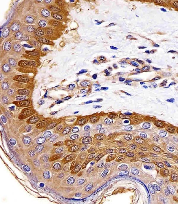
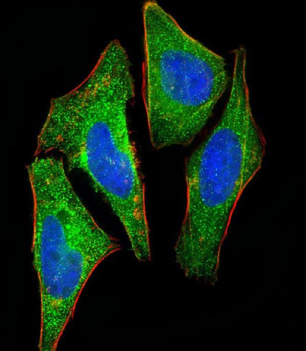


Comprehensive Human Protein Atlas IHC scoring per tissue (reliability: Enhanced — High consistency between antibody staining and RNA expression data.). Rows are taken directly from the HPA tissue chart — click any row's HPA link to view the source.
| Tissue | Cell type | Level | Evidence | Source |
|---|---|---|---|---|
| Bronchus | Respiratory epithelial cells | High | Protein (IHC) | HPA → |
| Esophagus | Squamous epithelial cells | High | Protein (IHC) | HPA → |
| Nasopharynx | Respiratory epithelial cells | High | Protein (IHC) | HPA → |
| Skin | Hair follicles | High | Protein (IHC) | HPA → |
| Cervix | Squamous epithelial cells | Medium | Protein (IHC) | HPA → |
| Tissue | Cell type | Level | Evidence | Source |
|---|---|---|---|---|
| Adipose tissue | Adipocytes | Not detected | Protein (IHC) | HPA → |
| Adrenal gland | Glandular cells | Not detected | Protein (IHC) | HPA → |
| Appendix | Glandular cells | Not detected | Protein (IHC) | HPA → |
| Bone marrow | Hematopoietic cells | Not detected | Protein (IHC) | HPA → |
| Breast | Adipocytes | Not detected | Protein (IHC) | HPA → |
Troubleshoot S100A2 staining in paraffin sections by checking retrieval, cell type, compartment and controls before comparing chromogenic signal across samples.
M03698 has IHC images from human lung and skin paraffin sections and an IF image from HeLa cells (M03698 image captions). Human is the only listed reactive species (M03698 catalog).
M03698 is listed for IHC-P and shown staining human lung and skin paraffin sections (M03698 catalog and IHC captions). M03698 is also listed for IF and shown staining paraformaldehyde-fixed, permeabilized HeLa cells (M03698 catalog and IF caption).
Which to pick: Choose M03698 for human tissue IHC: it is a mouse monoclonal antibody listed for IHC-P, and its own captions show staining in paraffin-embedded lung and skin sections with formaldehyde fixation reported (M03698 catalog and IHC captions). For IF/ICC, the same SKU is listed for IF and has an image from HeLa cells (M03698 catalog and IF caption). Its listed reactivity is Human, so the payload provides no basis to recommend it for another species (M03698 catalog).