This website uses cookies to ensure you get the best experience on our website.


- Table of Contents
Plan chromogenic S100A3 IHC in paraffin sections using hair cuticle cells as a positive control (HPA tissue IHC). Assess cytoplasmic staining and optimize the IHC-validated antibody within 1:100–1:300 (datasheet: IHC dilution).

Expected localisation, validated protocols, controls and antibodies — the at-a-glance facts below, then the full design guide.
| Expected localisation | Cytoplasmic in hair cuticle cells (HPA tissue IHC) | |
| Staining pattern | Hair cuticle cells show cytoplasmic staining (HPA tissue IHC) | |
| Antigen retrieval | Citrate pH 6.0 HIER, 95–98 °C, 20 min (rule: cytoplasmic / membrane antigen) | |
| Positive control | Hair | |
| Negative control | Appendix+4 more · see all |
| Fixation | Keep fixation consistent across sections (standard IHC practice; not target-specific) | |
| Caveat | Low staining in lung macrophages may complicate scoring (HPA tissue IHC) | |
| Regulation | Regulation not specified (UniProt) | |
| Isoform / epitope | No isoforms; residue 51 can be citrullinated (UniProt) |
The catalog antibody protocol is accompanied by published S100A3 IHC methods for colorectal cancer and liver tissue (PMC3829748; PMC5450779).
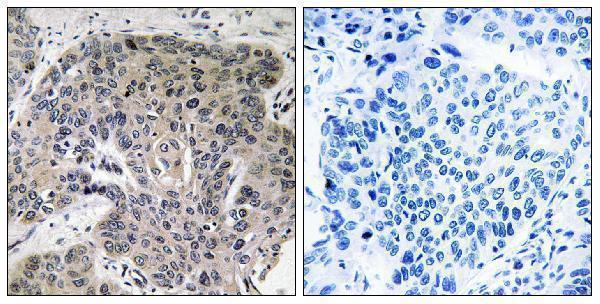
| Sample | Paraffin-embedded human lung carcinoma tissue; fixative not specified (datasheet A07744-1) |
| Fixation | Image fixative and duration unreported (datasheet A07744-1); verify before use. |
| Sectioning | 4–5 µm sections on charged slides (standard) |
| Deparaffinisation | Xylene, graded ethanol series to water (standard) |
| Antigen retrieval | Heat-induced epitope retrieval in citrate buffer, pH 6.0, 20 min at 95–98 °C (standard rule: cytoplasmic / membrane antigen) |
| Peroxidase block | 3% H2O2, 10 min, room temperature (standard) |
| Blocking | 10% normal serum of the secondary host, 30 min, room temperature (standard) |
| Primary antibody | Rabbit anti-S100A3, 1:100-1:300 (datasheet A07744-1) |
| Primary incubation | Overnight at 4 °C (standard) |
| Detection | HRP-polymer secondary, DAB chromogen 5–10 min (standard) |
| Counterstain | Hematoxylin, blue, dehydrate and mount (standard) |
| Expected result | S100A3-positive staining in cells in cuticle of hair (HPA tissue IHC: High). HPA tissue profile: Cytoplasmic expression mainly in cells of the hair cuticle layer. Also expressed in other tissues. No signal in the no-primary control. |
S100A3 should appear mainly in the cytoplasm of hair cuticle cells, where HPA reports high staining; UniProt places it in the cytoplasm and reports no transmembrane segment (HPA: High in hair cuticle cells; UniProt P33764: cytoplasm, no transmembrane segment). HPA rates its tissue IHC reliability Enhanced, while describing only medium consistency between staining and RNA expression (HPA: tissue IHC reliability).
| Strong cytoplasmic staining in hair cuticle cells, with recognizable hair structure. | This is the clearest expected positive pattern (HPA: High in hair cuticle cells; HPA: cytoplasmic tissue expression). Judge staining within the cuticle cells rather than treating color across the whole hair section as equivalent evidence (general IHC practice). |
| Predominantly nuclear staining, or a sharp membrane outline without convincing cytoplasmic staining. | This is discordant with the dominant tissue pattern (HPA: cytoplasmic tissue expression; UniProt P33764: cytoplasm). Check morphology and controls before calling it S100A3; HPA's additional plasma membrane location comes from ICC-IF and does not establish a membrane-only tissue IHC pattern (HPA: ICC-IF locations). |
| Strong staining in an HPA-listed negative cell population, such as appendix glandular cells or bone marrow hematopoietic cells. | Treat this as unexpected for those cells (HPA: Not detected in appendix glandular cells and bone marrow hematopoietic cells). Cross-reactivity or endogenous chromogen activity are possibilities, not diagnoses; compare an adjacent section processed without primary antibody (general IHC practice). |
| Weak color spread across tissue, including spaces or several unrelated cell populations, without a defined cuticle-cell pattern. | Diffuse background does not establish S100A3 expression. Review blocking, washes, chromogen development, and the no-primary control (general IHC practice). HPA reports some expression outside hair, so assess specific cell populations rather than requiring every other tissue to be blank (HPA: tissue IHC profile). |
| No detectable staining in a section containing identifiable hair cuticle cells. | An absent signal in the best supported positive site conflicts with the reported high cuticle-cell staining (HPA: High in hair cuticle cells). Confirm that cuticle cells are present, then review antibody dilution, retrieval, detection reagents, and control performance (general IHC practice). |
| Which cells give the strongest tissue reference? | Hair cuticle cells are the clearest positive reference (HPA: High in hair cuticle cells; UniProt P33764: inner endocuticle of hair fibers). The HPA tissue profile also reports expression elsewhere, so a positive call outside hair needs its own cell-level interpretation (HPA: tissue IHC profile). |
| How should low-level and negative examples be used? | HPA lists low staining in thyroid, parathyroid and adrenal glandular cells, respiratory epithelial cells of nasopharynx and bronchus, and lung macrophages (HPA: Low). Its listed negative examples refer to specified cells within tissues, not necessarily every cell in each organ (HPA: Not detected examples). |
| What does antibody validation support? | Both HPA042674 and HPA056140 are rated Enhanced for IHC and ICC (HPA: antibody validation). HPA defines Enhanced IHC as a pattern reproduced by independent antibodies or orthogonal data; its overall tissue assessment still notes medium staining–RNA consistency (HPA: validation and tissue reliability). |
| Do molecular features change the expected compartment? | UniProt describes a cytoplasmic protein without a transmembrane segment or signal peptide, and records a processed chain spanning residues 2–101 (UniProt P33764: location, topology, processing). These annotations support a cytoplasmic expectation; they do not specify a retrieval method or predict fixation sensitivity. |
| Do reported modifications predict staining changes? | UniProt records N-acetylalanine at residue 2 and PAD3-associated citrullination at residue 51 (UniProt P33764: modified residues). Without a mapped antibody epitope or modification-specific validation in the supplied evidence, do not infer that either modification raises or lowers IHC signal. |
| Q: What should an IF/ICC image show? | A: Mainly cytosolic signal, with additional Golgi and plasma membrane localization reported by HPA (HPA: ICC-IF subcellular locations). That cell-image observation can guide interpretation, but it does not supply an IF/ICC protocol or override the predominant cytoplasmic tissue IHC pattern (HPA: tissue IHC profile). |
| Situation | Likely cause | Next action |
|---|---|---|
| Hair is present, but the expected cuticle-cell signal is absent. | The section may lack intact, identifiable cuticle cells, or the IHC detection workflow may have failed; HPA reports high staining specifically in cuticle cells (HPA: High in hair cuticle cells). | Confirm cuticle morphology and a positive control, then check primary dilution, retrieval settings, secondary detection, and chromogen development against the chosen assay instructions (general IHC practice). |
| Color appears chiefly in nuclei or as a crisp membrane-only outline. | That distribution disagrees with the dominant cytoplasmic tissue pattern (HPA: tissue IHC profile; UniProt P33764: cytoplasm). HPA's additional membrane localization was observed in ICC-IF (HPA: ICC-IF locations). | Recheck cell boundaries and counterstain, compare with the no-primary control, and seek a reproducible cytoplasmic signal in hair cuticle cells before interpreting the tissue result (general IHC practice; HPA: High in hair cuticle cells). |
| Unexpected strong color appears in listed negative cells. | The signal conflicts with HPA observations for those particular cells (HPA: Not detected examples). Nonspecific binding or endogenous detection activity could produce misleading chromogenic color (general IHC practice). | Compare adjacent sections with and without primary antibody, review blocking and detection controls, and identify the stained cell type before assigning expression (general IHC practice). |
| The slide shows widespread weak haze. | Background from the staining workflow may obscure the cell pattern (general IHC practice). HPA's report of expression in other tissues does not make diffuse, poorly localized color a specific result (HPA: tissue IHC profile). | Check the no-primary control, washes, blocking, and chromogen development; score discrete cellular staining only after background is controlled (general IHC practice). |
| Low staining in lung macrophages or respiratory epithelial cells seems inconsistent with a hair-focused marker. | Hair cuticle cells are the high-staining reference, while HPA also lists low staining in lung macrophages and respiratory epithelial cells of nasopharynx and bronchus (HPA: High; HPA: Low). | Record intensity and cell identity separately, compare with a positive hair section and a no-primary control, and avoid applying a hair-level intensity threshold to every tissue (general IHC practice; HPA: tissue levels). |
| Two antibodies give different tissue patterns. | Both listed antibodies have Enhanced IHC status, yet the overall HPA tissue assessment reports medium consistency with RNA expression (HPA: antibody validation; HPA: tissue reliability). The supplied record does not identify the reason for a particular disagreement. | Compare matched sections, controls, staining conditions, and cell-level localization; report the disagreement rather than selecting one result solely from its validation label (general IHC practice). |


Comprehensive Human Protein Atlas IHC scoring per tissue (reliability: Enhanced — Medium consistency between antibody staining and RNA expression data. External characterization data supports antibody staining.). Rows are taken directly from the HPA tissue chart — click any row's HPA link to view the source.
| Tissue | Cell type | Level | Evidence | Source |
|---|---|---|---|---|
| Hair | Cells in cuticle | High | Protein (IHC) | HPA → |
| Tissue | Cell type | Level | Evidence | Source |
|---|---|---|---|---|
| Appendix | Glandular cells | Not detected | Protein (IHC) | HPA → |
| Bone marrow | Hematopoietic cells | Not detected | Protein (IHC) | HPA → |
| Breast | Adipocytes | Not detected | Protein (IHC) | HPA → |
| Caudate | Glial cells | Not detected | Protein (IHC) | HPA → |
| Cerebellum | Cells in granular layer | Not detected | Protein (IHC) | HPA → |
Troubleshoot S100A3 staining in paraffin sections by checking cuticle cell localization, section controls, and whether the signal survives specificity checks.
A07744-1 has a paraffin-section IHC figure and lists IF as an application; Human, Mouse, and Rat reactivity are listed (catalog IHC image alt; catalog applications/reactivity).
A07744-1 will render with an IHC figure from paraffin-embedded human lung carcinoma, including a peptide-blocked comparison (catalog IHC image alt). It lists IHC and IF applications and Human, Mouse, and Rat reactivity; no IF figure is supplied (catalog applications/reactivity/IF image alts).
Which to pick: For tissue IHC, choose A07744-1 based on its paraffin-section figure; the fixative is unreported (catalog IHC image alt). For IF/ICC, A07744-1 lists IF at 1:50, but supplies no IF figure or ICC validation (catalog applications/IF dilution/IF image alts). For cross-species work, A07744-1 lists Human, Mouse, and Rat reactivity; it is rabbit-hosted, with no clone or clonality reported (catalog reactivity/host/clone).