S1PR1 / Sphingosine 1-phosphate receptor 1 · IHC design guide

Design Immunohistochemistry for S1PR1

Plan paraffin IHC around S1PR1’s cytoplasmic and membranous tissue staining, including distinct expression in endothelial cells and fibroblasts (HPA tissue IHC). The catalog antibody lists an IHC starting concentration of 2.5 μg/mL (datasheet: A01502); interpret tissue patterns with the HPA’s medium staining–RNA agreement in mind (HPA tissue IHC).

Evidence assembled Oct 2026 · For research use; verify linked source records and product datasheet before use
Immunohistochemistry protocol sheet for S1PR1 (IHC for S1PR1): expected localisation Cytoplasmic and membranous in tissue (HPA tissue IHC); endosomal localization is expected (UniProt), antibody A01502, validated IHC image, and IHC protocol steps
Printable S1PR1 IHC protocol sheet — expected localisation Cytoplasmic and membranous in tissue (HPA tissue IHC); endosomal localization is expected (UniProt), antibody A01502, controls and protocol steps. Open the full S1PR1 IHC guide →

S1PR1 Immunohistochemistry Experimental Design Guide

Expected localisation, validated protocols, controls and antibodies — the at-a-glance facts below, then the full design guide.

Must know before staining
Expected localisation Cytoplasmic and membranous in tissue (HPA tissue IHC); endosomal localization is expected (UniProt)
Staining pattern Endothelial cells and fibroblasts; cytoplasmic/membranous staining (HPA tissue IHC)
Antigen retrieval Citrate pH 6.0 HIER, 95–98 °C, 20 min (rule: cytoplasmic / membrane antigen)
Positive control ⓘ Adrenal gland+4 more · see all
Negative control ⓘ Adipose tissue+3 more · see all
Important caveats
Reasons your staining may differ from the expected pattern.
Fixation Keep fixation consistent across sections. (standard IHC practice; not target-specific)
Caveat Tissue staining has medium RNA agreement; retesting is pending (HPA tissue IHC)
Regulation CD69 promotes receptor degradation (UniProt)
Isoform / epitope No annotated isoforms; extracellular versus cytoplasmic epitope matters (UniProt)
Section 1

Recommended S1PR1 IHC & IF Protocols

The catalog antibody’s IHC-P protocol is accompanied by three published S1PR1 chromogenic IHC protocols (PMC3612140; PMC12461039; PMC12891624).

Recommended immunohistochemistry (IHC-P) protocol parameters
SampleTissue sections; selected-image fixative not specified (standard IHC workflow)
FixationImage fixative and duration unreported (datasheet A01502); verify before use.
Sectioning4–5 µm sections on charged slides (standard)
DeparaffinisationXylene, graded ethanol series to water (standard)
Antigen retrievalHeat-induced epitope retrieval in citrate buffer, pH 6.0, 20 min at 95–98 °C (standard rule: cytoplasmic / membrane antigen)
Peroxidase block3% H2O2, 10 min, room temperature (standard)
Blocking10% normal serum of the secondary host, 30 min, room temperature (standard)
Primary antibodyRabbit anti-S1PR1, 2.5 μg/mL (datasheet A01502)
Primary incubationOvernight at 4 °C (standard)
DetectionHRP-polymer secondary, DAB chromogen 5–10 min (standard)
CounterstainHematoxylin, blue, dehydrate and mount (standard)
Expected resultS1PR1-positive staining in glandular cells of adrenal gland (HPA tissue IHC: High). HPA tissue profile: General cytoplasmic and membranous expression. Distinct expression in endothelial cells, fibroblast and in the brain. No signal in the no-primary control.
💡Decision noteStart with citrate pH 6.0 HIER at 95–98 °C for 20 min (page antigen-retrieval rule); use each paper’s stated retrieval when reproducing its protocol.
Section 2

What Is the Expected S1PR1 Staining Pattern?

S1PR1 is a 7-transmembrane receptor at the cell membrane and in endosomes (UniProt P21453 topology and subcellular annotation). In paraffin-section IHC, expect cytoplasmic and membranous staining, especially in endothelial cells and fibroblasts; other positive cell types depend on tissue (HPA: tissue IHC profile). HPA rates the tissue pattern Supported, with medium consistency against RNA expression and retesting pending (HPA: reliability description).

What am I looking at on my slide?
Vascular endothelial cells show membranous staining with some cytoplasmic signal.This fits S1PR1 at the cell membrane and in endosomes (UniProt P21453: subcellular annotation). Endothelial staining is part of the HPA tissue profile, including High staining in cerebral cortex endothelial cells (HPA: tissue IHC). Score the identified cells and compartments, rather than assigning a single intensity to the whole section (general IHC practice).
Staining is predominantly nuclear, with little discernible membrane or cytoplasmic signal.A nuclear dominant pattern is unsupported by the reported membrane and endosome locations or the tissue IHC profile (UniProt P21453: subcellular annotation; HPA: tissue IHC profile). Treat it as suspect and check the negative detection control and staining distribution before calling it S1PR1 (general IHC practice).
Strong staining appears in prostate glandular cells or adipocytes.HPA reports S1PR1 as Not detected in those specific cell populations (HPA: prostate and adipose tissue IHC). Consider antibody cross-reactivity or endogenous chromogenic activity; inspect the cell identity and a no-primary control (general IHC practice). This does not make every cell in either tissue a negative control (HPA: cell-specific tissue entries).
Brown signal spreads across stroma, lumens, or most cells without clear cellular boundaries.Diffuse deposition cannot establish the reported cytoplasmic and membranous pattern (HPA: tissue IHC profile). Background from detection chemistry or nonspecific antibody binding is possible (general IHC practice). Compare a no-primary control, then assess whether signal follows identifiable cells and remains above surrounding background (general IHC practice).
No staining is seen in a putative positive section.First verify the expected cell population: HPA reports High staining in cerebral cortex endothelial cells and bronchial respiratory epithelial cells, but Low staining in lung macrophages (HPA: tissue IHC entries). Absence in a low-staining population is less informative; if a high-staining population is present, investigate the IHC workflow and antibody performance (general IHC practice).
💡Expected S1PR1 appearanceCall a result positive when identifiable cells show membranous and cytoplasmic staining, particularly vascular endothelium or fibroblasts, with High signal possible in listed populations such as cerebral cortex endothelium (UniProt P21453: membrane/endosome location; HPA: tissue IHC profile and cerebral cortex entry); diffuse, nuclear dominant, or cell-free chromogen is suspect (general IHC practice).
How each factor affects the staining
Cell and tissue contextS1PR1 has low tissue RNA specificity, while observed protein intensity varies by cell population (HPA: RNA specificity and tissue IHC). Use cell-level expectations: bronchial respiratory epithelium is High, lung macrophages are Low, and prostate glandular cells are Not detected (HPA: tissue IHC entries).
Compartment and receptor movementSeven transmembrane segments support membrane localization, and ligand binding leads to internalization (UniProt P21453: topology and subcellular annotation). Cytoplasmic signal can therefore coexist with membrane staining (UniProt P21453: endosome annotation; HPA: tissue IHC profile). A static IHC section cannot by itself establish when or why internalization occurred (general IHC interpretation).
Antibody validationCAB010104 is listed as Supported for IHC; HPA075568 has ICC support but no listed IHC status (HPA: antibody validation entries). The tissue profile has medium consistency with RNA expression and is pending retesting (HPA: reliability description). Interpret unexpected staining with appropriate controls rather than treating the profile as definitive.
Does IF/ICC show the same pattern?HPA reports vesicles as the mainly observed, supported ICC-IF location (HPA: subcellular summary). This is compatible with the UniProt endosome annotation (UniProt P21453: subcellular annotation). Use the separate IF/ICC guide to evaluate that application; vesicular fluorescence alone does not validate a paraffin-section chromogenic IHC result.
Why is my staining missing, weak or wrong?
SituationLikely causeNext action
High-staining reference cells are present, but the section has no signal.The expected population may have been misidentified, or an IHC staining step may have failed (HPA: cell-specific tissue IHC entries; general IHC practice).Confirm the cells against tissue morphology, then check antibody application, detection reagents, and counterstain on a suitable positive section (general IHC practice). Cerebral cortex endothelial cells are a High HPA example (HPA: tissue IHC).
A proposed negative control contains scattered positive structures.HPA's Not detected entries apply to named cell populations, such as prostate glandular cells, rather than every structure in the section (HPA: tissue IHC entries).Score the specified cell population separately from vessels and neighboring tissue (general IHC practice). Pair the section with a no-primary control when assessing chromogenic background (general IHC practice).
Signal is nuclear dominant or lacks recognizable cell borders.That distribution does not match the reported membrane/endosome locations or general cytoplasmic and membranous IHC profile (UniProt P21453: subcellular annotation; HPA: tissue IHC profile).Inspect morphology and the no-primary control, and reassess whether the deposit tracks cells rather than nuclei or extracellular material (general IHC practice).
Brown deposit remains in the no-primary control.Antibody-independent chromogenic activity or detection background is possible (general IHC practice).Investigate the detection system and its background-control steps before attributing the deposit to S1PR1 (general IHC practice). Compare affected cells and structures with the full stain (general IHC practice).
Signal appears throughout the section, obscuring membrane detail.Excess nonspecific staining or detection background can mask a cellular pattern (general IHC practice).Check blocking, antibody dilution, wash steps, and chromogen development against the no-primary control (general IHC practice). Retain only conditions that resolve identifiable cells and compartments (general IHC practice).
Vesicular IF/ICC and membrane-rich IHC images appear different.HPA reports mainly vesicles in ICC-IF and general cytoplasmic and membranous expression in tissue IHC (HPA: subcellular summary and tissue IHC profile).Interpret each image against its own application and controls (general IHC/IF practice). Do not require identical compartment proportions across the two preparations; UniProt lists both membrane and endosome locations (UniProt P21453: subcellular annotation).

Sample controls for S1PR1 IHC & IF

🧪Run cerebral cortex first and look for staining in endothelial cells (High; HPA: cerebral cortex), then run prostate and assess its glandular cells as the negative tissue comparator (HPA: Not detected in prostate glandular cells). On the positive slide, use surrounding non-endothelial cells to assess background; the supplied HPA row does not establish them as S1PR1-negative (HPA: cerebral cortex endothelial cells).
Positive control tissue: Adrenal gland (Glandular cells, HPA High)
Negative control tissue: Adipose tissue (HPA Not detected)
ICC-IF cell lines (HPA subcellular resource): HPA ICC-IF images show S1PR1 in A-431, U-251MG, U2OS, BJ [Human fibroblast], with annotated localisation: Vesicles (supported) (HPA subcellular).
Technical controls: Include a no-primary (secondary-only) control, a host- and subclass-matched isotype control for a monoclonal primary or host-matched normal IgG for a polyclonal primary, and S1PR1-knockout tissue as a biological negative where available (standard IHC practice). Quench endogenous peroxidase and check vessel-associated blood pigment when interpreting chromogenic staining in cerebral cortex (standard IHC practice).
⚠️Feasibility: A target-specific fixation window or fixation effect is unreported in the supplied evidence; the selected A01502 mouse thymus IHC caption also leaves its fixative unstated (selected-SKU caption). Antigen-retrieval dependence is unreported, so optimize retrieval empirically for paraffin-section IHC (standard IHC practice). The evidence does not establish whether frozen sections or IF are easier; vesicular IF signal may be expected alongside membrane or endosomal localization (HPA: Vesicles; UniProt P21453 subcellular localization).

HPA tissue IHC evidence for S1PR1

Comprehensive Human Protein Atlas IHC scoring per tissue (reliability: Supported — Medium consistency between antibody staining and RNA expression data. Antibody staining in cells/structures not annotated, view images. Pending retesting.). Rows are taken directly from the HPA tissue chart — click any row's HPA link to view the source.

Positive expression · recommended positive controls

TissueCell typeLevelEvidenceSource
Adrenal gland Glandular cells High Protein (IHC) HPA →
Appendix Lymphoid tissue High Protein (IHC) HPA →
Bronchus Respiratory epithelial cells High Protein (IHC) HPA →
Cerebellum Bergmann glia - cytoplasm/membrane High Protein (IHC) HPA →
Cerebral cortex Endothelial cells High Protein (IHC) HPA →

Undetected expression · recommended negative controls

TissueCell typeLevelEvidenceSource
Adipose tissue Adipocytes Not detected Protein (IHC) HPA →
Caudate Glial cells Not detected Protein (IHC) HPA →
Hippocampus Glial cells Not detected Protein (IHC) HPA →
Prostate Glandular cells Not detected Protein (IHC) HPA →
Section 3

Advanced S1PR1 IHC Tips

Troubleshoot S1PR1 staining in paraffin sections by evaluating retrieval, compartment, cell identity and controls together (UniProt P21453; HPA tissue IHC).

How should I optimize retrieval when S1PR1 staining is weak in paraffin sections?
Use citrate pH 6.0 HIER at 95–98 °C for 20 min for S1PR1 paraffin IHC (page retrieval setting). Compare sections processed together, including a no-primary control and tissue with an expected endothelial signal, before changing retrieval conditions (UniProt P21453 tissue specificity; standard IHC practice). If staining remains weak, check that sections stayed immersed during heating and cooled consistently, then adjust retrieval duration in a small controlled series (standard IHC practice). Excessive retrieval can damage section morphology or increase diffuse staining, so judge any gain against cell boundaries and vascular detail (standard IHC practice). The catalog image reports 5 µg/mL in mouse thymus but gives no retrieval method to transfer (catalog IHC caption: A01502).
Could fixation explain faint or diffuse S1PR1 staining?
S1PR1-specific sensitivity to fixation is unknown because no target-specific fixation conditions are supplied (catalog IHC caption: A01502; provided evidence). The mouse thymus image reports antibody use at 5 µg/mL but does not state its fixative, so it cannot establish a fixation condition for paraffin sections (catalog IHC caption: A01502). Record fixation duration and processing history for each specimen, and compare matched sections before attributing a staining difference to the receptor (standard IHC practice). Inspect morphology and run a no-primary control to separate broad tissue-processing problems from antibody-associated staining (standard IHC practice). Keep retrieval and detection conditions constant during that comparison, changing one processing variable at a time (standard IHC practice).
Should S1PR1 appear at the membrane or inside cells?
Expect a mixture of membrane-associated and intracellular signal: S1PR1 is annotated at the cell membrane and endosome, and ligand binding leads to internalization (UniProt P21453 subcellular location). HPA describes general cytoplasmic and membranous tissue staining, while its ICC/IF summary places S1PR1 mainly in vesicles (HPA tissue IHC; HPA subcellular). In chromogenic sections, score convincing cell-border staining separately from granular cytoplasmic staining rather than calling every brown deposit membrane signal (standard IHC practice). Evaluate vascular cells in context, because endothelial expression is documented for this receptor (UniProt P21453 tissue specificity; HPA tissue IHC). A diffuse nuclear-only pattern does not match the supplied localization evidence and warrants review of controls and counterstain (UniProt P21453 subcellular location; standard IHC practice).
How can an unknown epitope affect S1PR1 IHC interpretation?
The record lists 0 alternative isoforms and a 382-residue receptor with 7 transmembrane segments (UniProt P21453 isoforms and topology). Its extracellular regions include residues 1–46, while the cytoplasmic tail spans 311–382; glycosylation is annotated at residues 30 and 36 (UniProt P21453 topology and glycosylation). The antibody epitope is not specified in the supplied evidence, so do not assign a weak stain to glycosylation, phosphorylation or an inaccessible membrane face (provided antibody evidence; UniProt P21453 modifications). Ask for epitope mapping if available, then compare retrieval conditions and staining controls on adjacent sections (standard IHC practice). Interpret a change in signal as an assay result until antibody specificity and the relevant epitope are established (standard IHC practice).
How should I check S1PR1 localization by multiplex IF?
Use IF/ICC as a separate localization experiment alongside paraffin IHC, with an endothelial marker such as CD31 to identify an expected S1PR1-expressing cell type (UniProt P21453 tissue specificity; standard IF practice). Choose fluorophores in spectral regions with low measured autofluorescence for the specimen, and include unstained and single-stain controls before interpreting overlap (standard IF practice). S1PR1 has extracellular loops and cytoplasmic regions, but the catalog antibody's epitope side is unspecified (UniProt P21453 topology; provided antibody evidence). Set permeabilisation according to the mapped epitope: intracellular epitopes require access through the membrane, whereas an accessible extracellular epitope may be examined without it (standard IF practice). Vesicular IF signal is plausible, but compare it with the membrane and endosomal annotations rather than forcing concordance with chromogenic intensity (HPA subcellular; UniProt P21453 subcellular location).
What should I check when S1PR1 IHC has widespread brown background?
First inspect a no-primary section and the distribution of stain relative to vessels, tissue edges and damaged areas (standard IHC practice). Block endogenous peroxidase before chromogenic detection, since its activity can produce DAB signal independent of the primary antibody (standard IHC practice). Titrate the primary antibody and optimize blocking and washes on matched sections, keeping exposure and development consistent across comparisons (standard IHC practice). The catalog mouse thymus image used 5 µg/mL, but its caption does not establish that concentration as optimal for other specimens or processing conditions (catalog IHC caption: A01502). Judge residual signal against expected endothelial staining and the no-primary control rather than treating uniform cytoplasmic color as specific S1PR1 (UniProt P21453 tissue specificity; standard IHC practice).
How should I score S1PR1 staining across paraffin sections? ⚠ ANSWER MARKED FOR VERIFICATION
Define the cell population and compartment before scoring, because endothelial expression is documented and tissue staining can be cytoplasmic or membranous (UniProt P21453 tissue specificity; HPA tissue IHC). For a defined cell population, report the percentage positive and an H-score from 0–300, calculated from the proportions at each intensity grade (standard IHC scoring practice). For vessels, positive-cell density per mm² can supplement intensity, but normalize it to sampled tissue area and report vessel density or endothelial-cell count when those differ between sections (standard IHC quantification practice). Score membrane and cytoplasmic patterns separately using one threshold set from controls, with blinded review when feasible (standard IHC practice). Record retrieval, detection and image settings so comparisons reflect staining differences rather than batch changes (standard IHC practice).
How can I distinguish true S1PR1 staining from artefact?
A credible pattern should fit cell identity and compartment: endothelial staining and membrane or cytoplasmic signal are supported, and receptor internalization can account for intracellular staining (UniProt P21453 tissue specificity and subcellular location; HPA tissue IHC). Compare suspicious regions with an adjacent section lacking primary antibody and examine whether color follows intact cell borders or discrete intracellular structures (standard IHC practice). Staining confined to cut edges, necrotic areas or damaged tissue is suspect, as is nuclear-only signal for this membrane and endosome-associated receptor (standard IHC practice; UniProt P21453 subcellular location). Persistent DAB signal in the no-primary control should prompt review of endogenous peroxidase blocking and detection reagents (standard IHC practice). HPA labels tissue IHC reliability Supported but notes medium RNA–staining consistency and pending retesting, so avoid treating one positive section as definitive specificity proof (HPA tissue IHC reliability).
Boster reagents

Best S1PR1 / Sphingosine 1-phosphate receptor 1 IHC Antibodies

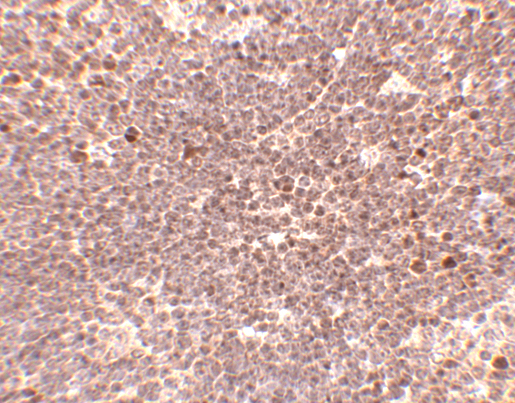
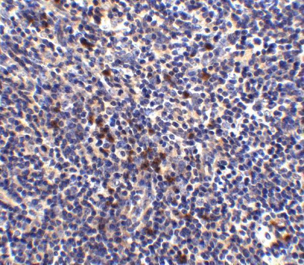
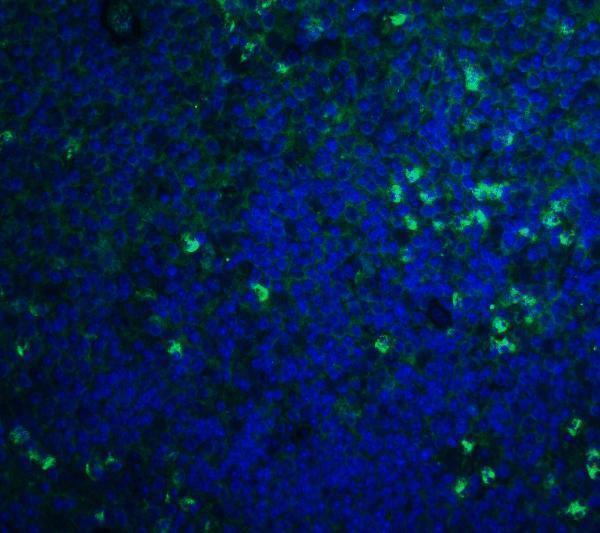
A01502 has IHC-P and IF applications and lists human, mouse, and rat reactivity (catalog applications/reactivity). Mouse thymus images provide IHC and IF data (A01502 image captions).

Real IHC data Immunohistochemistry of S1P1 in mouse thymus tissue with S1P1 antibody at 5 μg/mL.
Anti-S1P1 S1PR1 Antibody
Cat # A01502

A01502 will render with mouse thymus IHC images at 2.5 μg/mL and 5 μg/mL (A01502 IHC image captions). Its IF image shows mouse thymus tissue at 20 μg/mL, and its listed reactivity includes human, mouse, and rat (A01502 IF image caption; catalog reactivity).

Which to pick: For paraffin-section IHC, choose A01502: IHC-P is listed, and its own mouse thymus IHC captions show 2.5 μg/mL and 5 μg/mL; the fixative is unreported (catalog applications; A01502 IHC image captions). For IF, A01502 has a mouse thymus tissue image; ICC validation and clone status are unreported (A01502 IF image caption; catalog applications/clone field). For human or rat samples, A01502 lists reactivity in both species, while the supplied IHC and IF images show mouse thymus only (catalog reactivity; A01502 image captions).

Each figure is that product's own IHC / IF validation image from its datasheet.

References

  1. UniProt Consortium. UniProt entry P21453 (S1PR1_HUMAN, Sphingosine 1-phosphate receptor 1).
  2. Human Protein Atlas. S1PR1 tissue IHC expression (reliability: Supported).
  3. Human Protein Atlas. S1PR1 subcellular location (ICC-IF): Mainly localized to vesicles..
  4. Human Protein Atlas. S1PR1 antibody validation summary (2 antibodies).
  5. Sphingosine-1-phosphate receptor 1 in classical Hodgkin lymphoma: assessment of expression and role in cell migration. Laboratory investigation; a journal of technical methods and pathology 2013 — PMC3612140.
  6. Targeting sphingosine-1-phosphate receptor 1 alleviates neuropathic pain associated with pancreatic ductal adenocarcinoma in mice and inhibits tumor progression. Molecular pain 2025 — PMC12461039.
  7. Overexpression of sphingosine-1-phosphate receptor 1 and phospho-signal transducer and activator of transcription 3 is associated with poor prognosis in rituximab-treated diffuse large B-cell lymphomas. BMC cancer 2014 — PMC4265452.
  8. Expression of sphingosine-1-phosphate receptor 1 in the brain of fatal cerebral malaria. Scientific reports 2026 — PMC12891624.
  9. PubMed PMID:2160972 — UniProt-cited evidence.
  10. PubMed PMID:9409733 — UniProt-cited evidence.
  11. PubMed PMID:10982820 — UniProt-cited evidence.