SAA4 / Serum amyloid A-4 protein · IHC design guide

Design Immunohistochemistry for SAA4

Plan SAA4 paraffin IHC around the reported extracellular matrix and plasma staining (HPA tissue IHC). Start A07115 at 1:100–1:300 (datasheet: A07115), and account for the warning that the tissue stain targets protein from more than one gene (HPA tissue IHC).

Evidence assembled Oct 2026 · For research use; verify linked source records and product datasheet before use
Immunohistochemistry protocol sheet for SAA4 (IHC for SAA4): expected localisation Extracellular matrix and plasma (HPA tissue IHC), antibody A07115, validated IHC image, and IHC protocol steps
Printable SAA4 IHC protocol sheet — expected localisation Extracellular matrix and plasma (HPA tissue IHC), antibody A07115, controls and protocol steps. Open the full SAA4 IHC guide →

SAA4 Immunohistochemistry Experimental Design Guide

Expected localisation, validated protocols, controls and antibodies — the at-a-glance facts below, then the full design guide.

Must know before staining
Expected localisation Extracellular matrix and plasma (HPA tissue IHC)
Staining pattern Distinct matrix/plasma staining; pachytene spermatocytes high (HPA tissue IHC)
Antigen retrieval Citrate pH 6.0 HIER, 95–98 °C, 20 min (rule: cytoplasmic / membrane antigen)
Positive control ⓘ Testis
Negative control ⓘ Adipose tissue+4 more · see all
Important caveats
Reasons your staining may differ from the expected pattern.
Fixation Keep fixation consistent across sections (standard IHC practice; not target-specific)
Caveat Staining may include protein from another gene (HPA tissue IHC)
Regulation Constitutively expressed (UniProt)
Isoform / epitope No isoforms; mature chain 19–130 has no cytoplasmic domain (UniProt)
Section 1

Recommended SAA4 IHC & IF Protocols

The catalog antibody has an IHC-P protocol; the published options below cover first-trimester placenta (PMC4119475) and paired HCC tissues (PMC10834558).

Recommended immunohistochemistry (IHC-P) protocol parameters
SampleParaffin-embedded human lung carcinoma tissue; fixative not specified (datasheet A07115)
FixationImage fixative and duration unreported (datasheet A07115); verify before use.
Sectioning4–5 µm sections on charged slides (standard)
DeparaffinisationXylene, graded ethanol series to water (standard)
Antigen retrievalHeat-induced epitope retrieval in citrate buffer, pH 6.0, 20 min at 95–98 °C (standard rule: cytoplasmic / membrane antigen)
Peroxidase block3% H2O2, 10 min, room temperature (standard)
Blocking10% normal serum of the secondary host, 30 min, room temperature (standard)
Primary antibodyRabbit anti-SAA4, 1:100 - 1:300 (datasheet A07115)
Primary incubationOvernight at 4 °C (standard)
DetectionHRP-polymer secondary, DAB chromogen 5–10 min (standard)
CounterstainHematoxylin, blue, dehydrate and mount (standard)
Expected resultSAA4-positive staining in pachytene spermatocytes of testis (HPA tissue IHC: High). HPA tissue profile: Distinct positivity in extracellular matrix and plasma. No signal in the no-primary control.
💡Decision noteStart with citrate pH 6.0 HIER at 95–98 °C for 20 min (page antigen-retrieval setting); the HCC method also uses microwave citrate retrieval (PMC10834558: methods).
Section 2

What Is the Expected SAA4 Staining Pattern?

SAA4 is secreted, has no transmembrane segment, and is expressed by the liver and released into plasma (UniProt P35542). In tissue IHC, expect distinct extracellular matrix and plasma positivity and high staining in testicular pachytene spermatocytes (HPA tissue IHC). HPA rates this pattern Supported, but cautions that its antibodies may recognize proteins from more than one gene and that external verification is pending (HPA tissue IHC).

What am I looking at on my slide?
Distinct extracellular matrix or plasma staining, with strong staining in pachytene spermatocytes.This matches the reported tissue pattern and high testis signal (HPA tissue IHC). Because SAA4 is secreted, extracellular staining is plausible even away from its liver site of expression (UniProt P35542).
Predominantly crisp nuclear or membrane outlining signal replaces the extracellular pattern.Treat this as a possible artefact: SAA4 is secreted and has no transmembrane segment (UniProt P35542). Review morphology, counterstain, and controls before assigning this signal to SAA4 (general IHC practice).
Strong cellular staining appears in a cell population HPA reports as undetected.For example, HPA reports no detection in adrenal glandular cells or bone marrow hematopoietic cells (HPA tissue IHC). Consider antibody cross-reactivity or endogenous detection activity; repeat with appropriate controls (general IHC practice).
Broad, even chromogen obscures cells, matrix, and negative areas.This is diffuse background rather than the distinct extracellular pattern HPA describes (HPA tissue IHC). Check blocking, washes, antibody concentration, and the detection-only control as general IHC troubleshooting steps (general IHC practice).
No signal appears in testicular pachytene spermatocytes.The reported high-staining population is missing (HPA tissue IHC). Check section quality and the IHC-validated antibody's established IHC-P conditions before interpreting other negative fields (general IHC practice).
💡Expected SAA4 appearanceCall a result positive when extracellular matrix or plasma shows distinct staining and testicular pachytene spermatocytes show high signal; isolated nuclear or widespread uniform staining is suspect (HPA tissue IHC; UniProt P35542; general IHC practice).
How each factor affects the staining
Secreted distributionLiver expression and plasma secretion can separate the site of synthesis from the site of visible tissue protein (UniProt P35542). HPA specifically warns that RNA and protein tissue locations may differ for a secreted protein (HPA tissue IHC).
Processing and epitope interpretationThe precursor has a signal peptide at residues 1–18; the annotated mature chain spans residues 19–130 (UniProt P35542). If an antibody's epitope is known, compare it with that chain before interpreting mature-protein staining; no epitope is supplied here.
GlycosylationOne glycosylation site is annotated at residue 94 (UniProt P35542). Its presence alone does not establish a staining change or a specific antigen retrieval requirement; those effects cannot be inferred from this record.
IHC antibody evidenceHPA lists HPA060139 and CAB026061 as IHC Supported, while cautioning that the target may include protein from more than one gene and external verification is pending (HPA antibodies; HPA tissue IHC). Interpret unexpected cellular staining cautiously.
Can ICC/IF localisation be predicted?HPA summarizes SAA4 as secreted but supplies no main intracellular location or cell-line ICC-IF images; ICC status is unlisted for both supplied antibodies (HPA subcellular; HPA antibodies). Use the separate IF/ICC guide for that application.
Why is my staining missing, weak or wrong?
SituationLikely causeNext action
Testis control lacks the expected high pachytene spermatocyte signal.The reported positive population is absent (HPA tissue IHC); a technical failure is possible (general IHC practice).Check section identity and morphology, then review the catalog antibody's established IHC-P procedure and detection controls before scoring the run (general IHC practice).
Extracellular material stains, but a nearby cell population is negative.This can fit a secreted plasma protein: protein distribution need not match the cells expressing its RNA (UniProt P35542; HPA tissue IHC).Score extracellular and cellular compartments separately; use the reported testis pattern and documented negative cell types as references (HPA tissue IHC).
Adrenal glandular cells or bone marrow hematopoietic cells stain strongly.HPA reports both populations as not detected; cross-reactivity or endogenous detection activity is possible (HPA tissue IHC; general IHC practice).Inspect the detection-only control, compare morphology, and seek independent confirmation before calling the staining SAA4 (general IHC practice; HPA tissue IHC caveat).
Diffuse background makes the extracellular pattern unreadable.Excess antibody, insufficient washing, or detection background can obscure a distinct pattern (general IHC practice; HPA tissue IHC).Optimize antibody concentration, blocking, and washes, and inspect a detection-only control (general IHC practice). Do not score a field whose pattern remains obscured.
A sharp nuclear or membrane pattern dominates the slide.That localisation conflicts with the secreted, non-transmembrane annotation and HPA's extracellular pattern (UniProt P35542; HPA tissue IHC).Check the counterstain and controls, then verify the finding independently; do not assign an intracellular compartment from this IHC result alone (general IHC practice; HPA tissue IHC caveat).
An IF/ICC image is being used to decide where SAA4 should stain in paraffin IHC.HPA provides no cell-line ICC-IF images or specified intracellular location, while its tissue IHC profile describes extracellular and plasma signal (HPA subcellular; HPA tissue IHC).Use tissue IHC evidence for the paraffin-section call. Consult the separate IF/ICC guide for IF interpretation; the supplied ICC record does not establish an intracellular pattern (HPA subcellular).

Sample controls for SAA4 IHC & IF

🧪Run testis first: pachytene spermatocytes should stain (High; HPA: testis). Use adipose tissue as a negative comparator because adipocytes are Not detected (HPA: adipose tissue); on the testis slide, internal negative cells should lack chromogen while retaining counterstain, but HPA does not identify a specific negative cell type (HPA: testis).
Positive control tissue: Testis (Pachytene spermatocytes, HPA High)
Negative control tissue: Adipose tissue (HPA Not detected)
ICC-IF cell lines (HPA subcellular resource): HPA carries no ICC-IF cell line for SAA4; derive a cell-line control from the positive tissue's cell type (Pachytene spermatocytes) and confirm it by RNA or western blot first.
Technical controls: Include a no-primary (secondary-only) control and an isotype control matched to the primary antibody’s host species and clonality (standard IHC practice). Use peptide blocking as a biological specificity control (selected-SKU IHC caption: peptide-blocked image), and quench endogenous peroxidase in testis sections before chromogenic detection (standard IHC practice).
⚠️Feasibility: A target-specific fixation window or fixation effect is unreported; the selected-SKU paraffin-section caption does not state a fixative (selected-SKU IHC caption). Antigen-retrieval dependence is unreported, so optimize retrieval for the chosen IHC antibody rather than inferring a condition from HPA staining (HPA: testis). The supplied evidence does not establish that frozen sections or IF are easier, and it documents no testis-specific artefact; HPA lists no ICC-IF image-bearing cell lines (HPA: subcellular).

HPA tissue IHC evidence for SAA4

Comprehensive Human Protein Atlas IHC scoring per tissue (reliability: Supported — Caution, targets protein from more than one gene. Secreted protein, tissue location of RNA and protein is expected to differ. Pending external verification.). Rows are taken directly from the HPA tissue chart — click any row's HPA link to view the source.

Positive expression · recommended positive controls

TissueCell typeLevelEvidenceSource
Testis Pachytene spermatocytes High Protein (IHC) HPA →

Undetected expression · recommended negative controls

TissueCell typeLevelEvidenceSource
Adipose tissue Adipocytes Not detected Protein (IHC) HPA →
Adrenal gland Glandular cells Not detected Protein (IHC) HPA →
Appendix Glandular cells Not detected Protein (IHC) HPA →
Bone marrow Hematopoietic cells Not detected Protein (IHC) HPA →
Breast Adipocytes Not detected Protein (IHC) HPA →
Section 3

Advanced SAA4 IHC Tips

Troubleshoot SAA4 chromogenic staining in paraffin sections by tracking retrieval, extracellular distribution, antibody specificity, and matched controls (UniProt P35542; HPA tissue IHC).

How should I optimise retrieval when SAA4 staining is weak?
Use citrate buffer at pH 6.0 for HIER at 95–98 °C for 20 min on paraffin sections (page retrieval setting). The A07115 image shows paraffin-embedded lung carcinoma but gives no fixative, so record the specimen’s processing history when assessing retrieval (A07115 caption; standard IHC practice). If staining remains weak, compare shorter and longer heating intervals on adjacent sections before trying another buffer; hold antibody concentration and chromogen development constant (standard IHC practice). Assess extracellular staining separately from cellular staining because SAA4 is secreted and HPA reports extracellular matrix and plasma positivity (UniProt P35542; HPA tissue IHC).
Could fixation explain inconsistent SAA4 staining between paraffin blocks?
Target-specific fixation sensitivity is unknown: the A07115 caption identifies paraffin-embedded lung carcinoma but does not report its fixative (A07115 caption). Record fixative, fixation duration, processing history, and section thickness for each block, then compare sections using the same retrieval and detection conditions (standard IHC practice). If staining varies, inspect morphology and background alongside SAA4 signal before changing antibody concentration, because processing differences can affect IHC comparisons (standard IHC practice). Do not infer an SAA4-specific fixation effect from its secreted topology, glycosylation site, or HPA tissue staining pattern (UniProt P35542; HPA tissue IHC).
Which staining compartment should I expect for SAA4?
Prioritise extracellular and plasma-associated staining when reviewing SAA4 IHC: the protein is secreted and associates with HDL, while HPA reports extracellular matrix and plasma positivity (UniProt P35542; HPA tissue IHC). Its signal peptide spans residues 1–18, and the annotated mature chain spans 19–130, with no transmembrane segment (UniProt P35542). Mark vascular or extracellular signal separately from intracellular color in the scoring sheet so their distributions remain visible (standard IHC practice; HPA tissue IHC). Treat isolated membrane outlines or strong nuclear color cautiously and check them against controls and morphology before assigning SAA4 localisation (UniProt P35542; standard IHC practice).
How can I assess epitope specificity in SAA4 tissue staining?
Record the antibody’s stated immunogen or epitope before interpreting staining; no epitope sequence is supplied for A07115 in this payload (A07115 caption). SAA4 has one annotated mature chain, residues 19–130, a signal peptide at 1–18, one glycosylation site at residue 94, and no listed isoforms (UniProt P35542). The A07115 image includes a synthesized-peptide blocking comparison, which can assess peptide-competitive staining in that paraffin specimen (A07115 caption; standard IHC practice). Because HPA cautions that its tissue antibody targets proteins from more than one gene, require additional specificity evidence before assigning every positive structure uniquely to SAA4 (HPA tissue IHC).
How should I adapt the SAA4 localisation check for multiplex IF?
Use this entry to plan a separate IF/ICC validation; the A07115 evidence describes chromogenic staining of a paraffin-embedded specimen (A07115 caption). Pair SAA4 with a marker for the expected cell population and retain a channel that shows tissue boundaries, since secreted SAA4 signal can lie outside its producing cells (UniProt P35542; standard IF practice). Choose fluorophores after checking unstained tissue for autofluorescence, and include single-stain controls to assess channel bleed-through (standard IF practice). Match permeabilisation to the epitope’s accessible side: extracellular epitopes may need none, whereas intracellular epitopes require a tested permeabilisation step (standard IF practice; UniProt P35542 topology).
What should I check when diffuse SAA4 color obscures tissue detail?
First distinguish plausible extracellular signal from diffuse reagent background: SAA4 is secreted, and HPA describes positivity in extracellular matrix and plasma (UniProt P35542; HPA tissue IHC). Examine a section processed without primary antibody, and compare its color with the test section under identical development conditions (standard IHC practice). Check peroxidase quenching before chromogen development and adjust blocking, washes, or antibody concentration one variable at a time if control sections retain color (standard chromogenic IHC practice). Inspect folds, damaged edges, and necrotic areas separately, since these regions can complicate interpretation of diffuse chromogenic staining (standard IHC practice).
How should I score SAA4 when signal is extracellular and cellular? ⚠ ANSWER MARKED FOR VERIFICATION
Define regions of interest before scoring and report extracellular staining separately from positive cells, consistent with SAA4 secretion and HPA’s extracellular matrix and plasma profile (UniProt P35542; HPA tissue IHC). For cell-associated signal, report percentage of positive cells or an H-score; keep intensity categories and detection settings consistent across sections (standard IHC practice). For extracellular deposits, measure positive area or optical density per mm² of analysable tissue rather than counting unstained cells as negatives (standard IHC practice). Normalise to viable tissue area, document excluded vessels or necrosis, and apply identical thresholds to matched controls (standard IHC practice).
When is a positive SAA4 IHC result convincing?
Look for reproducible signal in anatomically interpretable extracellular or plasma-associated regions, consistent with SAA4 secretion and the HPA tissue profile (UniProt P35542; HPA tissue IHC). Compare the test section with a no-primary control and inspect the synthesized-peptide blocking comparison reported for A07115, while recognising that peptide competition alone cannot establish unique gene specificity (A07115 caption; standard IHC practice). Review unexpected nuclear staining, edge-heavy color, and necrotic regions as possible artefacts, and assess endogenous peroxidase with the detection controls (UniProt P35542 topology; standard IHC practice). Interpret a positive cell cautiously because HPA flags cross-gene targeting and notes that secreted protein location can differ from RNA location (HPA tissue IHC).
Boster reagents

Best SAA4 / Serum amyloid A-4 protein IHC Antibodies

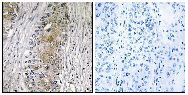
A07115 has a paraffin-section IHC image from human lung carcinoma (A07115 image caption) and lists IF/ICC applications and human, mouse, and rat reactivity (catalog: A07115); no IF image is supplied (catalog: A07115).

Real IHC data Immunohistochemistry analysis of paraffin-embedded human lung carcinoma, using SAA4 Antibody. The picture on the right is blocked with the synthesized peptide.
Anti-SAA4/Serum Amyloid A4 Antibody
Cat # A07115

The rendered card is A07115: its IHC image shows paraffin-embedded human lung carcinoma with a peptide-blocked comparison (A07115 image caption). A07115 also lists IHC, IF, and ICC applications and human, mouse, and rat reactivity; no IF image is supplied (catalog: A07115).

Which to pick: Choose A07115 for paraffin-section tissue IHC because its own image documents that preparation in human lung carcinoma; the fixative is unreported (A07115 image caption). For IF/ICC, A07115 is listed for both applications, while M07115 is a rabbit monoclonal listed for human IF/ICC; neither has an IF image in the payload (catalog: A07115, M07115). For mouse or rat samples, choose A07115 based on its listed reactivity; M07115 lists human reactivity only (catalog: A07115, M07115).

Each figure is that product's own IHC / IF validation image from its datasheet.

References

  1. UniProt Consortium. UniProt entry P35542 (SAA4_HUMAN, Serum amyloid A-4 protein).
  2. Human Protein Atlas. SAA4 tissue IHC expression (reliability: Supported).
  3. Human Protein Atlas. SAA4 subcellular location (ICC-IF): Secreted.
  4. Human Protein Atlas. SAA4 antibody validation summary (2 antibodies).
  5. Expression of serum amyloid A4 in human trophoblast-like choriocarcinoma cell lines and human first trimester/term trophoblast cells. Placenta 2014 — PMC4119475.
  6. Low SAA4 gene expression is associated with advanced HCC stage and a poor prognosis. Clinical and experimental medicine 2024 — PMC10834558.
  7. SAA4: An Underdog Within the Serum Amyloid a Superfamily?. International journal of molecular sciences 2026 — PMC13163884.
  8. PubMed PMID:1740433 — UniProt-cited evidence.
  9. PubMed PMID:7686132 — UniProt-cited evidence.
  10. PubMed PMID:1439582 — UniProt-cited evidence.