SAE1 / SUMO-activating enzyme subunit 1 · IHC design guide

Design Immunohistochemistry for SAE1

Plan SAE1 chromogenic IHC in paraffin sections using nuclear staining and high testis spermatogonia staining as benchmarks (HPA tissue IHC). This guide covers the catalog antibody’s 2–5 μg/ml IHC range (datasheet A04753-3) and scoring controls for presumed off-target binding (HPA tissue IHC).

Evidence assembled Oct 2026 · For research use; verify linked source records and product datasheet before use
Immunohistochemistry protocol sheet for SAE1 (IHC for SAE1): expected localisation Nuclear staining in tissue (HPA tissue IHC), antibody A04753-3, validated IHC image, and IHC protocol steps
Printable SAE1 IHC protocol sheet — expected localisation Nuclear staining in tissue (HPA tissue IHC), antibody A04753-3, controls and protocol steps. Open the full SAE1 IHC guide →

SAE1 Immunohistochemistry Experimental Design Guide

Expected localisation, validated protocols, controls and antibodies — the at-a-glance facts below, then the full design guide.

Must know before staining
Expected localisation Nuclear staining in tissue (HPA tissue IHC)
Staining pattern General nuclear staining across tissues (HPA tissue IHC)
Antigen retrieval EDTA pH 8.0 HIER, heat-mediated (datasheet A04753-3)
Positive control ⓘ Cerebellum+4 more · see all
Negative control ⓘ Heart muscle+3 more · see all
Important caveats
Reasons your staining may differ from the expected pattern.
Fixation Keep fixation consistent across sections (standard IHC practice; not target-specific); Matched tissue-IHC evidence does not establish the fixation claim. Validate the specimen-specific method before use. (selected-SKU IHC image A04753-3)
Caveat Presumed off-target binding can mislead scoring (HPA tissue IHC)
Regulation Rises in S phase; falls in G2 (UniProt)
Isoform / epitope Three isoforms; check epitope coverage (UniProt)
Section 1

Recommended SAE1 IHC & IF Protocols

Start with the catalog antibody’s IHC-P protocol, then compare the published glioma (PMC6659289) and colorectal biopsy (PMC10605852) workflows.

Recommended immunohistochemistry (IHC-P) protocol parameters
SampleParaffin-embedded human laryngeal carcinoma tissue; fixative not specified (datasheet A04753-3)
FixationImage fixative and duration unreported (datasheet A04753-3); verify before use.
Sectioning4–5 µm sections on charged slides (standard)
DeparaffinisationXylene, graded ethanol series to water (standard)
Antigen retrievalHeat retrieval: EDTA pH 8.0 (datasheet A04753-3); 20 min, 95–100 °C (standard)
Peroxidase block3% H2O2, 10 min, room temperature (standard)
Blocking10% goat serum (datasheet A04753-3)
Primary antibodyRabbit anti-SAE1, 2-5 μg/ml (datasheet A04753-3)
Primary incubationOvernight at 4 °C (datasheet A04753-3)
DetectionHRP-conjugated secondary, DAB chromogen (datasheet A04753-3)
CounterstainHematoxylin, blue, dehydrate and mount (standard)
Expected resultSAE1-positive staining in processes in granular layer of cerebellum (HPA tissue IHC: High). HPA tissue profile: General nuclear expression. No signal in the no-primary control.
💡Decision noteTry heat-mediated EDTA retrieval at pH 8.0 first (datasheet A04753-3); the colorectal study used pH 6.0 retrieval with a different antibody (PMC10605852).
Section 2

What Is the Expected SAE1 Staining Pattern?

SAE1 should show predominantly nuclear staining in tissue IHC, with high staining reported in testicular spermatogonia and cerebellar granular-layer processes (HPA tissue IHC: Supported; general nuclear expression). SAE1 is a nuclear protein without a transmembrane segment (UniProt Q9UBE0: subcellular location; topology). HPA also reports nucleoplasmic and cytosolic signal in ICC-IF; that observation should not be treated as a tissue IHC intensity standard (HPA ICC-IF: main locations).

What am I looking at on my slide?
Nuclear staining in spermatogonia, with variable staining across other cells.This fits HPA's general nuclear IHC profile and high spermatogonial staining (HPA tissue IHC: Supported; testis, High). Compare nuclei within the same section before calling a field negative: SAE1 expression can change through the cell cycle (UniProt Q9UBE0: protein level rises in S phase and falls in G2).
Strong cytoplasmic or membrane-like staining with little nuclear staining.Treat this as a compartment mismatch in IHC and investigate background or antibody specificity (HPA tissue IHC: general nuclear expression; UniProt Q9UBE0: nucleus, no transmembrane segment). Cytosolic ICC-IF signal is reported, so cytoplasmic staining alone does not prove an artefact (HPA ICC-IF: cytosol enhanced).
Prominent staining of cardiomyocytes or cholangiocytes.These specific cell populations were not detected in the HPA tissue IHC examples; strong signal warrants a cross-reactivity or detection-background check (HPA tissue IHC: heart muscle cardiomyocytes and liver cholangiocytes, Not detected). Do not interpret either entire tissue as an established SAE1-negative control.
Brown signal spread across stroma, lumens or the whole section without cell boundaries.This does not match the reported general nuclear profile (HPA tissue IHC). In chromogenic IHC, diffuse colour can arise from nonspecific reagent binding or detection background; examine a no-primary control and the counterstain before assigning cellular SAE1 positivity (general IHC practice).
No nuclear signal in a testis section containing spermatogonia.The result conflicts with the HPA high-staining example (HPA tissue IHC: spermatogonia, High), but one failed section does not establish absent SAE1. Confirm that the expected cells are present, then review the antibody's IHC-P conditions, retrieval, dilution and detection controls (general IHC practice).
💡Expected SAE1 appearanceCall a positive IHC result when nuclei stain in the appropriate cells, especially with strong spermatogonial signal; diffuse extracellular colour or dominant membrane-like staining is suspect (HPA tissue IHC: general nuclear expression; spermatogonia, High; UniProt Q9UBE0: no transmembrane segment).
How each factor affects the staining
Cell population and comparator choiceHPA reports High staining in spermatogonia and cerebellar granular-layer processes, Medium staining in skin granular-layer cells, and Not detected staining in cardiomyocytes (HPA tissue IHC). These labels apply to the named cells or structures, not every cell in their tissues; use morphology to select the comparison.
Cell-cycle variationSAE1 protein expression rises during S phase and drops in G2 (UniProt Q9UBE0: tissue specificity). This supports biological variation between cells; IHC staining intensity alone cannot assign an individual cell's cycle phase (general IHC interpretation).
Isoforms and N-terminal processingThree isoforms and chains beginning at residue 1 or 2 are listed (UniProt Q9UBE0: isoforms; processing). Without an antibody epitope in this payload, their effect on this assay cannot be predicted; avoid attributing an unexpected pattern to a particular isoform or processed chain.
IF/ICC: where should SAE1 appear?HPA reports enhanced nucleoplasmic and cytosolic locations in ICC-IF (HPA ICC-IF: main locations). Interpret that result on the separate IF/ICC guide page; for paraffin-section chromogenic IHC, HPA describes general nuclear expression (HPA tissue IHC). The two applications need their own controls and scoring.
Why is my staining missing, weak or wrong?
SituationLikely causeNext action
Expected spermatogonial nuclei are unstained.The assay may have insufficient detectable signal; the payload does not identify a SAE1-specific fixation or retrieval failure (HPA tissue IHC: spermatogonia, High).Verify cell identity and section quality, then check the catalog antibody's IHC-P instructions, retrieval and detection controls; change one general IHC variable at a time (general IHC practice).
Most cells show nuclear colour, including a comparison population.SAE1 has general nuclear expression and low tissue specificity at RNA level, so widespread nuclear staining is plausible (HPA tissue IHC).Score the named cell populations and relative intensity. Do not use a whole heart or liver section as a blanket negative: only cardiomyocytes and cholangiocytes were listed as Not detected (HPA tissue IHC).
Cytoplasm dominates while nuclei remain pale.The distribution conflicts with the tissue IHC profile, although cytosol is reported by ICC-IF (HPA tissue IHC: general nuclear expression; HPA ICC-IF: cytosol enhanced).Check a no-primary section for detection background and compare nuclear counterstain and cell boundaries; retain the application-specific distinction when reporting the result (general IHC practice).
Brown precipitate appears outside cells or across the section.Nonspecific binding or chromogenic detection activity can create background (general IHC practice). This appearance is not established as SAE1 staining by the HPA nuclear profile (HPA tissue IHC).Review blocking, wash and detection steps, and examine a no-primary control processed alongside the sample (general IHC practice).
Cardiomyocytes or cholangiocytes stain strongly.The observed result differs from the named HPA examples; cross-reactivity or detection background is possible, but the discrepancy alone cannot identify its cause (HPA tissue IHC: both cell types, Not detected).Confirm morphology, compare a no-primary control and check whether nuclear staining persists in a supported positive population (general IHC practice; HPA tissue IHC: spermatogonia, High).
Cerebellar granular-layer signal seems inconsistent with a nuclear call.HPA lists High staining in granular-layer processes while summarizing tissue IHC as general nuclear expression (HPA tissue IHC: cerebellum; profile).Record process and nuclear staining separately, and avoid forcing the reported process signal into a nuclear score; assess diffuse background with an appropriate control (general IHC practice).

Sample controls for SAE1 IHC & IF

🧪Run testis first and score SAE1 staining in spermatogonia (HPA: High in spermatogonia cells); use skeletal muscle myocytes as the negative tissue (HPA: Not detected in myocytes). On the positive slide, cells outside the scored spermatogonia should show only control-level background if used as internal negatives; HPA does not identify a confirmed negative cell type within testis (HPA: testis row).
Positive control tissue: Cerebellum (Processes in granular layer, HPA High)
Negative control tissue: Heart muscle (HPA Not detected)
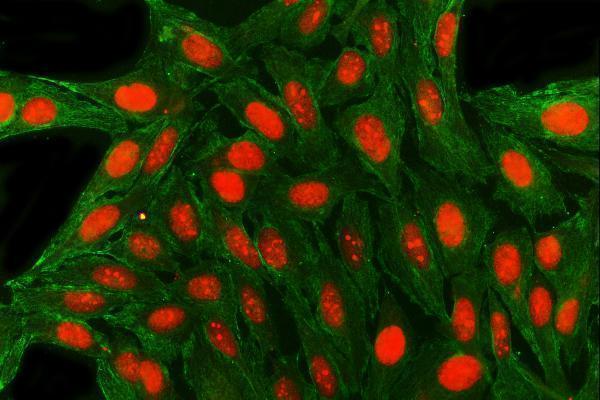
ICC-IF cell lines (HPA subcellular resource): HPA ICC-IF images show SAE1 in A-431, U2OS, HeLa BAC 5380, HeLa , NIH 3T3, with annotated localisation: Nucleoplasm (enhanced), Cytosol (enhanced) (HPA subcellular).
Technical controls: Include no-primary (secondary-only) and host-species- and isotype-matched IgG controls, plus an SAE1-knockout specimen if available as a biological specificity control (standard IHC practice). Quench endogenous peroxidase and inspect the testis section for pigment or other DAB-like background before scoring (standard chromogenic IHC practice).
⚠️Feasibility: A target-specific fixation window or fixation effect is unreported, and the selected A04753-3 paraffin-section caption does not report a fixative (caption: fixative not stated). The reported IHC-P workflow uses heat-mediated retrieval in EDTA at pH 8.0, but whether retrieval is essential is unreported (caption: EDTA retrieval). No supplied evidence establishes that frozen sections or IF are easier; crowded spermatogenic nuclei can complicate chromogenic nuclear scoring, so compare staining with the counterstain and controls (standard IHC practice).

HPA tissue IHC evidence for SAE1

Comprehensive Human Protein Atlas IHC scoring per tissue (reliability: Supported — High consistency between antibody staining and RNA expression data. Presumed off target binding observed and disregarded.). Rows are taken directly from the HPA tissue chart — click any row's HPA link to view the source.

Positive expression · recommended positive controls

TissueCell typeLevelEvidenceSource
Cerebellum Processes in granular layer High Protein (IHC) HPA →
Skin Cells in granular layer Medium Protein (IHC) HPA →
Testis Spermatogonia cells High Protein (IHC) HPA →
Adrenal gland Glandular cells Medium Protein (IHC) HPA →
Appendix Lymphoid tissue Medium Protein (IHC) HPA →

Undetected expression · recommended negative controls

TissueCell typeLevelEvidenceSource
Heart muscle Cardiomyocytes Not detected Protein (IHC) HPA →
Liver Cholangiocytes Not detected Protein (IHC) HPA →
Ovary Ovarian stroma cells Not detected Protein (IHC) HPA →
Skeletal muscle Myocytes Not detected Protein (IHC) HPA →
Section 3

Advanced SAE1 IHC Tips

Troubleshoot SAE1 staining in paraffin sections by checking retrieval, nuclear localisation, controls and cell-specific scoring before interpreting DAB signal.

How should I optimize retrieval when SAE1 staining is weak in paraffin sections?
Begin with heat-mediated retrieval in EDTA pH 8.0 for SAE1 paraffin IHC (datasheet A04753-3). Keep the antibody at the caption's 2 μg/ml and overnight incubation at 4°C while comparing adjacent sections, so retrieval is the main variable (datasheet A04753-3; standard IHC practice). If signal remains weak, compare shorter and longer heating, such as 10 and 20 minutes, and inspect tissue morphology and background alongside nuclear staining (standard IHC practice; HPA tissue IHC: general nuclear expression). Record cooling and wash conditions for each run, because inconsistent handling can obscure the effect of retrieval (standard IHC practice).
Could fixation explain variable SAE1 staining between paraffin specimens?
SAE1-specific fixation sensitivity is unknown because the selected paraffin-section caption does not report a fixative (datasheet A04753-3). Record each specimen's fixative and fixation duration, then compare matched sections using EDTA pH 8.0 retrieval and 2 μg/ml primary antibody to limit processing differences (datasheet A04753-3; standard IHC practice). In a controlled pilot, compare routinely fixed specimens at documented time points, such as 6 and 24 hours, while scoring morphology and nuclear signal (standard IHC practice; HPA tissue IHC: general nuclear expression). Do not treat a staining difference as an established SAE1 fixation effect until matched material and controls reproduce it (standard IHC practice).
How should I assess nuclear and cytoplasmic SAE1 staining?
Score nuclear DAB first, because SAE1 is annotated in the nucleus and tissue IHC shows general nuclear expression (UniProt Q9UBE0: nucleus; HPA tissue IHC: general nuclear expression). Record cytoplasmic staining separately: cellular IF data place SAE1 in the nucleoplasm and cytosol, but that observation alone does not validate every cytoplasmic IHC pattern (HPA subcellular: nucleoplasm and cytosol; standard IHC practice). Compare both compartments with the no-primary control and with intact, well-preserved cells on the same section (standard IHC practice). If nuclear signal disappears while diffuse cytoplasmic DAB rises, recheck EDTA pH 8.0 retrieval and detection background before assigning a biological shift (datasheet A04753-3; standard IHC practice).
Can this IHC stain distinguish SAE1 isoforms or processing states?
Do not assign an isoform from this stain without an epitope map: SAE1 has 3 annotated isoforms, and the supplied caption does not identify the antibody's binding region (UniProt Q9UBE0: isoforms; datasheet A04753-3). The record also describes chains beginning at residue 1 or 2, plus modifications at residues 12 and 198 (UniProt Q9UBE0: processing and modified residues). Check whether the documented immunogen overlaps variable sequence or the processed N terminus before proposing isoform selectivity (UniProt Q9UBE0: isoforms and processing; standard IHC practice). If compartmental staining differs, seek epitope documentation and an independent antibody or perturbation control rather than attributing the pattern to a particular isoform (standard IHC practice).
How can IF help resolve an ambiguous SAE1 IHC pattern?
On the separate IF/ICC workflow, co-stain with a validated marker of the expected cell type; spermatogonia are one high-staining population reported in tissue IHC (HPA tissue IHC: high in spermatogonia; standard IF practice). Choose a far-red SAE1 fluorophore when tissue autofluorescence complicates shorter wavelengths, and inspect single-channel and secondary-only controls (standard IF practice). SAE1 lacks a transmembrane segment and is reported in the nucleoplasm and cytosol, so permeabilise the plasma and nuclear membranes when assessing access to a nuclear epitope (UniProt Q9UBE0: topology; HPA subcellular: nucleoplasm and cytosol; standard IF practice). A brief 0.1% detergent trial can test access, but optimise it against cell morphology and background rather than assuming the IHC conditions transfer to IF (standard IF practice).
What should I check when SAE1 DAB staining is diffuse or widespread?
Run a no-primary section to reveal detection-system background, and inspect pigment and tissue edges before changing antibody concentration (standard IHC practice). The selected assay used 10% goat serum blocking, 2 μg/ml primary antibody, a peroxidase-linked secondary and DAB; these are starting conditions for this catalog antibody (datasheet A04753-3). Include a peroxidase-blocking step and verify its effect with the no-primary control, since endogenous enzyme can produce DAB signal (standard IHC practice). If background persists, titrate the primary around the documented concentration and compare nuclear signal against off-target staining, which the tissue resource reports for its antibody assessment (datasheet A04753-3; HPA tissue IHC: presumed off-target binding).
How should I quantify SAE1 staining across paraffin sections? ⚠ ANSWER MARKED FOR VERIFICATION
Define the target cell population and score intact nuclei in comparable regions, consistent with SAE1's nuclear annotation and general nuclear tissue pattern (UniProt Q9UBE0: nucleus; HPA tissue IHC: general nuclear expression). Report both the percentage of positive cells and a nuclear H-score, calculated as 1×% weak + 2×% moderate + 3×% strong, with a range of 0–300 (standard IHC practice). Use the total number of viable target-cell nuclei in each region as the denominator, and record cytoplasmic staining separately (standard IHC practice). Keep retrieval, DAB development and scoring thresholds consistent across sections; document the number of regions and cells counted before comparing groups (standard IHC practice).
How can I distinguish credible SAE1 staining from artefact?
Credible staining should include a reproducible nuclear component in intact cells, while cytosolic signal warrants separate evaluation because cellular IF also reports SAE1 there (UniProt Q9UBE0: nucleus; HPA tissue IHC: general nuclear expression; HPA subcellular: cytosol). Compare cell identities with reference patterns: spermatogonia stain highly, whereas cardiomyocytes were not detected in the supplied tissue data (HPA tissue IHC: spermatogonia and cardiomyocytes). Exclude edge staining, necrotic areas and DAB signal persisting without primary antibody; check endogenous peroxidase blocking when these appear (standard IHC practice). Finally, staining measures detectable SAE1 antigen and cannot by itself establish SUMO E1 activity or exclude off-target binding (UniProt Q9UBE0: heterodimer function; HPA tissue IHC: presumed off-target binding).
Boster reagents

Best SAE1 / SUMO-activating enzyme subunit 1 IHC Antibodies

The IHC-validated SAE1 antibody has paraffin-section images from human, mouse and rat tissues (catalog IHC captions); IF images cover human, mouse and rat samples (catalog IF captions).

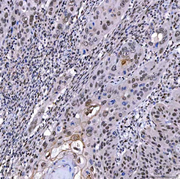
Real IHC data IHC analysis of SAE1 using anti-SAE1 antibody (A04753-3). SAE1 was detected in a paraffin-embedded section of human laryngeal carcinoma tissue. Heat mediated antigen retrieval was performed in EDTA buffer (pH 8.0, epitope retrieval solution). The tissue section was blocked with 10% goat serum. The tissue section was then incubated with 2 μg/ml rabbit anti-SAE1 Antibody (A04753-3) overnight at 4°C. Peroxidase Conjugated Goat Anti-rabbit IgG was used as secondary antibody and incubated for 30 minutes at 37°C. The tissue section was developed using HRP Conjugated Rabbit IgG Super Vision Assay Kit (Catalog # SV0002) with DAB as the chromogen.
Anti-SAE1 Antibody ®
Cat # A04753-3

The rendered card is A04753-3, with IHC images from human laryngeal carcinoma and duodenum, mouse testis and rat testis paraffin sections (A04753-3 IHC captions). It also has IF images from human cells and esophageal squamous carcinoma, mouse testis and rat testis (A04753-3 IF captions).

Which to pick: Choose A04753-3 for tissue IHC: its own paraffin-section captions document staining at 2 μg/ml after heat retrieval in EDTA, pH 8.0 (A04753-3 IHC captions); the fixative is unreported (A04753-3 IHC captions). For IF/ICC, A04753-3 has image evidence, while rabbit monoclonal M04753-2 lists ICC/IF for human and rat but has no IF image in this payload (A04753-3 IF captions; M04753-2 catalog applications and reactivity). For human, mouse and rat work, A04753-3 lists all three species for IHC and IF; M04753-2 lists human and rat and does not list IHC (catalog applications and reactivity).

Each figure is that product's own IHC / IF validation image from its datasheet.

References

  1. UniProt Consortium. UniProt entry Q9UBE0 (SAE1_HUMAN, SUMO-activating enzyme subunit 1).
  2. Human Protein Atlas. SAE1 tissue IHC expression (reliability: Supported).
  3. Human Protein Atlas. SAE1 subcellular location (ICC-IF): Mainly localized to the nucleoplasm and cytosol..
  4. Human Protein Atlas. SAE1 antibody validation summary (2 antibodies).
  5. SAE1 May Play a Pro-Carcinogenic Role in Pancreatic Adenocarcinoma: A Comprehensive Study Integrating Multiple Pieces of Evidence. IET systems biology 2025 — PMC12041129.
  6. SAE1 promotes human glioma progression through activating AKT SUMOylation-mediated signaling pathways. Cell communication and signaling : CCS 2019 — PMC6659289.
  7. Upregulated SAE1 Drives Tumorigenesis and Is Associated with Poor Clinical Outcomes in Breast Cancer. The breast journal 2024 — PMC11227943.
  8. SUMO-Activating Enzyme Subunit 1 Is Associated with Poor Prognosis, Tumor Progression, and Radio-Resistance in Colorectal Cancer. Current issues in molecular biology 2023 — PMC10605852.
  9. PubMed PMID:9920803 — UniProt-cited evidence.
  10. PubMed PMID:10217437 — UniProt-cited evidence.
  11. PubMed PMID:10187858 — UniProt-cited evidence.